BASIGIN is a receptor essential for erythrocyte invasion by Plasmodium falciparum (original) (raw)
. Author manuscript; available in PMC: 2012 Jun 22.
Published in final edited form as: Nature. 2011 Nov 9;480(7378):534–537. doi: 10.1038/nature10606
Abstract
Erythrocyte invasion by Plasmodium falciparum is central to the pathogenesis of malaria. Invasion requires a series of extracellular recognition events between erythrocyte receptors and ligands on the merozoite, the invasive form of the parasite. None of the few known receptor-ligand interactions involved1-4 are required in all parasite strains suggesting that the parasite is able to access multiple redundant invasion pathways5. Here, we show that we have identified a receptor-ligand pair that is essential for erythrocyte invasion in all tested P. falciparum strains. By systematically screening a library of erythrocyte proteins, we have found that the Ok blood group antigen, BASIGIN, is a receptor for PfRh5, a parasite ligand that is essential for blood stage growth6. Erythrocyte invasion was potently inhibited by soluble BASIGIN or by BASIGIN knockdown, and invasion could be completely blocked using low concentrations of anti-BASIGIN antibodies; importantly, these effects were observed across all laboratory-adapted and field strains tested. Furthermore, Ok(a−) erythrocytes, which express a BASIGIN variant that has a weaker binding affinity for PfRh5, exhibited reduced invasion efficiencies. Our discovery of a cross-strain dependency on a single extracellular receptor-ligand pair for erythrocyte invasion by P. falciparum provides a focus for novel anti-malarial therapies.
Amongst the many P. falciparum merozoite proteins that are believed to have a role in erythrocyte invasion, most attention has focussed on two major parasite protein families: the EBAs and Rhs7. Although erythrocyte receptors have been identified for some of them (members of the glycophorin family are receptors for three EBAs1-3and Complement receptor 1 (CD35) has recently been identified as a receptor for PfRh44) none of these receptor-ligand pairs are essential in all parasite strains tested. PfRh5 is unique amongst the _EBA_s and _Rh_s because it cannot be deleted in any P. falciparum strain and is therefore apparently essential for parasite growth in blood stage culture5,6. Both native and recombinant PfRh5 have been previously shown to bind erythrocytes through an unknown glycosylated receptor that is resistant to chymotrypsin, trypsin and neuraminidase treatment6,8,9.
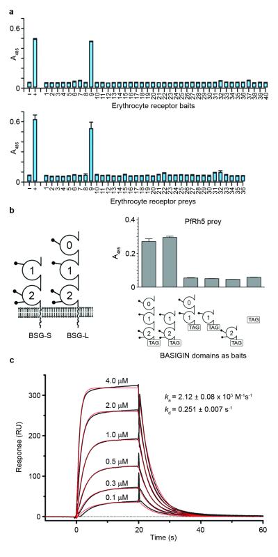
To identify an erythrocyte receptor for PfRh5, we employed a systematic screening approach by first compiling a library of abundant cell surface and secreted proteins expressed by human erythrocytes based on published proteomics data10. Proteins for which the entire ectodomain was expected to be expressed as a soluble recombinant protein were selected (Supplementary Table 1), and expressed by mammalian cells (Supplementary Fig. 1). The 40 proteins within the erythrocyte ectodomain protein library were then systematically screened using the AVEXIS assay11 for interactions with a recombinant PfRh5 protein, also produced by mammalian cells. The AVEXIS assay (AVidity-based EXtracellular Interaction Screen) is designed to detect direct low affinity protein interactions between ectodomain fragments expressed as either biotin-tagged baits or highly avid pentameric ß-lactamase-tagged preys12,13. The PfRh5 prey interacted with a single erythrocyte receptor bait (Fig. 1a, top panel) corresponding to the Ok blood group antigen, BASIGIN (BSG, also known as CD147, EMMPRIN and M614). The same single interaction was identified in the reciprocal bait-prey orientation (Fig. 1a, lower panel).
Figure 1. BSG is an erythrocyte receptor for PfRh5.
(a) PfRh5 was screened as either a prey (top panel) or a bait (bottom panel) against an erythrocyte receptor protein library using AVEXIS. BSG (protein 9) was identified as a receptor for PfRh5 in both bait-prey orientations. (b) Domain structure of the BSG isoforms (left); lollipops represent potential N-linked glycosylation sites. BSG regions were expressed as baits and used to map the PfRh5 binding site to the two membrane-proximal domains. Bar charts show mean ± SEM; n = 3. (c) Biophysical analysis of the PfRh5-BSG-S interaction using SPR. The indicated concentrations of purified PfRh5 were injected over immobilised BSG, and biophysical parameters derived from a 1:1 binding model (red line).
BSG is a member of the immunoglobulin superfamily (IgSF) and has been implicated in many biological functions including embryo implantation, spermatogenesis15 and retinal development16. BSG exists in both long (three IgSF domains, BSG-L) and short (two IgSF domains, BSG-S) splice isoforms (Fig. 1b) and although BSG-L was used in the screen, BSG-S is thought to be the major isoform expressed on erythrocytes. Binding experiments using domain deletions established that PfRh5 could interact with BSG-S and this required both domains since neither of the two BSG-S IgSF domains were individually able to bind PfRh5 (Fig. 1b, Supplementary Fig. 2). We showed that PfRh5 directly interacted with BSG-S and BSG-L using purified proteins and surface plasmon resonance (SPR). Both kinetic (Fig. 1c) and equilibrium (Supplementary Fig. 3) binding parameters for the interaction were derived using a 1:1 binding model and were in excellent agreement (Supplementary Table 2). These parameters are typical of extracellular protein interactions measured using this technique17. Removal of glycans from BSG either by mutating all predicted glycosylation motifs or by enzymatic treatment did not affect PfRh5 binding (Supplementary Fig. 4), suggesting the PfRh5 binding site is solely located in the BSG protein core. BSG is also known to be resistant to trypsin and chymotrypsin treatment18 consistent with previous PfRh5-erythocyte binding studies6,8,9.
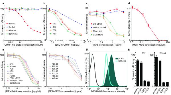
To determine whether the PfRh5-BSG interaction was required for invasion, we added purified pentamerised soluble BSG-S into invasion assays to specifically compete with the membrane-bound receptor. We found that BSG-S strongly inhibited invasion in a dose-dependent manner relative to controls which included each of the two non-binding BSG-S IgSF domains added individually (Fig. 2a). Strong inhibition was also observed across multiple strains (Fig. 2b) or when soluble BSG-L was added (Supplementary Fig. 5) although this was slightly weaker for the 3D7 strain. Soluble forms of BSG consisting of the extracellular regions are known to have biological effects such as upregulation of matrix metalloproteases19. To rule out an indirect effect of exogenous BSG on invasion, we added to invasion assays two independent purified anti-BSG monoclonal antibodies (MEM-M6/6 and TRA-1-85) which could both block the PfRh5-BSG interaction in vitro (data not shown). These high affinity reagents gave a potent invasion blocking effect that was saturable at very low antibody concentrations (IC50 ~ 0.5 μg/ml), consistent with binding and occluding a specific surface receptor of typical abundance (~104 to 106 molecules per cell20) (Fig. 2c). Preadsorbtion of the MEM-M6/6 antibody with soluble monomeric BSG specifically relieved the inhibition, ruling out any indirect effect of the antibody on non-BSG targets; furthermore, MEM-M6/6 did not affect intra-erythrocytic P. falciparum development (Supplementary Fig. 6). Invasion was quantified using flow cytometry and a fluorescent DNA dye to stain parasites21. Using this assay, apparent invasion could not be eliminated, with efficiencies reduced to a maximum of 80-90%, even at much higher concentrations of antibody (up to 1.5 mg/ml of MEM-M6/6 – data not shown); however, direct observation of parasites using Giemsa-stained thin smears revealed that this residual staining in cytometry assays was due to extracellular parasites and debris in the culture. Using microscopy-based assays, we found that MEM-M6/6 concentrations of 10 μg/ml or more was sufficient to prevent all detectable invasion (Fig. 2d).
Figure 2. Soluble BSG, anti-BSG antibodies and BSG knockdown potently block erythrocyte invasion.
(a) Erythrocyte invasion was inhibited by purified pentamerised BSG-S-Cd4d3+4-COMP-His ectodomains but not by the two non-binding BSG-S domains added individually or Cd4d3+4-COMP-His (control); strain = Dd2. (b) Cross-strain inhibition of invasion using pentamerised BSG-S. (c) Anti-BSG monoclonal antibodies, TRA-1-85 and MEM-M6/6, potently inhibited invasion of erythrocytes; strain = 3D7. (d) MEM-M6/6 concentrations ≥10 μg/ml prevented all detectable invasion by microscopic observation of cultures; strain = 3D7. (e, f) MEM-M6/6 inhibited invasion of synchronised P. falciparum culture-adapted lines (e) and unsynchronised field isolates (f). (g) Cell surface BSG is reduced in erythrocytes differentiated from hematopoietic stem cells transduced with lentiviruses containing shRNA targeting BSG (light blue line) relative to a control virus (pLKO, shaded); black line represents secondary antibody alone. (h) 3D7 and W2mef invasion was inhibited in BSG knockdown erythrocytes. A and B are replicates. Invasion efficiencies are mean ± SEM, n = 3.
P. falciparum isolates can vary widely in their ability to invade erythrocytes treated with different receptor-modifying enzymes such as trypsin, chymotrypsin and neuraminidase, revealing differential dependencies on erythrocyte receptors for invasion. To determine if BSG was a critical invasion receptor across P. falciparum lines that use different invasion pathways, we tested the ability of MEM-M6/6 to block erythrocyte invasion on nine culture-adapted strains representing seven different PfRh5 sequence variants (Supplementary Table 3). We observed that the invasion of all lines was potently inhibited by MEM-M6/6 (Fig. 2e). To show that the dependency on BSG was not an unusual feature of culture-adapted lines, we also tested six freshly-isolated P. falciparum strains from Senegal22 and again observed a potent inhibitory effect (Fig. 2f). Assays with the field isolates were carried out with unsynchronised parasites, decreasing the overall inhibitory effect because not all parasites had reinvaded over the course of the assay. All six Senegal isolates, however, were inhibited by MEM-M6/6 to the same extent as an unsynchronised culture-adapted line, W2mef, tested at the same time. This demonstrated that freshly-isolated field strains have the same dependency on BSG as laboratory-adapted lines (Fig. 2f).
To independently confirm the essentiality of BSG as a P. falciparum invasion receptor, we used a genetic approach by differentiating erythrocytes from hematopoietic stem cells transduced with lentiviruses containing either an shRNA targeting BSG or a scrambled shRNA control (pLKO). _BSG_-targeted erythrocytes showed a reproducible knockdown to approximately 50 to 60% of cell surface BSG levels relative to the pLKO control (Fig. 2g) and expressed markers indicative of complete erythrocyte maturation (Supplementary Fig. 7). The invasion of both the 3D7 and W2mef P. falciparum strains into BSG-knockdown erythrocytes was significantly reduced compared to the control (18% versus 94% for 3D7 and 14% versus 103% for W2mef, Fig. 2h). By contrast, previous knockdown of GYPA, the major surface sialoglycoprotein, significantly inhibited the W2mef but not the 3D7 strain23. The inhibition of erythrocyte invasion by multiple P. falciparum strains using soluble BSG, anti-BSG monoclonal antibodies, or knockdown of BSG surface expression suggests that BSG is a critical host receptor for P. falciparum invasion.
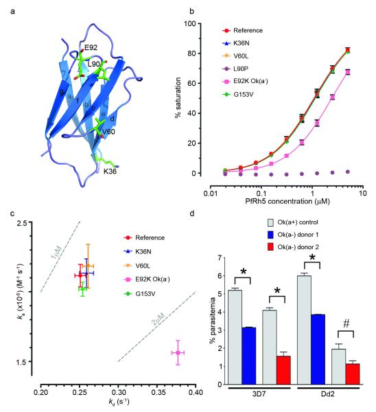
Malaria is thought to have been a strong selective pressure in human evolutionary history and given the apparently essential roles of PfRh5 and BSG in P. falciparum invasion we sought to determine if any human populations contained genetic variants in BSG that might affect PfRh5 binding and invasion. Five nonsynonymous single nucleotide polymorphisms (SNPs) have been described within the BSG-S IgSF domains (Supplementary Table 4, Fig. 3a). These variants were expressed and the biophysical PfRh5 binding parameters determined using SPR. Equilibrium measurements showed that two variants had lower binding affinity compared to the BSG reference sequence: L90P and E92K (Fig. 3b, Supplementary Table 2). L90P did not interact with PfRh5 and binding profiles of several anti-BSG monoclonal antibodies suggested local misfolding of the membrane-distal IgSF domain (Supplementary Fig. 8). No verification or population frequency data for this SNP are currently available preventing further biological interpretation of this variant. E92K had a two-fold lower affinity for PfRh5 (Fig. 3b) and a comparative kinetic analysis demonstrated that this was due to both a slower association and a faster dissociation rate (Fig. 3c, Supplementary Table 2). The E92 residue is solvent exposed and located within the loop connecting the F-G ß-strands close to the glycan-free GFC ß-sheet, consistent with a possible PfRh5 binding interface (Fig. 3a). E92K is the variant responsible for the Ok(a−) blood group, which has been described in eight Japanese families14. Ok(a−) erythrocytes from two unrelated donors showed reduced invasion with both 3D7 and Dd2 P. falciparum strains relative to Ok(a+) controls (Fig. 3d, Supplementary Fig. 9), correlating with the reduced affinity of the Ok(a−) variant for PfRh5. The extreme rarity and restriction of the Ok(a−) blood group to Japanese individuals suggest that this specific allele has not played a major role in conferring resistance to malaria. It is possible that other BSG polymorphisms, as yet unknown, have evolved in some malaria-exposed populations as a mechanism of resistance to P. falciparum. The search for functional polymorphisms of BSG needs to go beyond gene coding regions as the results of our knockdown experiments suggest that expression levels of BSG at the erythrocyte surface influence the ability of the parasite to invade. The Duffy variant which confers resistance to P. vivax is also a non-coding regulatory polymorphism that suppresses expression of the invasion receptor by erythrocytes. Our ability to address this problem is currently limited by the lack of data on genome variation among the many different ethnic groups that are exposed to P. falciparum malaria, but will be greatly enhanced by the 1000 Genomes Project, MalariaGEN and other genetic studies that are now in progress in Africa and other malaria-endemic regions of the world24-26. Inter-population comparisons of haplotype length and frequency provide a potentially powerful way of addressing this problem27, and there is preliminary evidence that a region of chromosome 19 encompassing BSG and several neighbouring genes has undergone recent positive selection in West Africa, but a considerable amount of further work is needed to determine whether this is causally related to the role of BSG as a malaria invasion receptor (MalariaGEN consortium, unpublished data).
Figure 3. The Ok(a−) BSG variant has reduced binding affinity for PfRh5 and Ok(a−) erythrocytes have reduced merozoite invasion frequencies.
(a) Schematic of the membrane distal IgSF domain of BSG-S showing the location of naturally-occurring variants. (b) Equilibrium binding isotherms of PfRh5 binding to BSG-S variants. (c) Association (_k_a) and dissociation (_k_d) rate constants of PfRh5 binding to BSG-S and variants. Means ± SEM; n = 3. (d) Invasion of 3D7 and Dd2 strains in Ok(a−) blood cells are reduced relative to the Ok(a+) control. Mean ± SEM, n = 3; *, P ≤ 0.0003; #, P = 0.0349, unpaired one-tailed t test. A repeat is shown in Supplementary Fig. 9.
In summary, we have applied a systematic protein interaction screening approach (AVEXIS) to identify BSG as an erythrocyte receptor for PfRh5. Importantly, we were able to prevent all detectable erythrocyte invasion by every P. falciparum strain that we tested using only modest concentrations of anti-BSG antibodies. These observations, coupled with the inability to delete PfRh56, lead us to conclude that the interaction between BSG and PfRh5 is essential for parasite entry, and may perform a fundamentally different function to the other EBA and Rh proteins, which are involved in redundant, partially overlapping invasion pathways. The dependence on a single receptor-ligand pair across many P. falciparum strains may provide new possibilities for therapeutic intervention.
Methods summary
Recombinant protein production and interaction screening
Protein production, purification, AVEXIS assays and SPR were performed essentially as described11 except the type II proteins which were expressed with an N-terminal Cd4d3+4-biotin tag and a mouse antibody signal peptide. PfRh5 was expressed as above except that a non-endogenous signal peptide was added and the threonines in potential N-linked glycan sequons were mutated to alanine to prevent inappropriate glycosylation. All constructs were chemically synthesized and codon optimised for mammalian expression (Geneart AG). Purified pentameric proteins used in invasion assays were made by replacing the β-lactamase reporter in the prey plasmid with a hexa-his tag, purified and buffer exchanged into RPMI prior to use. BSG variants were produced by site directed mutagenesis.
P. falciparum culture, lentiviral transduction and invasion assays
P. falciparum parasite strains were routinely cultured in human O+ erythrocytes at 5% hematocrit. Use of erythrocytes from human donors for P. falciparum culture was approved by the NHS Cambridgeshire 4 Research Ethics Committee. Ok(a−) and control Ok(a+) blood was obtained from donors in Japan with informed consent, shipped on ice and experiments performed within 72 hours. Invasion assays were carried out as described previously21 using the two-colour assay for the Ok(a−) experiment. Lentiviral transduction of HSCs was performed as previously described23.
Methods
Recombinant protein production
Proteins selected for expression included all type I, type II, GPI-linked receptors and secreted proteins. Some multipass transmembrane proteins were also included where there was an extracellular N-terminus preceded by a signal peptide (Supplementary Table 1). Individual domains of human BSG were produced by identifying domain boundaries using the structure of the BSG extracellular region28,29 and amplifying these regions using primers with flanking NotI and AscI restriction enzyme sites to facilitate cloning. BSG-d0+1 and BSG-d1 C-terminal domain truncation boundary amino acid sequence = HGPP. BSG-d2 was cloned into the same vector as PfRh5 to add an exogenous signal peptide required for protein secretion and encompassed the sequence between PPRV.. to ..RSHL. Glycosylation sites were removed in BSG by mutating codons encoding all three asparagines in glycosylation motifs to aspartic acid. To remove N-linked glycans from soluble recombinant BSG, 500 units of PNGaseF (New England Biolabs) were added to 10 μl of a spent tissue culture supernatant and incubated for 15 minutes at 37 °C. Sialic acid residues were removed by adding 1.6 milli-units of Vibrio cholerae neuraminidase (Sigma) to 10 μl of a spent tissue culture supernatant and incubated for 15 minutes at 37 °C.
Interaction screening by AVEXIS
For the AVEXIS assay, bait and prey protein preparations were normalised to activities that have been previously shown to detect transient interactions (monomeric half-lives less than 0.1 second) with a low false positive rate11. Biotinylated baits dialysed against HBS were immobilised in the wells of a streptavidin-coated 96-well microtitre plate (NUNC). Normalised preys were added, incubated for 2 hours at room temperature, washed 3x HBS/0.1% Tween-20, 1x HBS. 125 μg/ml of nitrocefin was added, and absorbance values measured at 485 nm on a Pherastar plus (BMG laboratories). Controls were essentially as described12 and included: the Cd4d3+4 tag alone as a negative control bait, a biotinylated anti-Cd4 (anti-prey) antibody as a prey capture positive control. A positive control interaction consisting of the rat Cd200 bait detected using the rat Cd200R prey used at the threshold level and both 1:10 and 1:100 dilutions was included on each plate. The negative (−) and positive (+) control interactions shown in Figure 1a are the rat Cd200R prey used at the screening threshold probed against the Cd4d3+4 (−) or rat Cd200 (+) baits.
P. falciparum culture, characterisation and invasion assays
All P. falciparum parasite strains were routinely cultured in human O+ erythrocytes at 5% hematocrit in complete medium (RPMI-1640 containing 10% human serum), under an atmosphere of 1% O2, 3% CO2, and 96% N2. To confirm their identity, laboratory-adapted strains were genotyped by PCR within polymorphic regions of the msp1 and msp2 genes30. Parasite cultures were synchronized in early stages with 5% (w/v) D-sorbitol (Sigma). Use of erythrocytes from human donors for P. falciparum culture was approved by the NHS Cambridgeshire 4 Research Ethics Committee. Ok(a−) blood was obtained from donors in Japan with informed consent, and shipped on ice. For each sample, a control Ok(a+) sample was collected at the same time under identical conditions. All experiments were performed within 72 hours of collection.
Invasion assays were carried out in round-bottom 96-well plates, with a culture volume of 100 μL per well at a hematocrit of 2%. Parasites in trophozoite stage were mixed with pentamerized BSG-S-Cd4d3+4-COMP-His ectodomains or with anti-BSG monoclonal antibodies and incubated in the plates for 24 hours at 37 °C inside a static incubator culture chamber (VWR), gassed with 1% O2, 3% CO2, and 96% N2. At the end of the incubation period, red blood cells (RBC) were harvested and parasitized RBC (pRBC) were stained with 2 μM Hoechst 33342 (Invitrogen), as described previously21. Invasion assays using Ok(a−) blood and control Ok(a+) blood were carried out following the two-colour flow cytometric assay described in21. Briefly, Ok(a−) blood and control Ok(a+) blood were labelled with 10 μM DDAO-SE (Invitrogen). RBC were resuspended to 2% hematocrit, mixed with pRBC (ring stage) and incubated in 96-well plates for 48 hours as described above. At the end of the incubation period, RBC were harvested and pRBC were stained with 2 μM Hoechst 33342. Standard blood smear microscopy was performed to determine parasitemia. Briefly, a small aliquot of the culture was smeared on a glass slide, fixed with 100% methanol and stained with Field’s Stain (Pro-Lab Diagnostics). Parasitemia was determined by counting the number of parasitized red blood cells (pRBC) per 2,000 total red blood cells (RBC) examined by oil immersion with a Leica DME microscope (Leica Microsystems). All parasitemia represented were the average of three replicates. Lentiviral-delivered shRNA sequences were: BSG; TRC clone ID (TRCN0000006736) hairpin sequence: GAAGTCGTCAGAACACATCAACTCGAGTTGATGTGTTCTGACGACTTC, pLKO scrambled control; (Addgene plasmid 1864) hairpin sequence: CCTAAGGTTAAGTCGCCCTCGCTCGAGCGAGGGCGACTTAACCTTAGG; loop region indicated in bold. Detailed Standard Operating Procedures for all invasion assays are available at http://www.sanger.ac.uk/research/
Flow cytometry
Stained samples were examined with a 355 nm UV laser (20 mW) and a 633 nm red laser (17 mW) on a BD LSRII flow cytometer (BD Biosciences). Hoechst 33342 (Invitrogen) was excited using the UV laser and detected with a 450/50 filter, while DDAO-SE (Invitrogen) was excited using the red laser and detected with a 660/20 filter. BD FACS Diva (BD Biosciences) was used to collect 100,000 events for each sample. FSC and SSC voltages of 423 and 198, respectively, and a threshold of 2,000 on FSC were applied to gate the erythrocyte population. The data collected were further analyzed with FlowJo (Tree Star). All experiments were carried out in triplicate. GraphPad Prism (GraphPad Software) was used to plot the generated parasitemia data.
PfRh5 cloning and sequencing
Total RNA was extracted from 3D7 and FCR3 schizonts using the QIAamp RNA Blood Mini Kit (Qiagen). Isolated RNA was treated with TURBO™ DNase (Ambion) and reverse transcribed to cDNA using the High-Capacity™ cDNA Archive Kit (Applied Biosystems) following the manufacturer’s instructions. A 10 μl aliquot of cDNA was used as a template in a standard PCR reaction, using the primers Rh5-F (5′-ATGATAAGAATAAAAAAAAAATTAATTTTGACCATT-3′) and Rh5-R (5′-TCATTGTGTAAGTGGTTTATTTTTTTTATATGTTTG-3′). Amplified fragments were subcloned into pCR2.1-TOPO, using the TOPO TA Cloning Kit (Invitrogen) and three clones from each strain were sequenced and analysed.
Antibodies
Antibodies were obtained from the following suppliers: anti-rat Cd4d3+4 (OX68) (AbD Serotec), anti-CD59 (AbD Serotec), mouse IgG1 control (Abcam). Anti-BSG monoclonal antibodies used were: 8J251 (Lifespan Biosciences), MEM-M6/1 (Abcam) and TRA-1-85 (R&D systems). MEM-M6/6 was provided as an ascitic fluid and was a generous gift of Professor Vaclav Horejsi (Institute of Molecular Genetics, Czech Republic); the antibody was purified using a HiTrap protein G column (GE Healthcare) as described31 and exchanged into RPMI.
Surface plasmon resonance
Surface plasmon resonance studies were performed using a Biacore T100 instrument. Briefly, biotinylated bait proteins were captured on a streptavidin-coated sensor chip (Biacore, GE Healthcare). Approximately 150RU of the negative control bait (biotinylated rat Cd4d3+4) were immobilised in the flow cell used as a reference and approximate molar equivalents of the query protein immobilised in other flow cells. Purified analyte proteins were separated by gel filtration just prior to use in SPR experiments to remove small amounts of protein aggregates which are known to influence kinetic binding measurements32. Increasing concentrations of purified proteins were injected at high flow rates (100 μl/min) to minimise rebinding effects for kinetic studies or at 10 μl/min for equilibrium analysis. Although essentially all the bound PfRh5 dissociated during the wash out phase (see Fig. 1c), the surface was “regenerated” with a pulse of 2M NaCl at the end of each cycle. Duplicate injections of the same concentration in each experiment were superimposable demonstrating no loss of activity after regenerating the surface. Both kinetic and equilibrium binding data were analysed in the manufacturer’s Biacore T100 evaluation software (Biacore). Equilibrium binding measurements were taken once equilibrium had been reached using reference-subtracted sensorgrams. Both the kinetic and equilibrium binding studies involving BSG-S and variants were performed three times using independent protein preparations of both PfRh5 and the BSG proteins, and once for BSG-L and its variants. All experiments were performed at 37 °C.
Enzyme-linked immunosorbant assay (ELISA)
Biotinylated ectodomains were immobilized on streptavidin-coated plates (Nunc) for one hour before being incubated for 90 minutes with 10 μg/ml primary antibody. The plates were washed in HBS/0.1% Tween-20 (HBST) before incubation with an appropriate secondary antibody conjugated to alkaline phosphatase (Sigma). Plates were washed 3x HBST and 1x HBS before adding 100 μl _p_-nitrophenyl phosphate (Sigma 104 alkaline phosphatase substrate) at 1 mg/ml. Optical density measurements were taken at 405 nm on a Pherastar plus (BMG laboratories). The whole procedure was performed at room temperature.
Supplementary Material
1
2
Acknowledgements
We are deeply grateful to the Ok(a−) blood donors. We thank Vaclav Horejsi for monoclonal antibodies and Damien Ahr for technical assistance. This work was supported by the Wellcome Trust grant numbers [077108] (G.J.W.) and [089084] (J.C.R.) and National Institutes of Health [R01AI057919] (M.T.D.). A.K.B. is supported by a Center for Disease Control grant R36 CK000119-01 and an Epidemiology of Infectious Disease and Biodefense Training Grant 2T32 AI007535-12.
Footnotes
Supplementary information is linked to the online version of the paper at www.nature.com/nature.
Author contributions C.C. compiled the erythrocyte protein library and identified the PfRh5-BSG interaction. L.Y.B. led the P. falciparum functional validation, with support from M.T. S.J.B. performed the biochemical and biophysical characterization of the interaction. A.K.B. performed the lentiviral knock-down and parasite invasion experiments under the direction of M.T.D. M.U. provided the Ok(a−) blood samples and matching controls. O.N. and S.M. supervised the collection and culturing of field strains. D.P.K. performed genetic analysis on the BSG and PfRh5 loci. G.J.W. and J.C.R. conceived and supervised the project, and wrote the manuscript.
Competing financial interests: C.C., L.Y.B., S.J.B., J.C.R. and G.J.W. are named on a patent application relating to this work.
References
- 1.Maier AG, et al. Plasmodium falciparum erythrocyte invasion through glycophorin C and selection for Gerbich negativity in human populations. Nature Med. 2003;9:87–92. doi: 10.1038/nm807. [DOI] [PMC free article] [PubMed] [Google Scholar]
- 2.Mayer DC, et al. Glycophorin B is the erythrocyte receptor of Plasmodium falciparum erythrocyte-binding ligand, EBL-1. Proc. Natl Acad. Sci. USA. 2009;106:5348–5352. doi: 10.1073/pnas.0900878106. [DOI] [PMC free article] [PubMed] [Google Scholar]
- 3.Sim BK, et al. Receptor and ligand domains for invasion of erythrocytes by Plasmodium falciparum. Science. 1994;264:1941–1944. doi: 10.1126/science.8009226. [DOI] [PubMed] [Google Scholar]
- 4.Tham WH, et al. Complement receptor 1 is the host erythrocyte receptor for Plasmodium falciparum PfRh4 invasion ligand. Proc. Natl Acad. Sci. USA. 2010;107:17327–17332. doi: 10.1073/pnas.1008151107. [DOI] [PMC free article] [PubMed] [Google Scholar]
- 5.Cowman AF, Crabb BS. Invasion of red blood cells by malaria parasites. Cell. 2006;124:755–766. doi: 10.1016/j.cell.2006.02.006. [DOI] [PubMed] [Google Scholar]
- 6.Baum J, et al. Reticulocyte-binding protein homologue 5 - an essential adhesin involved in invasion of human erythrocytes by Plasmodium falciparum. Int. J. Parasitol. 2009;39:371–380. doi: 10.1016/j.ijpara.2008.10.006. [DOI] [PubMed] [Google Scholar]
- 7.Iyer J, et al. Invasion of host cells by malaria parasites: a tale of two protein families. Mol. Microbiol. 2007;65:231–249. doi: 10.1111/j.1365-2958.2007.05791.x. [DOI] [PubMed] [Google Scholar]
- 8.Hayton K, et al. Erythrocyte binding protein PfRH5 polymorphisms determine species-specific pathways of Plasmodium falciparum invasion. Cell Host Microbe. 2008;4:40–51. doi: 10.1016/j.chom.2008.06.001. [DOI] [PMC free article] [PubMed] [Google Scholar]
- 9.Rodriguez M, et al. PfRH5: a novel reticulocyte-binding family homolog of Plasmodium falciparum that binds to the erythrocyte, and an investigation of its receptor. PLoS ONE. 2008;3:e3300. doi: 10.1371/journal.pone.0003300. [DOI] [PMC free article] [PubMed] [Google Scholar]
- 10.Pasini EM, et al. In-depth analysis of the membrane and cytosolic proteome of red blood cells. Blood. 2006;108:791–801. doi: 10.1182/blood-2005-11-007799. [DOI] [PubMed] [Google Scholar]
- 11.Bushell KM, et al. Large-scale screening for novel low-affinity extracellular protein interactions. Genome Res. 2008;18:622–630. doi: 10.1101/gr.7187808. [DOI] [PMC free article] [PubMed] [Google Scholar]
- 12.Martin S, et al. Construction of a large extracellular protein interaction network and its resolution by spatiotemporal expression profiling. Mol. Cell. Proteomics. 2010;9:2654–2665. doi: 10.1074/mcp.M110.004119. [DOI] [PMC free article] [PubMed] [Google Scholar]
- 13.Sollner C, Wright GJ. A cell surface interaction network of neural leucine-rich repeat receptors. Genome Biol. 2009;10:R99. doi: 10.1186/gb-2009-10-9-r99. [DOI] [PMC free article] [PubMed] [Google Scholar]
- 14.Spring FA, et al. The Oka blood group antigen is a marker for the M6 leukocyte activation antigen, the human homolog of OX-47 antigen, basigin and neurothelin, an immunoglobulin superfamily molecule that is widely expressed in human cells and tissues. Eur. J. Immunol. 1997;27:891–897. doi: 10.1002/eji.1830270414. [DOI] [PubMed] [Google Scholar]
- 15.Igakura T, et al. A null mutation in basigin, an immunoglobulin superfamily member, indicates its important roles in peri-implantation development and spermatogenesis. Dev. Biol. 1998;194:152–165. doi: 10.1006/dbio.1997.8819. [DOI] [PubMed] [Google Scholar]
- 16.Fadool JM, Linser PJ. 5A11 antigen is a cell recognition molecule which is involved in neuronal-glial interactions in avian neural retina. Dev. Dyn. 1993;196:252–262. doi: 10.1002/aja.1001960406. [DOI] [PubMed] [Google Scholar]
- 17.Wright GJ. Signal initiation in biological systems: the properties and detection of transient extracellular protein interactions. Mol. Biosyst. 2009;5:1405–1412. doi: 10.1039/b903580j. [DOI] [PMC free article] [PubMed] [Google Scholar]
- 18.Williams BP, et al. Biochemical and genetic analysis of the OKa blood group antigen. Immunogenetics. 1988;27:322–329. doi: 10.1007/BF00395127. [DOI] [PubMed] [Google Scholar]
- 19.Guo H, et al. Stimulation of matrix metalloproteinase production by recombinant extracellular matrix metalloproteinase inducer from transfected Chinese hamster ovary cells. J. Biol. Chem. 1997;272:24–27. [PubMed] [Google Scholar]
- 20.Anstee DJ. The nature and abundance of human red cell surface glycoproteins. J. Immunogenet. 1990;17:219–225. doi: 10.1111/j.1744-313x.1990.tb00875.x. [DOI] [PubMed] [Google Scholar]
- 21.Theron M, Hesketh RL, Subramanian S, Rayner JC. An adaptable two-color flow cytometric assay to quantitate the invasion of erythrocytes by Plasmodium falciparum parasites. Cytometry A. 2010;77:1067–1074. doi: 10.1002/cyto.a.20972. [DOI] [PMC free article] [PubMed] [Google Scholar]
- 22.Neafsey DE, et al. Genome-wide SNP genotyping highlights the role of natural selection in Plasmodium falciparum population divergence. Genome Biol. 2008;9:R171. doi: 10.1186/gb-2008-9-12-r171. [DOI] [PMC free article] [PubMed] [Google Scholar]
- 23.Bei AK, Brugnara C, Duraisingh MT. In vitro genetic analysis of an erythrocyte determinant of malaria infection. J. Infect. Dis. 2010;202:1722–1727. doi: 10.1086/657157. [DOI] [PMC free article] [PubMed] [Google Scholar]
- 24.Durbin RM, et al. A map of human genome variation from population-scale sequencing. Nature. 2011;467:1061–1073. doi: 10.1038/nature09534. [DOI] [PMC free article] [PubMed] [Google Scholar]
- 25.Jallow M, et al. Genome-wide and fine-resolution association analysis of malaria in West Africa. Nature Genet. 2009;41:657–665. doi: 10.1038/ng.388. [DOI] [PMC free article] [PubMed] [Google Scholar]
- 26.Teo YY, Small KS, Kwiatkowski DP. Methodological challenges of genome-wide association analysis in Africa. Nature Rev. Genet. 2010;11:149–160. doi: 10.1038/nrg2731. [DOI] [PMC free article] [PubMed] [Google Scholar]
- 27.Sabeti PC, et al. Genome-wide detection and characterization of positive selection in human populations. Nature. 2007;449:913–918. doi: 10.1038/nature06250. [DOI] [PMC free article] [PubMed] [Google Scholar]
Methods references
- 28.Schlegel J, et al. Solution characterization of the extracellular region of CD147 and its interaction with its enzyme ligand cyclophilin A. J. Mol. Biol. 2009;391:518–535. doi: 10.1016/j.jmb.2009.05.080. [DOI] [PMC free article] [PubMed] [Google Scholar]
- 29.Yu XL, et al. Crystal structure of HAb18G/CD147: implications for immunoglobulin superfamily homophilic adhesion. J. Biol. Chem. 2008;283:18056–18065. doi: 10.1074/jbc.M802694200. [DOI] [PubMed] [Google Scholar]
- 30.Snounou G, Beck HP. The use of PCR genotyping in the assessment of recrudescence or reinfection after antimalarial drug treatment. Parasitol. Today. 1998;14:462–467. doi: 10.1016/s0169-4758(98)01340-4. [DOI] [PubMed] [Google Scholar]
- 31.Crosnier C, Staudt N, Wright GJ. A rapid and scalable method for selecting recombinant mouse monoclonal antibodies. BMC Biol. 2010;8:76. doi: 10.1186/1741-7007-8-76. [DOI] [PMC free article] [PubMed] [Google Scholar]
- 32.van der Merwe PA, Barclay AN. Analysis of cell-adhesion molecule interactions using surface plasmon resonance. Curr. Opin. Immunol. 1996;8:257–261. doi: 10.1016/s0952-7915(96)80065-3. [DOI] [PubMed] [Google Scholar]
Associated Data
This section collects any data citations, data availability statements, or supplementary materials included in this article.
Supplementary Materials
1
2


