Classical Macrophage Activation Up-Regulates Several Matrix Metalloproteinases through Mitogen Activated Protein Kinases and Nuclear Factor-κB (original) (raw)
Abstract
Remodelling of the extracellular matrix (ECM) and cell surface by matrix metalloproteinases (MMPs) is an important function of monocytes and macrophages. Recent work has emphasised the diverse roles of classically and alternatively activated macrophages but the consequent regulation of MMPs and their inhibitors has not been studied comprehensively. Classical activation of macrophages derived in vitro from un-fractionated CD16+/− or negatively-selected CD16− macrophages up-regulated MMP-1, -3, -7, -10, -12, -14 and -25 and decreased TIMP-3 steady-state mRNA levels. Bacterial lipopolysaccharide, IL-1 and TNFα were more effective than interferonγ except for the effects on MMP-25, and TIMP-3. By contrast, alternative activation decreased MMP-2, -8 and -19 but increased MMP -11, -12, -25 and TIMP-3 steady-state mRNA levels. Up-regulation of MMPs during classical activation depended on mitogen activated protein kinases, phosphoinositide-3-kinase and inhibitor of κB kinase-2. Effects of interferonγ depended on janus kinase-2. Where investigated, similar effects were seen on protein concentrations and collagenase activity. Moreover, activity of MMP-1 and -10 co-localised with markers of classical activation in human atherosclerotic plaques in vivo. In conclusion, classical macrophage activation selectively up-regulates several MMPs in vitro and in vivo and down-regulates TIMP-3, whereas alternative activation up-regulates a distinct group of MMPs and TIMP-3. The signalling pathways defined here suggest targets for selective modulation of MMP activity.
Introduction
The matrix metalloproteinases (MMPs) are a group of structurally-related enzymes that have a catalytic Zn2+ ion and are subject to inhibition by complexing with tissue inhibitors of metalloproteinases (TIMPs) [1]. The enzymes have overlapping specificities for a large spectrum of ECM components. A few MMPs (including MMPs-1, -8, -13, -14 and -19) can cleave fibrillar collagens, whereas others cleave denatured collagens, proteoglycan core proteins and elastin [1]. Several MMPs that attach to cell surface proteins and the so-called membrane-type MMPs (MMP-14 to -17, -25, and -26) that are intrinsic membrane proteins, mediate pericellular proteolysis. MMPs may also cleave cell surface and soluble proteins or release factors sequestered in the ECM [1]. Finally, several of the MMPs have the ability to cleave and activate the pro-forms of other MMPs [1].
Through their effects of the ECM, MMPs promote the egress of leukocytes from bone marrow and their invasion into foci of inflammation [2]. Moreover, cleavage of matrix and non-matrix proteins, including several mediators of inflammation [3], affects proliferation, migration and death of leucocytes [2], [4]. For this reason there is great interest in the regulation of MMP production in monocytes and macrophages. Much recent work has focussed on the diversity of macrophage behaviour. At one extreme, macrophages may be classically activated by Toll-like receptor ligands and pro-inflammatory mediators, including tumour necrosis factor-α (TNFα), interleukin-1 (IL-1) and interferonγ (IFNγ); at the other they may be alternatively activated by distinct mediators, including IL-4 and IL-13 [5], [6]. During inflammation, for example, classically activated macrophages effectively clear infectious organisms and also orchestrate angiogenesis and the ingress of connective tissue cells to form a granuloma, events that could depend on ECM remodelling by MMPs [2]. During subsequent healing, alternatively activated macrophages may encourage connective tissue cells to reform the ECM [5], [6], which also requires tightly-regulated proteolysis [2]. In chronic inflammatory states including persistent infections, auto-immune diseases and situations of repeated physical or biological injury remodelling of the ECM by MMPs can be more extensive and irreversible [7]. In extreme cases, the ECM may lose its structural integrity leading to mechanical failure. Examples include periodontal disease [8], arthritides [9] and the complications of tuberculosis [10]. In advanced atherosclerosis, MMPs can contribute to plaque rupture and myocardial infarction [11], which is the leading cause of death in advanced societies.
Defining the spectrum and mechanisms of MMP production from macrophages might help develop therapies for all these pathologies. Two previous studies surveyed the MMP and TIMP system in monocytes [12], [13] but their pattern of expression in macrophages and the effects of classical and alternative activation have not been previously reported. We therefore conducted a comprehensive in vitro study on the regulation of MMPs and TIMPs in macrophages and the signalling pathways involved and then validated some major conclusions in human atherosclerotic plaques in vivo.
Materials and Methods
Reagents included Ficoll-Paque Plus (GE healthcare, Little Chalfont, Bucks, UK) RPMI, FBS, DPBS (Invitrogen, Paisley, UK) MACS monocyte isolation kit II (Miltenyi Biotec, Bisley, Surrey, UK). QuantiTect Reverse Transcription Kit and Quanti Tect SYBR Green PCR Kit (Qiagen, Crawley, W. Sussex, UK). SC514 was obtained from Cayman Chemicals (Cambridge Bioscience, Cambridge, UK), whereas LY 294002, PD98059, SB20380, SP600125 and JAK inhibitor IV, [3-amino-5-(N-tert-butylysulfonamido-4-phenyl)-indazole] (ABSPI), were obtained from Merck Chemicals (Beeston, Notts, UK); all were dissolved in DMSO. Recombinant human interferonγ (IFNγ), interleukin-4 (IL-4) and macrophage colony stimulating factor (MCSF) were from R&D systems (Abingdon, Oxon, UK). Fatty acid free bovine serum albumin (BSA) for cell treatments was obtained from Roche (Welwyn Garden City, Herts, UK). LPS (E.coli 026:B6) and all other reagents and primers were purchased from Sigma-Aldrich (Gillingham, Dorset, UK). The following antibodies were used: MAPKs, AKT (S473), NF-κBp65(S536P), STAT-6 (Y641P) and STAT1(total and Y701P), were from New England Biolabs (Herts, UK), MMP-14 (AB8221), TIMP-3 (MAB3318) and GAPDH (MAB374) (Millipore, Watford, UK), MMP-10 (MAB9101)and CD206 (AF2534) from R&D, MMP-12 from Abcam (ab38935, Cambridge, UK), COX-2 (SC-19999) and IκBα (SC-371) from Santa Cruz (Heidleberg, Germany) and HRP-labelled secondary antibodies from Sigma-Aldrich.
Monocytes were isolated from buffy coats from healthy blood donors, which were collected from National Blood Transfusion Service (Bristol, UK) or from heparinised blood of healthy volunteers after written informed consent under National Research Ethics Service approval from Frenchay Research Ethics Committee reference 09/H0107/22 and South West 4 Research Ethics Committee reference 10/HO102/72, respectively. Unselected CD16+/− monocytes cells were isolated using Ficoll-Paque Plus, cleared of erythrocytes and allowed to adhere to plastic for 2 hours. CD16− monocytes were purified by negative selection using MACS monocyte isolation kit II according to the manufacturer's instructions. Monocyte maturation was performed in RPMI 1640 containing 10% FCS and 20 ng/mL of MCSF for 7 days and the medium was replaced on day 4. To polarize macrophages, complete RPMI media with 5% foetal bovine serum was supplemented with recombinant human IFNγ (20 ng/mL) and LPS (100 ng/mL) or interleukin-4 (20 ng/mL) [14]. Conditioned medium and cell extracts for RNA and protein were then obtained and subjected to real time quantitative PCR western blotting or zymography as described previously [13]. Primer sets are recorded in Table 1. MMP-1 total and active was captured by antibody binding and quantified using the Fluorokine™E kit from R&D, according to the manufacturer's instructions
Table 1. Primers for quantitative RT-PCR.
| Primer | Sequence | Annealing temp (°C) | Fragment size (bp) | |
|---|---|---|---|---|
| 18S | Forward | CTCGATGCTCTTAGCTGAGT | 56 | 300 |
| Reverse | CTTCAAACCTCCGACTTTCG | |||
| MMP-1 | Forward | TGTCACACCTCTGACATTCACCAA | 58 | 162 |
| Reverse | AAATGAGCATCCCCTCCAATACCT | |||
| MMP-2 | Forward | CCATTGAACAAGAAGGGGAACTTG | 60 | 182 |
| Reverse | GGATACCCCTTTGACGGTAAGGAC | |||
| MMP-3 | Forward | CGCCTGTCTCAAGATGATATAAAT | 60 | 152 |
| Reverse | CTGACAGCATCAAAGGACAA | |||
| MMP-7 | Forward | GCTTTAAACATGTGGGGCAAAGAG | 60 | 286 |
| Reverse | CAGAGGAATGTCCCATACCCAAAG | |||
| MMP-8 | Forward | AAAAGCATATCAGGTGCCTTTCCA | 60 | 194 |
| Reverse | CAGCCACATTTGATTTTGCTTCAG | |||
| MMP-9 | Forward | GAGGCGCTCATGTACCC | 58 | 300 |
| Reverse | CGATGGCGTCGAAGATG | |||
| MMP-10 | Forward | CAAAATCTGTTCCTTCGGGATCTG | 58 | 299 |
| Reverse | GATGCCTCTTGGATAACCTGCTTG | |||
| MMP-11 | Forward | GGGCTGAGTGCCCGCAACCGAC | 60 | 236 |
| Reverse | TCCCCATGCCAGTACCTGGCGAAGT | |||
| MMP-12 | Forward | TTACCCCCTTGAAATTCAGCAAGA | 60 | 225 |
| Reverse | CGTGAACAGCAGTGAGGAACAAGT | |||
| MMP-14 | Forward | GGAGACAAGCATTGGGTGTT | 58 | 343 |
| Reverse | GGTAGCCCGGTTCTACCTTC | |||
| MMP-17 | Forward | ACGAGGCCTGGACCTTCCGCTCCT | 60 | 216 |
| Reverse | ACTCCCGCACACCGTACAGCTGCC | |||
| MMP-19 | Forward | CCGAGTCTACTTCTTCAAGGGCAA | 60 | 180 |
| Reverse | GTGGTATCCAAGGTTATGCCCGTA | |||
| MMP-23 | Forward | GGGACCACTTCAACCTCACCTACA | 60 | 175 |
| Reverse | GGGTAGAAGCCTATCCGGAGGTC | |||
| MMP-25 | Forward | GGGCGTGGACTGGCTGACTCGCTA | 60 | 166 |
| Reverse | ACGCATGGTGGCCACTGTCCCTGG | |||
| TIMP-1 | Forward | GATACTTCCACAGGTCCCACAACC | 60 | 160 |
| Reverse | CAGCCAACAGTGTAGGTCTTGGTG | |||
| TIMP-2 | Forward | GAAGGAAGTGGACTCTGGAAACGA | 60 | 234 |
| Reverse | ATGAAGTCACAGAGGGTGATGTGC | |||
| TIMP-3 | Forward | CTTCCGAGAGTCTCTGTGGCCTTA | 60 | 230 |
| Reverse | CTCGTTCTTGGAAGTCACAAAGCA | |||
| 36B4 | Forward | GCCAGCGAAGCCACGCTGCTGAAC | 60 | 76 |
| Reverse | CGAACACCTGCTGGATGACCAGCCC | |||
| COX-2 | Forward | GCCATGGGGTGGACTTAAATCATA | 60 | 168 |
| Reverse | CAGGGACTTGAGGAGGGTAGATCA | |||
| CD206 | Forward | CGGTGACCTCACAAGTATCCACAC | 58 | 216 |
| Reverse | TTCATCACCACACAATCCTCCTGT | |||
| MAF-1 | Forward | GAGGGACGCGTACAAGGAGAAATA | 60 | 266 |
| Reverse | TAGGTGGTTCTCCATGACTGCAAA | |||
| SOCS-3 | Forward | CCCCCAGAAGAGCCTATTACATCT | 60 | 149 |
| Reverse | GTACTGGTCCAGGAACTCCCGAAT | |||
| IKK2 | Forward | TGAGGATGAGAAGACTGTTGTCCG | 60 | 295 |
| Reverse | CGTGAAACTCTGGTCTTGTTCCCT | |||
| IκB (Hu) | Forward | CTACTGGACGACCGCCACGACAGC | 60 | 91 |
| Reverse | CGAGGCGGATCTCCTGCAGCTCCTTG | |||
| IκB (Pig) | Forward | CTGGTGTCGCTCTTGTTGAAGTGT | 60 | 208 |
| Reverse | GCAGCTCATCCTCTGTGAACTCTG |
Human coronary artery specimens were collected from cadaveric heart donors to the Bristol Coronary Artery Biobank under National Research Ethics Service approval from Frenchay Research Ethics Committee reference 08/H0107/48. The left and right coronary arteries were dissected within 48 hours of death and pressure fixed at 100 mmHg with 4% paraformaldehyde for 24 hours at 4°C. After paraffin embedding, serial 3 or 5 µm sections were used for immunocytochemistry using the methods described previously [15] and the following antibodies; CD68, Dako M0876; MMP-1, Millipore MAB3307; MMP10, R& D MAB9101; COX-2, Abcam AB15191 and NF-κB, Abcam, ab31481. For dual staining antibodies were labelled to yield either a red fluorescent product at the site of the antigen (Alexifluor 594TM) or a green fluorescent product at the site of the antigen (Alexifluor 488TM). DAPI was then used in order to fluorescently label nuclei blue. The specificity of the immunostaining was demonstrated by inclusion of a negative control using isotype-specific non-immune serum or IgG.
Means were compared by ANOVA followed by Student's t-test with Bonferroni correction for multiple experimental conditions. Normally distributed data is reported as mean±SD or SEM, as indicated. Non-normally distributed data was log transformed and, if normalised, tested as above. In this case values are reported as log weighted means with 95% confidence limits in parenthesis.
Results
Expression of mRNAs for MMPs and TIMPs in un-stimulated, classically and alternatively activated macrophages
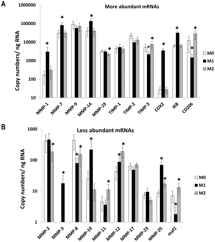
Based on copy numbers of mRNA, MMPs -7, -9, -14, -19 and TIMP-1, -2 and -3 were constitutively expressed at high levels in un-stimulated human macrophages differentiated with M-CSF from un-fractionated populations of CD16+/− monocytes (Fig. 1A). The mRNAs for other MMPs studied were expressed at lower levels (Fig. 1B) and MMP-13 mRNA was undetectable.
Figure 1. Effects of classical and alternative macrophage activation on MMP and TIMP mRNA levels.
Steady state mRNA levels were measured by qPCR in M-CSF differentiated macrophages derived from unselected human CD16+/− monocytes (M0) or classically activated with LPS and IFNγ (M1) or alternatively activated with IL-4 (M2) for 18 hours. Panels A and B show more and less abundant mRNAs, respectively. Values are means ± SEM * p<0.05 vs M0 (n = 7).
Macrophages in a pro-inflammatory milieu are likely to be classically activated, and to simulate this we initially used LPS and IFNγ to produce so-called M1 macrophages (Fig. 1A, B). By contrast, during resolution of inflammation macrophages may be alternatively activated, which we simulated with IL-4 to produce M2 macrophages (Fig. 1A, B). Based on previous studies [14], cyclo-oxygenase-2 (COX-2) and IκBα were included as positive controls for classical activation and the mannose receptor (CD206) and maf1 for alternative activation. Consistent with these expectations we found that classical-activation of macrophages (M1) up-regulated COX-2 and IκBα but down-regulated CD206 and maf1 mRNAs (Fig. 1A, B). By contrast, alternative activation of macrophages (M2) up-regulated CD206 and maf1 mRNAs (Fig. 1A, B). Classical activation (M1) also up-regulated mRNAs for MMP-1, -3, -7, -10, -12, -14 and -25 and down-regulated MMP-8 and TIMP-3 mRNAs (Fig. 1A, B). Note the scales are logarithmic and that fold changes are given in Table 2. Alternative activation with IL-4 (M2 conditions) increased mRNAs for MMPs-11, -12 and -25 and TIMP-3 and down-regulated MMPs-2, -8 and -19 (Fig. 1A, B and Table 2). As a result MMP-1, -2, -3, -7, -10, -14 and -25 were increased significantly in classically versus alternatively activated macrophages (Table 2). Conversely, MMPs-11 and -12 and TIMP-3 were increased significantly in alternatively versus classically activated macrophages, whereas other MMPs and TIMPs were similar in M1 and M2 macrophages (Table 2).
Table 2. Comparison of quantitative RT-PCR results among macrophage phenotypes from adhered mononuclear cells and CD16- monocytes (n = 7).
| M1/M2 ratio | M1/Control ratio | M2/Control ratio | ||||||||||
|---|---|---|---|---|---|---|---|---|---|---|---|---|
| Gene | mean | lower 95%CI | upper 95% CI | p value | mean | lower 95%CI | upper 95% CI | p value | mean | lower 95%CI | upper 95% CI | p value |
| MMP-1 | 5.7 | 2.1 | 14.9 | 0.016 | 9.5 | 4.5 | 20.2 | 0.002 | 1.7 | 1.2 | 2.4 | 0.055 |
| 4.9 | 2.7 | 8.9 | 0.002 | 7.4 | 1.8 | 31.3 | 0.032 | 1.5 | 0.6 | 3.8 | 0.397 | |
| MMP-2 | 4.2 | 1.8 | 9.6 | 0.014 | 1.3 | 0.9 | 1.7 | 0.197 | 0.3 | 0.2 | 0.5 | 0.007 |
| 5.1 | 1.3 | 19.3 | 0.047 | 1.4 | 0.6 | 3.0 | 0.434 | 0.3 | 0.1 | 1.4 | 0.168 | |
| MMP-3 | 324.6 | 42.7 | 2467.0 | 0.003 | 232.6 | 49.8 | 1086.1 | 0.001 | 0.7 | 0.4 | 1.3 | 0.363 |
| 1875.2 | 808.2 | 4351.4 | <0.001 | 1632.5 | 744.1 | 3581.6 | <0.001 | 0.9 | 0.7 | 1.1 | 0.374 | |
| MMP-7 | 3.2 | 2.3 | 4.3 | <0.001 | 3.6 | 2.4 | 5.4 | 0.001 | 1.1 | 0.9 | 1.5 | 0.325 |
| 3.5 | 2.1 | 6.1 | <0.001 | 3.4 | 1.8 | 6.7 | <0.001 | 0.9 | 0.7 | 1.3 | 0.792 | |
| MMP-8 | 0.6 | 0.4 | 0.8 | 0.017 | 0.2 | 0.1 | 0.3 | <0.001 | 0.3 | 0.2 | 0.5 | 0.005 |
| 0.3 | 0.03 | 2.3 | 0.577 | 0.3 | 0.1 | 0.6 | 0.011 | 0.3 | 0.2 | 0.6 | 0.020 | |
| MMP-9 | 0.6 | 0.2 | 2.1 | 0.453 | 0.6 | 0.3 | 1.1 | 0.147 | 0.9 | 0.5 | 1.7 | 0.834 |
| 3.9 | 0.6 | 24.5 | 0.186 | 2.6 | 0.2 | 36.8 | 0.506 | 0.7 | 0.2 | 2.2 | 0.509 | |
| MMP-10 | 24.6 | 3.9 | 157.0 | 0.009 | 12.2 | 2.4 | 60.5 | 0.016 | 0.5 | 0.1 | 2.3 | 0.400 |
| 15.7 | 6.0 | 40.9 | 0.001 | 9.2 | 1.7 | 48.4 | 0.037 | 0.6 | 0.2 | 2.2 | 0.444 | |
| MMP-11 | 0.3 | 0.2 | 0.5 | 0.006 | 0.8 | 0.5 | 1.4 | 0.540 | 2.4 | 1.8 | 3.2 | 0.002 |
| 0.3 | 0.2 | 0.4 | 0.001 | 1.1 | 0.5 | 2.4 | 0.840 | 3.8 | 2.0 | 7.3 | 0.006 | |
| MMP-12 | 0.5 | 0.3 | 0.7 | 0.002 | 2.5 | 0.8 | 7.4 | 0.142 | 5.1 | 1.7 | 15.4 | 0.020 |
| 0.3 | 0.1 | 1.0 | 0.090 | 3.6 | 1.4 | 9.5 | 0.038 | 11.3 | 4.3 | 29.4 | 0.002 | |
| MMP-14 | 10.7 | 2.2 | 52.7 | 0.025 | 8.0 | 1.6 | 40.1 | 0.041 | 0.8 | 0.4 | 1.3 | 0.328 |
| 5.6 | 2.3 | 13.9 | 0.008 | 9.3 | 1.1 | 77.2 | 0.079 | 1.7 | 0.2 | 16.6 | 0.680 | |
| MMP-17 | 0.7 | 0.5 | 1 | 0.084 | 0.8 | 0.5 | 1.2 | 0.374 | 1.2 | 0.9 | 1.5 | 0.245 |
| 1.3 | 0.4 | 4.2 | 0.653 | 0.6 | 0.4 | 1.1 | 0.173 | 0.5 | 0.1 | 2.4 | 0.426 | |
| MMP-19 | 1.2 | 0.8 | 1.8 | 0.382 | 0.8 | 0.6 | 1.1 | 0.216 | 0.7 | 0.6 | 0.8 | 0.001 |
| 3.1 | 0.7 | 13.6 | 0.171 | 0.9 | 0.8 | 1.0 | 0.056 | 0.3 | 0.1 | 1.3 | 0.146 | |
| MMP-23 | 1.4 | 0.4 | 5.2 | 0.646 | 0.8 | 0.4 | 1.9 | 0.694 | 0.6 | 0.3 | 1.3 | 0.227 |
| 2.8 | 0.4 | 22.5 | 0.366 | 1.2 | 0.8 | 1.7 | 0.417 | 0.4 | 0.1 | 3.0 | 0.419 | |
| MMP-25 | 3.2 | 1.5 | 6.8 | 0.020 | 7.1 | 3.6 | 14 | 0.001 | 2.2 | 1.6 | 3.1 | 0.004 |
| 2.6 | 1.4 | 5.1 | 0.015 | 10.9 | 6.5 | 18.6 | <0.001 | 4.2 | 2.0 | 8.7 | 0.005 | |
| TIMP-1 | 0.8 | 0.4 | 1.7 | 0.549 | 0.9 | 0.6 | 1.3 | 0.530 | 1.1 | 0.7 | 1.6 | 0.641 |
| 1.0 | 0.7 | 1.5 | 0.888 | 0.9 | 0.7 | 1.1 | 0.216 | 0.8 | 0.5 | 1.5 | 0.553 | |
| TIMP-2 | 1.2 | 0.3 | 3.9 | 0.811 | 0.5 | 0.3 | 0.7 | 0.008 | 0.4 | 0.1 | 1.7 | 0.269 |
| 1.0 | 0.5 | 2.2 | 0.918 | 0.9 | 0.4 | 2.0 | 0.813 | 0.9 | 0.8 | 1.0 | 0.074 | |
| TIMP-3 | 0.3 | 0.1 | 0.5 | 0.004 | 0.4 | 0.3 | 0.7 | 0.007 | 1.6 | 1.2 | 2.2 | 0.014 |
| 0.2 | 0.1 | 0.5 | 0.023 | 0.5 | 0.1 | 2.1 | 0.395 | 3.4 | 1.4 | 8.6 | 0.035 | |
| maf1 | 0.1 | 0.03 | 0.2 | 0.001 | 0.1 | 0.1 | 0.3 | 0.001 | 1.7 | 1.2 | 2.5 | 0.033 |
| 0.1 | 0.03 | 0.1 | <0.001 | 0.2 | 0.1 | 0.5 | 0.013 | 3.5 | 2.2 | 5.6 | 0.002 | |
| CD206 | 0.05 | 0.03 | 0.1 | <0.001 | 0.2 | 0.1 | 0.6 | 0.015 | 5.3 | 3.0 | 9.1 | 0.001 |
| 0.2 | 0.1 | 0.5 | 0.008 | 0.4 | 0.2 | 0.9 | 0.064 | 2.2 | 0.6 | 8.1 | 0.289 | |
| COX2 | 140.6 | 85.4 | 231.2 | <0.001 | 114.0 | 67.6 | 192.2 | <0.001 | 0.8 | 0.6 | 1.1 | 0.183 |
| 177.3 | 124.8 | 251.7 | <0.001 | 137.2 | 62.9 | 299.3 | <0.001 | 0.8 | 0.4 | 1.5 | 0.482 | |
| IκB | 3.9 | 2.3 | 6.7 | <0.001 | 3.9 | 2.4 | 6.6 | <0.001 | 1 | 0.8 | 1.2 | 0.928 |
| 4.4 | 1.9 | 10.3 | 0.001 | 3.2 | 1.7 | 6.3 | 0.001 | 0.7 | 0.4 | 1.3 | 0.306 |
A major population of CD16− monocytes and several minor CD16+ populations occur in human blood [16]. We observed similar levels of MMPs and TIMPs in un-stimulated macrophages derived from CD16+/− populations of adhered mononuclear cells or negatively selected CD16− monocytes (Table 2). Furthermore, similar effects of classical and alternative activation were seen with macrophages differentiated from mixed CD16+/− or CD16− monocytes (Table 2). Occasionally significant differences in one or other of CD16+/− and CD16− monocytes were merely trends in the other preparation (Table 2). Hence, none of these MMPs or TIMP mRNAs appeared to be exclusively expressed or up-regulated in macrophages derived from CD16+ monocytes.
Effects of LPS and IFNγ separately and comparison with other classical activators, IL-1 and TNFα
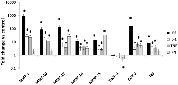
LPS on its own was a strong activator of COX-2, IκBα and MMPs-1, -10, -12, -14 and -25 but did not affect TIMP-3 (Fig. 2). LPS also up-regulated MMP-3 from undetectable starting values (results not shown). Maximal concentrations of IL-1 and TNFα were weaker activators than LPS (Fig. 2). IFNγ increased COX-2 and MMPs-12 and -14 mRNAs significantly, albeit to a lesser extent than other classical activators. IFNγ was more effective than the other classical activators tested in increasing MMP-25 and decreasing TIMP-3 mRNAs (Fig. 2).
Figure 2. Effects of classical activators on MMP and TIMP mRNA levels.
Steady state mRNA levels were measured by qPCR in M-CSF differentiated macrophages derived from unselected human CD16+/− monocytes. Cells were kept un-stimulated or treated with LPS (100 ng/mL), IL-1α (20 ng/mL) TNFα (10 ng/mL), or IFNγ (100 ng/mL) for 18 hours. Steady state mRNA levels were measured and the ratio between treated cells and M-CSF alone was calculated. Values are means ± SEM * p<0.05, n = 7.
Role of MAP kinases, and NF-κB in MMP and TIMP up-regulation
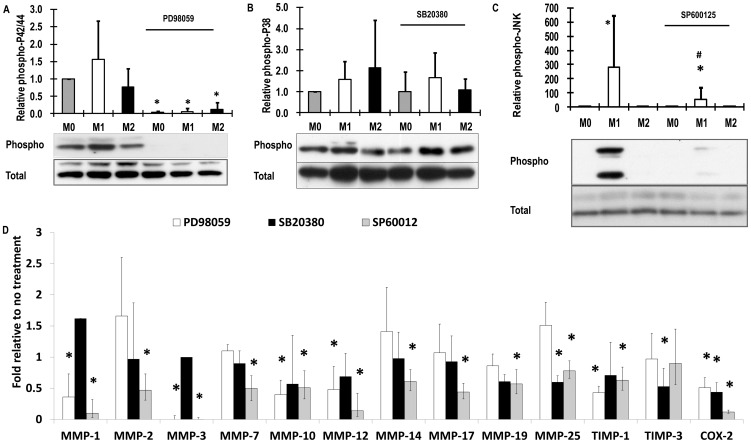
We reported previously that constitutive expression of MMPs and TIMPs from M-CSF differentiated macrophages (M0 conditions) was not affected by inhibitors of ERK, p38 or JNK MAP kinases or IKK2 [13]. Nevertheless, production of many inflammatory mediators from macrophages depends on activation of MAP kinases leading to activation of the activator protein-1 (AP-1) transcription factor and of inhibitor of κB kinase-2 (IKK2) leading to activation of NF-κB. We therefore used well characterised, selective inhibitors to investigate whether the same is true for MMPs up-regulated in classically-activated macrophages. M-CSF differentiated macrophages had constitutive levels of phospho-ERK1/2 and p38 MAP kinase that were not further increased by classical or alternative activation (Fig. 3A, B). However, phospho-JNK was stimulated under M1 conditions (Fig. 3C). Selective inhibition of MEK with PD98059 and JNK with SP600125 inhibited ERK1/2 and JNK phosphorylation, respectively (Fig. 3A, C). SB20380 did not inhibit the p38 phosphorylation (Fig. 3B), as expected, since it is a direct inhibitor of p38 kinase activity. After induction by LPS and IFNγ (M1 conditions) MEK inhibitor PD98059 decreased mRNA levels of positive control gene COX-2 and MMPs-1, -3, -10, and -12 and TIMP-1 (Fig. 3D). JNK inhibitor SP600125 reduced mRNA levels of all these proteins plus MMPs-2, -14, -17, -19 and -25 (Fig. 3D). By contrast, p38 inhibitor SB20380 only reduced levels of COX-2, MMP-25 and TIMP-3 mRNA (Fig. 3D).
Figure 3. Activation of MAP kinases and effects of inhibitors.
M-CSF differentiated macrophages derived from unselected human CD16+/− monocytes (M0) were pre-treated for 45 min with 10 µM PD98059, SB20380, SP600125 or vehicle (0.1% v/v DMSO) and then some were classically activated with LPS and IFNγ (M1). Phospho and total proteins were measured by western blotting. Levels were normalised to total proteins levels. (A) ERK1/2 with and without PD98059, (B) p38 MAPK with and without SB20380, (C) JNK with and without SP600125, (D) Steady-state mRNA levels of MMPs and other genes were measured after 18 hours of stimulation with LPS and IFNγ. Values are log weighted means and 95% confidence intervals * p<0.05 compared to without inhibitor (n = 7).
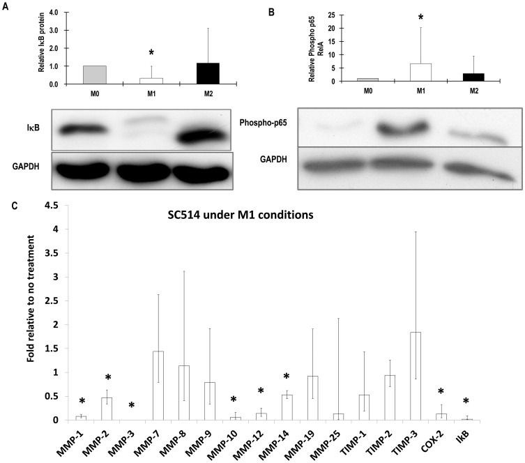
As expected, M1 treatment stimulated NF-κB activity based on measuring early increases in phospho-IκBα (Fig. 4A) and phospho-p65RelA (Fig. 4B) and later on increased IκBα mRNA levels (Fig. 4C). As expected, IKK2 inhibitor SC514 prevented the rise in IκBα mRNA levels (Fig. 4C). SC514 also decreased expression of the control gene COX-2 and MMPs-1, -2, -3, -10, -12, and -14 (Fig. 4C).
Figure 4. Activation of NF-κB and effects of SC514.
M-CSF differentiated macrophages derived from unselected human CD16+/− monocytes (M0) were pre-treated for 45 min with 40 µM SC514 or vehicle (0.1% v/v DMSO) and then some were classically activated with LPS and IFNγ (M1) for 45 minutes. Total IκB proteins and phospho- p65 RelA were measured by western blotting. Levels were normalised to GAPDH as shown. Values are means ± SD * p<0.05 compared to M0 (n = 5). (A) IκB protein (lower band) and phospho- IκB (upper band), (B) Phosho-p65 RelA, (C) Steady-state mRNA levels of MMPs and other genes were measured after 18 hours of stimulation with LPS and IFNγ. Values are log weighted means and 95% confidence intervals * p<0.05 compared to without inhibitor (n = 7).
Role of phosphoinostide-3-kinase (PI-3K)
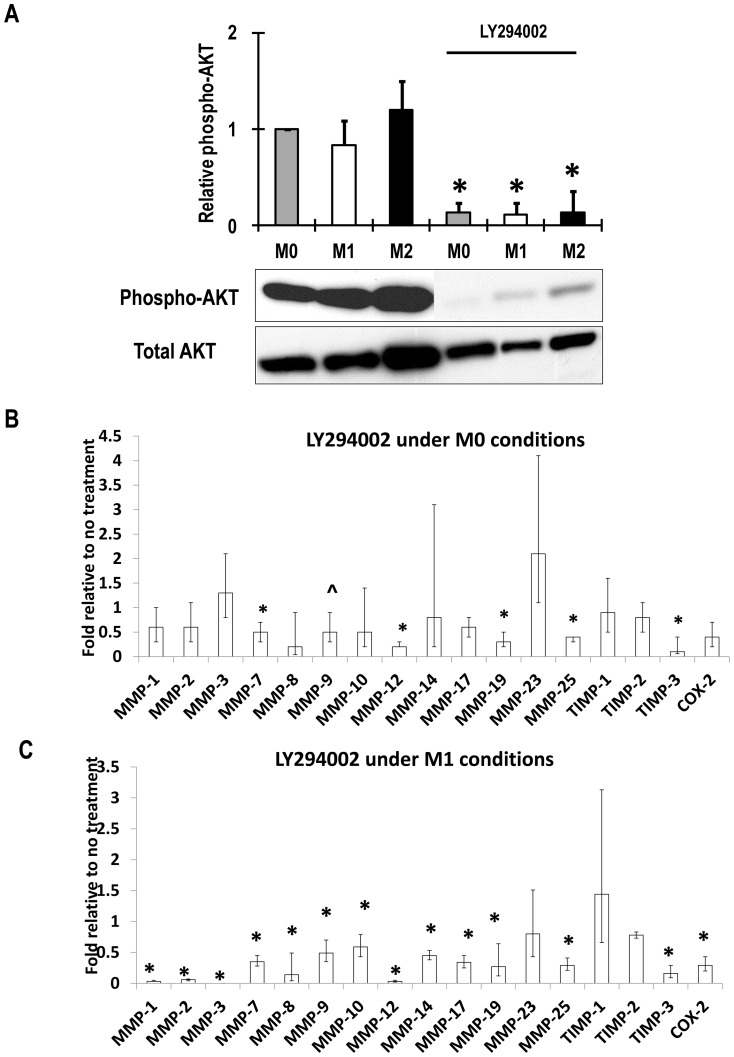
Macrophages differentiated in M-CSF had constitutive levels PI-3K activity based on Akt phosphorylation and these were reduced by LY294002 (Fig. 5A). PI-3K inhibitor LY 294002 decreased constitutive mRNA levels of MMP-7, -12, -19 and -25 and TIMP-3 and tended to decrease expression of MMP-9 (p = 0.074), but did not affect the other constitutively expressed MMPs and TIMPs (Fig. 5B). Under M1 conditions LY294002 inhibited all the MMPs, including MMP-9, and TIMP-3 that were inhibited under M0 conditions. LY294002 also inhibited the up-regulation of MMPs-1, -2, -3, -10, and -14 and the M1 marker gene COX-2 (Fig. 5C).
Figure 5. Activation of PI-3K and effects of LY294002.
M-CSF differentiated macrophages derived from unselected human CD16+/− monocytes (M0) were pre-treated for 45 min with 10 µM LY294002 and then some were classically activated with for 45 minutes LPS and IFNγ. (A) PI-3-kinase activity as phospho-AKT with and without LY294002 measured by western blotting. Values normalised to total AKT are means ± SD * p<0.05 compared to without inhibitor. (B) Steady-state mRNA levels of MMPs and other genes were measured after 18 hours of no stimulation (M0 conditions). (C) Steady-state mRNA levels of MMPs and other genes were measured after 18 hours of stimulation with LPS and IFNγ (M1 conditions). Panels B and C values are log weighted means and 95% confidence intervals * p<0.05 compared to without inhibitor (n = 7).
Expression of MMP proteins in un-stimulated, classically and alternatively activated macrophages
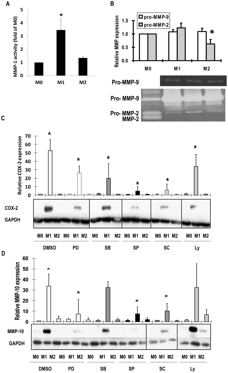
If good assays were available, we confirmed the mRNA expression results at the protein level. For example, using a highly sensitive antibody capture method we showed that MMP-1 activity was relatively low constitutively but increased by classical activation (Fig. 6A). By zymography, gelatinase activity of MMP-2 was much less than MMP-9 and was further suppressed under M2 conditions, consistent with the mRNA data (Fig. 6B). By western blotting, COX-2 and MMP-10 were undetectable in un-stimulated macrophages but were increased in classically activated macrophages (Fig. 6C, D). Moreover, COX-2 protein depended on ERK1/2, p38, JNK, IKK2 and PI-3K (6C), whereas MMP-10 protein depended on ERK1/2, JNK and IKK2 (Fig. 6D), consistent with the mRNA findings (Figs. 3, 4, 5). MMP-12 protein levels were below the limits of detection in un-stimulated and classically-activated macrophages but were stimulated after alternative activation (results not shown), consistent with the greater increase in MRNA levels (Fig. 1B).
Figure 6. Effects of classical and alternative macrophage activation on MMP protein levels.
M-CSF differentiated macrophages derived from unselected human CD16+/− monocytes (M0) or classically activated with LPS and IFNγ (M1) or alternatively activated with IL-4 (M2) for 48 hours. Where indicated cells were pre-treated with 10 µM PD98059 (PD), SB20380 (SB), SP600125 (SP), or LY 294002 (Ly) or 40 µM SC514 (SC) or vehicle (0.1% v/v DMSO). (A) After antibody capture MMP-1 activity was measured with a quenched fluorescent substrate. (B) Zymography for MMP-2 and MMP-9 in conditioned medium. (C) COX-2 in conditioned medium by western blotting. (D) MMP-10 in conditioned medium by western blotting. Values are means ± SD * p<0.05 vs M0 n = 5.
Role of the JAK-2 STAT-1 pathway in MMP and TIMP responses to IFNγ
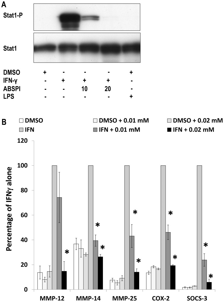
Some but not all actions of IFNγ are mediated through janus kinase-2 (JAK-2) [17]. IFNγ activated JAK-2 in macrophages based on STAT-1 phosphorylation and this was inhibited by the low molecular weight inhibitor ABSPI (Fig. 7A). The effects of IFNγ to increase MMP-12, -14 and -25, COX-2 and another positive control gene, SOCS-3, were all inhibited by the JAK-2 inhibitor ABSPI (Fig. 7B). The mRNA levels of MMP-1, -9 and -10 that did not change with IFNγ, were not affected by the JAK-2 inhibitor (not shown), which confirmed the specificity of this treatment.
Figure 7. Effects of a JAK-2 inhibitor.
M-CSF differentiated macrophages derived from unselected human CD16+/− monocytes were untreated or classically activated with IFNγ. (A) Phospho and total STAT-1 were measured after 45 minutes of IFNγ with and without JAK-2 inhibitor, 10 or 20 µM ABSPI as indicated, or vehicle (0.1% v/v DMSO). (B) Steady-state mRNA levels of MMPs and other genes were measured after 18 hours of stimulation as in panel A. Values are means ± SEM * p<0.05 compared to without inhibitor (n = 4).
Association of MMPs-1 and -10 with classical macrophage activation in atherosclerotic plaques in vivo
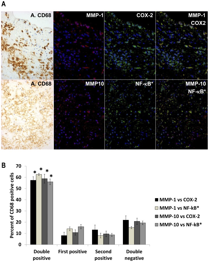
Our major in vitro findings included that MMP-1 and MMP-10 are up-regulated together with COX-2, the marker of classically activated macrophages, through activation of NF-κB. We took advantage of tissues available from the Bristol Coronary Biobank to validate these findings in vivo. Using dual fluorescence immunocytochemistry we showed that over 80% of cells in areas of plaque with abundant CD68 positive macrophages were either double positive for MMP-1 or MMP-10 and COX-2, or else double negative (Fig. 8). Furthermore, over 80% of cells were either double positive for MMP-1 or MMP-10 and nuclear localised NF-κB, or else double negative (Fig. 8).
Figure 8. Co-localisation of MMPs with markers of classical activation in human atherosclerotic plaques.
(A) Serial sections were stained by peroxidase with anti-CD68 (A. CD68) and by dual immuno-fluorescence with anti-MMP-1 or anti-MMP-10 (red) together with anti-COX-2 or p65RelA (green) as shown, and the images were superimposed digitally. Nuclear localised p65RelA (NF-kB*) was detected because of the shift in nuclear counterstain colour from dark blue (DAPI alone) to sky blue (blue plus green) in the superimposed image. (B) Areas rich in macrophages identified from the peroxidase stain were identified in the serial section. Cells in the whole field were counted and a percentage of each staining pattern calculated. Values are means ± SEM, n = 6, * p<0.05.
Discussion
Major findings
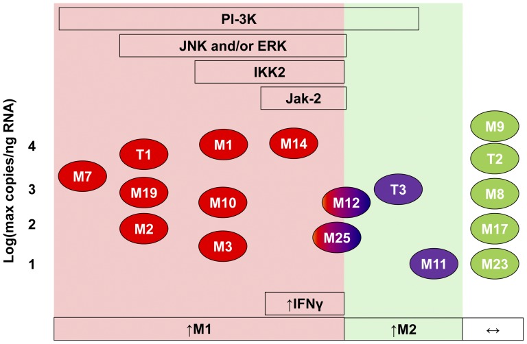
Our study is the first to comprehensively survey the MMP system in classically and alternatively activated human macrophages. We demonstrated differential regulation of several MMPs and TIMP-3, including, most excitingly, MMPs -10, -14, -19 and -25 and TIMP-3, which have received little previous attention in the context of inflammation. Moreover we have elucidated the signalling pathways responsible. Fig. 9 sets out a map of the major differences in MMP and TIMP expression and the underlying signalling pathways. In Fig. 9 genes up-regulated during classical activation have red symbols and those down-regulated during alternative activation have blue symbols. A few exceptional genes, MMPs -12 and -25, show up-regulation by both classical and alternative activation and are therefore given red-blue symbols. Genes in the pink zone of the map are up-regulated by classical activators both in vitro and in at least one model of human inflammation, the atherosclerotic plaque. Satisfyingly, this group of MMPs depended for their expression on ERK and JNK MAP kinases, PI-3K, and NF-κB, which have been associated previously with classical macrophage activation [5], [18]. The changes mediated selectively by IFNγ rather than other classical activators were all dependent upon JAK-2, which is another new finding. Only MMPs-11 and -12 and TIMP-3 are more up-regulated during alternative activation (Fig. 9 green zone). In the white zone are the MMP and TIMP genes that show no or minor regulation under the conditions of our experiments. Another novel aspect of our study is to consider the impact CD16− and CD16+ monocyte populations [16]. We did not find any major consequences of deleting the CD16+ population.
Figure 9. A map of regulation of the MMP system in human macrophages.
Pink zone: genes up-regulated by classical activators LPS and IFNγ as shown in the boxes below. Green zone: genes up-regulated during alternative activation (IL-4). White zone: no or minor regulation. Red symbols: up-regulated during classical activation. Blue symbols: up-regulated during alternative activation. Red-blue symbols: similar regulation by classical and alternative activation. Green symbols: scarcely regulated. The boxes above indicate dependence of MMP or TIMP activity on the specified kinases or transcription factors.
Comparison with previous studies
From previous literature [12], [13] and analysis of expression databases we focussed on those MMPs and TIMPs that have detectable copy numbers of mRNA in human macrophages. In addition we studied MMP-13 and MMP-23 that were identified at high levels in mouse macrophages. Our initial studies used the protocol and resulting marker genes for classical activation with LPS and IFNγ (M1) and for alternative activation with IL-4 (M2) reported in the genomic study of Martinez and colleagues [14]. No MMPs or TIMPs were reported to have a significant, more than 4-fold ratio of expression in M1/M2 conditions in that study [14]. We succeeded, however, because the larger number of replicates we used for our qRT-PCR study mitigated the quite high variability in different blood preparations (Fig. 1, Table 2).
The signalling pathways controlling MMP expression have been previously reviewed [19] and several have been shown to depend on MAP kinases and NF-κB in other cell types [20]. We significantly extended this literature by showing that dependence on ERK1/2 and/or JNK MAP kinases and NF-κB activating kinase, IKK2, is common to all those MMPs and TIMPs that are stimulated by classical macrophage activation, except MMP-7. Interestingly, basal expression of several MMPs (especially MMP-1, -3, and -10) remained very low in the absence of classical activation, despite constitutive ERK1/2 and p38 MAP kinase. This indicates that additional activation of JNK and IKK2 is necessary in concert for MMP up-regulation. Similar conclusions have been reached in several other cell types [20]. The promoters for all the MMPs that are up-regulated by classical activation or decreased by IL-4 during differentiation contain proximal AP-1 or SP-1 transcription factor binding sites [19]. Moreover, the cognate transcription factors depend on MAP kinase activity [21]. Dependency of MMP promoters on NF-κB is less well documented since only MMP-1, -9 and -14 have defined proximal binding sites [19]. The observation in our study that MMP-9 is not induced by classical macrophage activation and that its constitutive production is independent of NF-κB activity is consistent with previous work in human primary macrophages and rabbit foam cell macrophages [22].Nevertheless, well-characterised AP-1 and NF-κB binding sites occur in the MMP-9 promoter and are responsible for synergistic induction of MMP-9 by growth factors and inflammatory cytokines in several other cell types [23], [24]. The basis for the unusual pattern of regulation in macrophages requires further study.
The relative effectiveness of different classical activators on MMP expression levels has not been systematically studied previously. We found here that for most MMPs, the effects of LPS and cytokines were much greater than those of IFNγ. Nevertheless, significant effects of IFNγ were seen on stimulation of MMPs-14 and -25 and inhibition of TIMP-3. MMP-14 and TIMP-3 have been shown to have counter-regulatory roles in macrophage invasion, proliferation and apoptosis [15] and it will be interesting to investigate the role of IFNγ in this switch. Signalling from IFNγ has been shown previously to proceed along JAK-2 dependent and independent pathways [25]. We showed here that all the effects on the MMP system we observed are JAK-2 dependent.
Functional relevance
There is much literature to support a role for MMPs in various forms of inflammation [2], [3], [8]. For example, Libby's group showed based on histological studies in man and animal experiments, that release of several collagenases form macrophages plays a key role in atherosclerotic plaque rupture (reviewed in [26]). Based on copy numbers of mRNA, we show here that the membrane-type1 MMP, MMP-14, is the most abundantly-expressed collagenase of un-stimulated human macrophages (Fig. 1, 9). Our data from mRNA and protein measurements demonstrated that MMP-14 is further increased by classical activation, which appears to be a novel finding (see review in [4]). MMP-14 has been shown to mediate ICAM-dependent migration of monocytes across endothelial cell layers [27] and to promote the invasion of macrophages and foam cell macrophages through matrigel [15]. The secreted collagenase MMP-1 is also greatly up-regulated upon classical activation and this led to an increase in collagenase activity (Fig. 6A). Furthermore we showed that MMP-1 co-localised with COX-2, the marker of classical macrophage activation, in human atherosclerotic plaques. By contrast, the so-called neutrophil collagenase, MMP-8, was down-regulated under M1 conditions in our study. Although abundant in mouse macrophages (reviewed in [4]), we could not detect MMP-13 expression in human macrophages when using 3 different sets of validated PCR primers, in agreement with a previous study [12]. MMP-19 is also considered a collagenase, although from mouse knockout studies [28], tenascin-C appears to be an important substrate in vivo. MMP-19 has been previously identified as a cell-surface associated MMP in monocytes stimulated by adhesion [29] or chemokine CCR5 but not LPS [30]. We now add that it is abundant in un-stimulated macrophages. Knockout of MMP-19 in mice leads to complex phenotypes owing to a defect in thymocyte maturation [31]. Nevertheless, future studies to identify its function in inflammatory conditions including atherosclerosis appear warranted.
The gelatinases, MMP-2 and MMP-9 have unique gelatin binding domains inserted into their catalytic domains [1]. We show here that MMP-9 is much more abundant than MMP-2 in differentiated human macrophages based on mRNA levels and zymography.
Among the stromelysins, MMP-3 has been quite widely studied in the context of inflammation [2] but role of MMP-10 is much less appreciated. MMP-10 is stimulated in human brain microglia by amyloid beta peptide [32] and recent transcriptomic studies identified MMP-10 as up-regulated among other genes during inflammation in human chondrocytes and adipocytes [33], [34]. MMP-10 can activate the pro-form of MMP-1 [35] and hence the similar substantial up-regulation of MMP-1 and -10 in vitro and their co-localisation in atherosclerotic plaques (Fig. 8) could be significant for collagen degradation. Indeed cleavage of collagen has long been known to co-localise with MMP-1 expression in human atherosclerotic plaques [36].
The matrilysin MMP-7, which lacks a C terminal haemopexin domain, was abundant in un-stimulated macrophages and further up-regulated during classical activation, consistent with its previously identified roles in inflammation [2].
Interestingly, we found that the much less studied membrane-type metalloproteinase MMP-25 (MT6-MMP) is induced by classical and alternative activation, although the effects of classical activation are stronger. MMP-25 was originally cloned and named leukolysin because of its restriction to blood leukocytes and bone marrow [37]. The alpha-1-proteinase inhibitor [38] and myelin basic protein [39] have been identified as efficient substrates of MMP-25 and a role for the enzyme has been proposed in autoimmune multiple sclerosis [39]. MMP-25 was shown to be up-regulated by LPS in mouse macrophages [39] but regulation in humans has not apparently been reported. Our results prompt more research into this MMP.
In great contrast to the other MMPs, the matrix metalloelastase, MMP-12, appears to be more readily up-regulated in alternatively than classically-activated macrophages. A prominent role for MMP-12 in alternative activated macrophages has been implied previously during aneurysm formation in mice [40].
TIMP-1, -2 and -3 were expressed at relatively high copy numbers in un-stimulated macrophages in vitro and TIMP-3 was down-regulated by classical activation, which implies a further increase in the ratio of MMP gene expression to that of TIMPs. Conversely, TIMP-3 was increased by alternative activation, which appears to be a novel finding (see review in [4]).
Conclusion
In summary, Fig. 9 maps out the divergent regulation of the MMPs and TIMPs during classical and alternative activation of human macrophages. Furthermore, it demonstrates how differential dependence on ERK1/2 and JNK MAP kinases, NF-κB activating kinase IKK2, PI-3K and JAK-2 activation underlie this regulation. Our mechanistic studies should help future work aimed at modulating MMP expression and thereby preserving the ECM in diseases such as rheumatoid arthritis, periodontal disease atherosclerosis and aneurysms, which result from excessive classical or alternative macrophage activation.
Acknowledgments
We thank Dr. Sue Finerty for expert technical assistance.
Funding Statement
This work was supported by British Heart Foundation CH95/001 and RG04/009 and the National Health Research Institute (UK) Bristol Biomedical Research Unit in Cardiovascular Medicine. W-C Huang held Fellowships from the Yen Tjing Ling Medical Foundation and Medical Foundation in Memory of Dr. Deh-Lin Cheng, Taiwan. The funders had no role in study design, data collection and analysis, decision to publish, or preparation of the manuscript.
References
- 1.Nagase H, Visse R, Murphy G (2006) Structure and function of matrix metalloproteinases and TIMPs. Cardiovasc Res 69: 562–573. [DOI] [PubMed] [Google Scholar]
- 2.Parks WC, Wilson CL, Lopez-Boado YS (2004) Matrix metalloproteinases as modulators of inflammation and innate immunity. Nat Rev Immunol 4: 617–629. [DOI] [PubMed] [Google Scholar]
- 3.Manicone AM, McGuire JK (2008) Matrix metalloproteinases as modulators of inflammation. Semin Cell Dev Biol 19: 34–41. [DOI] [PMC free article] [PubMed] [Google Scholar]
- 4.Newby AC (2008) Metalloproteinase expression in monocytes and macrophages and its relationship to atherosclerotic plaque instability. Arterioscler Thromb Vasc Biol 28: 2108–2114. [DOI] [PubMed] [Google Scholar]
- 5.Gordon S, Taylor PR (2005) Monocyte and macrophage heterogeneity. Nat Rev Immunol 5: 953–964. [DOI] [PubMed] [Google Scholar]
- 6.Martinez FO, Helming L, Gordon S (2008) Alternative Activation of Macrophages: An Immunologic Functional Perspective. Annu Rev Immunol 27: 451–483. [DOI] [PubMed] [Google Scholar]
- 7.Page-McCaw A, Ewald AJ, Werb Z (2007) Matrix metalloproteinases and the regulation of tissue remodelling. Nat Rev Mol Cell Biol 8: 221–233. [DOI] [PMC free article] [PubMed] [Google Scholar]
- 8.Sorsa T, Tjaderhane L, Konttinen YT, Lauhio A, Salo T, et al. (2006) Matrix metalloproteinases: contribution to pathogenesis, diagnosis and treatment of periodontal inflammation. Ann Med 38: 306–321. [DOI] [PubMed] [Google Scholar]
- 9.Murphy G, Knauper V, Atkinson S, Butler G, English W, et al. (2002) Matrix metalloproteinases in arthritic disease. Arthritis Res 4 Suppl 3S39–49. [DOI] [PMC free article] [PubMed] [Google Scholar]
- 10.Elkington PT, Ugarte-Gil CA, Friedland JS (2011) Matrix metalloproteinases in tuberculosis. Eur Respir J 38: 456–464. [DOI] [PubMed] [Google Scholar]
- 11.Newby AC (2005) Dual role of matrix metalloproteinases (matrixins) in intimal thickening and atherosclerotic plaque rupture. Physiol Rev 85: 1–31. [DOI] [PubMed] [Google Scholar]
- 12.Bar-Or A, Nuttall RK, Duddy M, Alter A, Kim HJ, et al. (2003) Analyses of all matrix metalloproteinase members in leukocytes emphasize monocytes as major inflammatory mediators in multiple sclerosis. Brain 126: 2738–2749. [DOI] [PubMed] [Google Scholar]
- 13.Reel B, Sala-Newby GB, Huang W-C, Newby AC (2011) Diverse patterns of cyclooxygenase-independent metalloproteinase gene regulation in human monocytes. Br J Pharmacol 163: 1679–1690. [DOI] [PMC free article] [PubMed] [Google Scholar]
- 14.Martinez FO, Gordon S, Locati M, Mantovani A (2006) Transcriptional profiling of the human monocyte-to-macrophage differentiation and polarization: new molecules and patterns of gene expression. J Immunol 177: 7303–7311. [DOI] [PubMed] [Google Scholar]
- 15.Johnson JL, Sala-Newby GB, Ismail Y, Aguilera CM, Newby AC (2008) Low tissue inhibitor of metalloproteinases 3 and high matrix metalloproteinase 14 levels defines a subpopulation of highly invasive foam-cell macrophages. Arterioscler Thromb Vasc Biol 28: 1647–1653. [DOI] [PMC free article] [PubMed] [Google Scholar]
- 16.Ziegler-Heitbrock L, Ancuta P, Crowe S, Dalod M, Grau V, et al. (2010) Nomenclature of monocytes and dendritic cells in blood. Blood 116: e74–80. [DOI] [PubMed] [Google Scholar]
- 17.Schroder K, Hertzog PJ, Ravasi T, Hume DA (2004) Interferon-gamma: an overview of signals, mechanisms and functions. J Leukoc Biol 75: 163–189. [DOI] [PubMed] [Google Scholar]
- 18.Lopez-Pelaez M, Soria-Castro I, Bosca L, Fernandez M, Alemany S (2011) Cot/tpl2 activity is required for TLR-induced activation of the Akt p70 S6k pathway in macrophages: Implications for NO synthase 2 expression. Eur J Immunol 41: 1733–1741. [DOI] [PubMed] [Google Scholar]
- 19.Clark IM, Swingler TE, Sampieri CL, Edwards DR (2008) The regulation of matrix metalloproteinases and their inhibitors. Int J Biochem Cell Biol 40: 1362–1378. [DOI] [PubMed] [Google Scholar]
- 20.Vincenti MP, Brinckerhoff CE (2002) Transcriptional regulation of collagenase (MMP-1, MMP-13) genes in arthritis: integration of complex signaling pathways for the recruitment of gene-specific transcription factors. Arthritis Res 4: 157–164. [DOI] [PMC free article] [PubMed] [Google Scholar]
- 21.Chakraborti S, Mandal M, Das S, Mandal A, Chakraborti T (2003) Regulation of matrix metalloproteinases: An overview. Mol Cell Biochem 253: 269–285. [DOI] [PubMed] [Google Scholar]
- 22.Chase A, Bond M, Crook MF, Newby AC (2002) Role of nuclear factor-kB activation in metalloproteinase-1, -3 and -9 secretion by human macrophages in vitro and rabbit foam cells produced in vivo. Arterioscler Thromb Vasc Biol 22: 765–771. [DOI] [PubMed] [Google Scholar]
- 23.Bond M, Baker AH, Newby AC (1999) Nuclear factor kappa-B activity is essential for matrix metalloproteinase-1 and -3 upregulation in rabbit dermal fibroblasts. Biochem Biophys Res Comm 264: 561–567. [DOI] [PubMed] [Google Scholar]
- 24.Bond M, Chase AJ, Baker AH, Newby AC (2001) Inhibition of transcription factor NF-kB reduces matrix metalloproteinase-1, -3 and -9 production by vascular smooth muscle cells. Cardiovasc Res 50: 556–565. [DOI] [PubMed] [Google Scholar]
- 25.Harvey EJ, Li N, Ramji DP (2007) Critical role for casein kinase 2 and phosphoinositide-3-kinase in the interferon-gamma-induced expression of monocyte chemoattractant protein-1 and other key genes implicated in atherosclerosis. Arterioscler Thromb Vasc Biol 27: 806–812. [DOI] [PubMed] [Google Scholar]
- 26.Dollery CM, Libby P (2006) Atherosclerosis and proteinase activation. Cardiovasc Res 69: 625–635. [DOI] [PubMed] [Google Scholar]
- 27.Sithu SD, English WR, Olson P, Krubasik D, Baker AH, et al. (2007) Membrane-type 1-matrix metalloproteinase regulates intracellular adhesion molecule-1 (ICAM-1)-mediated monocyte transmigration. J Biol Chem 282: 25010–25019. [DOI] [PubMed] [Google Scholar]
- 28.Gueders MM, Hirst SJ, Quesada-Calvo F, Paulissen G, Hacha J, et al. (2010) Matrix metalloproteinase-19 deficiency promotes tenascin-C accumulation and allergen-induced airway inflammation. Am J Respir Cell Mol Biol 43: 286–295. [DOI] [PubMed] [Google Scholar]
- 29.Mauch S, Kolb C, Kolb B, Sadowski T, Sedlacek R (2002) Matrix metalloproteinase-19 is expressed in myeloid cells in an adhesion-dependent manner and associates with the cell surface. J Immunol 168: 1244–1251. [DOI] [PubMed] [Google Scholar]
- 30.Locati M, Deuschle U, Massardi ML, Martinez FO, Sironi M, et al. (2002) Analysis of the gene expression profile activated by the CC chemokine ligand 5/RANTES and by lipopolysaccharide in human monocytes. J Immunol 168: 3557–3562. [DOI] [PubMed] [Google Scholar]
- 31.Beck IM, Ruckert R, Brandt K, Mueller MS, Sadowski T, et al. (2008) MMP19 is essential for T cell development and T cell-mediated cutaneous immune responses. PLoS One 3: e2343. [DOI] [PMC free article] [PubMed] [Google Scholar]
- 32.Walker DG, Link J, Lue LF, Dalsing-Hernandez JE, Boyes BE (2006) Gene expression changes by amyloid beta peptide-stimulated human postmortem brain microglia identify activation of multiple inflammatory processes. J Leukoc Biol 79: 596–610. [DOI] [PubMed] [Google Scholar]
- 33.Andreas K, Lubke C, Haupl T, Dehne T, Morawietz L, et al. (2008) Key regulatory molecules of cartilage destruction in rheumatoid arthritis: an in vitro study. Arthritis Res Ther 10: R9. [DOI] [PMC free article] [PubMed] [Google Scholar]
- 34.O'Hara A, Lim FL, Mazzatti DJ, Trayhurn P (2009) Microarray analysis identifies matrix metalloproteinases (MMPs) as key genes whose expression is up-regulated in human adipocytes by macrophage-conditioned medium. Pflugers Arch 458: 1103–1114. [DOI] [PubMed] [Google Scholar]
- 35.Montero I, Orbe J, Varo N, Beloqui O, Monreal JI, et al. (2006) C-reactive protein induces matrix metalloproteinase-1 and -10 in human endothelial cells: implications for clinical and subclinical atherosclerosis. J Am Coll Cardiol 47: 1369–1378. [DOI] [PubMed] [Google Scholar]
- 36.Sukhova GK, Schonbeck U, Rabkin E, Schoen FJ, Poole AR, et al. (1999) Evidence for increased collagenolysis by interstitial collagenases-1 and-3 in vulnerable human atheromatous plaques. Circulation 99: 2503–2509. [DOI] [PubMed] [Google Scholar]
- 37.Pei D (1999) Leukolysin/MMP25/MT6-MMP: a novel matrix metalloproteinase specifically expressed in the leukocyte lineage. Cell Res 9: 291–303. [DOI] [PubMed] [Google Scholar]
- 38.Nie J, Pei D (2004) Rapid inactivation of alpha-1-proteinase inhibitor by neutrophil specific leukolysin/membrane-type matrix metalloproteinase 6. Exp Cell Res 296: 145–150. [DOI] [PubMed] [Google Scholar]
- 39.Shiryaev SA, Remacle AG, Savinov AY, Chernov AV, Cieplak P, et al. (2009) Inflammatory proprotein convertase-matrix metalloproteinase proteolytic pathway in antigen-presenting cells as a step to autoimmune multiple sclerosis. J Biol Chem 284: 30615–30626. [DOI] [PMC free article] [PubMed] [Google Scholar]
- 40.Shimizu K, Shichiri M, Libby P, Lee RT, Mitchell RN (2004) Th2-predominant inflammation and blockade of IFN-gamma signaling induce aneurysms in allografted aortas. J Clin Invest 114: 300–308. [DOI] [PMC free article] [PubMed] [Google Scholar]
