RIPK3 contributes to TNFR1-mediated RIPK1 kinase-dependent apoptosis in conditions of cIAP1/2 depletion or TAK1 kinase inhibition (original) (raw)
Abstract
Receptor-interacting protein kinase (RIPK) 1 and RIPK3 have emerged as essential kinases mediating a regulated form of necrosis, known as necroptosis, that can be induced by tumor necrosis factor (TNF) signaling. As a consequence, inhibiting RIPK1 kinase activity and repressing RIPK3 expression levels have become commonly used approaches to estimate the contribution of necroptosis to specific phenotypes. Here, we report that RIPK1 kinase activity and RIPK3 also contribute to TNF-induced apoptosis in conditions of cellular inhibitor of apoptosis 1 and 2 (cIAP1/2) depletion or TGF-_β_-activated kinase 1 (TAK1) kinase inhibition, implying that inhibition of RIPK1 kinase activity or depletion of RIPK3 under cell death conditions is not always a prerequisite to conclude on the involvement of necroptosis. Moreover, we found that, contrary to cIAP1/2 depletion, TAK1 kinase inhibition induces assembly of the cytosolic RIPK1/Fas-associated protein with death domain/caspase-8 apoptotic TNF receptor 1 (TNFR1) complex IIb without affecting the RIPK1 ubiquitylation status at the level of TNFR1 complex I. These results indicate that the recruitment of TAK1 to the ubiquitin (Ub) chains, and not the Ub chains per se, regulates the contribution of RIPK1 to the apoptotic death trigger. In line with this, we found that cylindromatosis repression only provided protection to TNF-mediated RIPK1-dependent apoptosis in condition of reduced RIPK1 ubiquitylation obtained by cIAP1/2 depletion but not upon TAK1 kinase inhibition, again arguing for a role of TAK1 in preventing RIPK1-dependent apoptosis downstream of RIPK1 ubiquitylation. Importantly, we found that this function of TAK1 was independent of its known role in canonical nuclear factor-_κ_B (NF-_κ_B) activation. Our study therefore reports a new function of TAK1 in regulating an early NF-_κ_B-independent cell death checkpoint in the TNFR1 apoptotic pathway. In both TNF-induced RIPK1 kinase-dependent apoptotic models, we found that RIPK3 contributes to full caspase-8 activation independently of its kinase activity or intact RHIM domain. In contrast, RIPK3 participates in caspase-8 activation by acting downstream of the cytosolic death complex assembly, possibly via reactive oxygen species generation.
Keywords: RIPK3, TAK1, CYLD, apoptosis, necroptosis, Nec-1
Tumor necrosis factor (TNF) is a pleiotropic cytokine that controls a variety of cellular responses, including proliferation, differentiation, inflammatory cytokine production, survival and death.1 TNF signals by binding and activating two cell surface receptors, TNF receptor (TNFR) 1 and TNFR2,2 but most of its biological activities have been associated with TNFR1.3 In most cell types, TNFR1 activation does not induce cell death but instead leads to the transcriptional upregulation of genes encoding pro-survival and pro-inflammatory molecules.4 This function is mediated by the assembly of a plasma membrane-bound multiprotein signaling complex, referred to as TNFR1 complex I,5 that contains TNF receptor-associated death domain (TRADD), receptor-interacting protein kinase 1 (RIPK1, also known as RIP1), TNF receptor-associated factor 2, cellular inhibitor of apoptosis protein 1 and 2 (cIAP1/2) and the linear ubiquitin (Ub) chain assembly complex.6 Within this complex, RIPK1 and other proteins are rapidly conjugated with Ub chains of various types.7, 8, 9, 10 These Ub chains act as scaffolds for the recruitment and activation of the TAK1-binding protein 2/3 (TAB2/3)–TGF-_β_-activated kinase 1 (TAK1) complex and the inhibitor of nuclear factor-_κ_B (NF-κ_B) kinase (IKK) complex (NEMO-IKK_α_-IKK_β), which subsequently leads to the activation of the mitogen-activated protein kinases and canonical NF-_κ_B signaling pathways that collectively drive transcription of genes that prevent cell death and sustain inflammation. Accordingly, when the NF-_κ_B response is inhibited, either genetically or by the use of the general translation inhibitor cycloheximide (CHX), TNFR1 ligation switches from a pro-survival to a pro-apoptotic response. This switch occurs via a mechanism that involves internalization of complex I and assembly of a cytoplasmic caspase-8-inducing death complex, known as TNFR1 complex IIa, which contains TRADD, Fas-associated protein with death domain (FADD) and procaspase-8.5, 11 Under these conditions, TNF-mediated death was shown not to depend on RIPK1.12, 13
cIAP1/2 are required for TNF-induced canonical NF-_κ_B activation.10, 14, 15, 16 Consequently, their depletion, obtained either genetically or by the use of Smac Mimetics (SM), also induces a switch to apoptosis. Intriguingly, in the absence of cIAP1/2, TNF-induced death was shown to rely on RIPK1 and not on TRADD,12, 15, 17, 18, 19 suggesting that cIAP1/2 additionally regulate an NF-_κ_B-independent cell death checkpoint in the TNFR1 pathway. To discriminate the RIPK1-containing cytosolic death complex obtained in cIAP1/2-depleted conditions from complex IIa, it has been defined as complex IIb.11 The molecular mechanism accounting for the differential assembly of complex IIa versus IIb is poorly understood, but suggested to rely on RIPK1 ubiquitylation status in complex I. Indeed, cIAP1/2 act as direct ubiquitin ligases for RIPK18, 14, 15, 20, 21 and cIAP1/2-mediated RIPK1 ubiquitylation in complex I is believed to prevent the transition of RIPK1 from complex I to complex II.12, 15, 22, 23 A notion supported by the fact that repression of the RIPK1 deubiquitinase cylindromatosis (CYLD) inhibits recruitment of RIPK1 to complex IIb.12
In addition to apoptosis, TNF signaling can also induce necroptosis, a regulated form of necrosis that prevails in caspase-8-inhibited conditions, and whose physiological relevance has recently been demonstrated by in vivo studies.24 TNF-mediated necroptosis relies on the assembly of another cytosolic death complex, known as the necrosome, which consists of FADD, RIPK1 and RIPK3.25, 26, 27 Because both RIPK1 and RIPK3 kinase activities were shown to be crucial for necroptosis induction, necrostatin-1 (Nec-1), an inhibitor of RIPK1 kinase activity,28 is commonly used as a chemical tool to inhibit necroptosis. As an alternative approach, RIPK3 depletion has become a gold standard to estimate the contribution of necroptosis in a defined phenotype.29
In this study, we report on a new NF-_κ_B-independent but kinase-dependent function of TAK1 in protecting cells from complex IIb-mediated apoptotic cell death. Because TAK1 kinase inhibition induces complex IIb assembly without affecting RIPK1 ubiquitylation in complex I, our results suggest that the recruitment of TAK1 to the Ub chains conjugated to RIPK1 and not the Ub chains per se regulates the death potential of RIPK1. In addition, we demonstrate that the use of Nec-1 or the depletion of RIPK3 protects cells from TNF-mediated RIPK1-dependent apoptosis, both in conditions of cIAP1/2 depletion or TAK1 kinase inhibition.
Results
TAK1 kinase activity protects cells from TNF-induced RIPK1 kinase-dependent apoptosis independently of NF-_κ_B activation
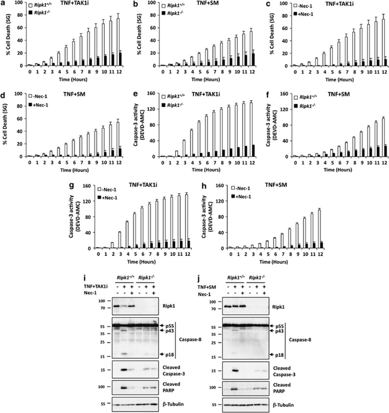
TNFR1 engagement has been reported to mediate both RIPK1-independent and -dependent apoptosis based on the cellular context.12, 15, 17 The kinase activity of TAK1 has a crucial role in the activation of the mitogen-activated protein kinases and canonical NF-_κ_B pathways downstream of TNFR1.30, 31 As a consequence, TAK1-deficient cells were shown to succumb by apoptosis upon single TNF stimulation.31, 32, 33 However, whether these cells die apoptotically in a RIPK1-independent or -dependent manner is unclear. Because TAK1 recruitment to complex I is reported to occur downstream of RIPK1 ubiquitylation, we speculated that TAK1 kinase inhibition would sensitize cells to TNF-induced apoptosis in a RIPK1-independent manner. Surprisingly, we found that RIPK1-deficiency protected immortalized MEFs against death induced by TNF in the presence of the TAK1 kinase inhibitor NP-009245 (TNF+TAK1i) similarly as against co-treatment with the SM CmpA34 (TNF+SM; Figures 1a and b, Supplementary Figures S1A, B). Of note, at the concentrations used, cell death occurred more rapidly with the TAK1 kinase inhibitor than with the SM. As negative control, we treated the MEFs with TNF in the presence of low concentration of CHX (TNF+CHX) and, as previously reported,13 observed that _Ripk1_−/− MEFs were not protected but sensitized to this apoptotic trigger (Supplementary Figure S2). Interestingly, we found that inhibiting RIPK1 kinase activity by Nec-1-protected Ripk1+/+ MEFs to either TNF+TAK1i or TNF+SM as efficiently as the RIPK1 deficiency itself (Figures 1c and d, Supplementary Figures S1A and B). As RIPK1 kinase activity is mainly associated with necroptotic cell death, we confirmed protection to apoptosis, and not necroptosis, by measuring caspase-3 activity by DEVDase assay (Figures 1e–h, Supplementary Figures S1C and D, 3), by monitoring processing of caspase-8, caspase-3 and PARP (Figures 1i and j) and by analyzing the cell death morphology (Supplementary Figure S4). Of note, although apoptosis induction upon TNF+TAK1i or TNF+SM treatment had been confirmed, we found that inhibiting caspase activity by the use of the pan caspase inhibitor zVAD-fmk (Z-Val-Ala-DL-Asp-fluoromethylketone) did not protect cells from death but instead induced a switch to RIPK1/3-dependent necroptosis in both conditions (Supplementary Figures S4, S5A and SB and data not shown). To exclude an off-target effect of the TAK1 kinase inhibitor, we confirmed the requirement of RIPK1 kinase activity for TNF-mediated apoptosis in the absence of functional TAK1 using Tak1+/+ and _Tak1_−/− MEFs treated with TNF in the absence or presence of Nec-1 (Supplementary Figure S5C). Of note, the TAK1 kinase inhibitor did not sensitize _Tak1_−/− MEFs to apoptosis induced by TNF, demonstrating the TAK1-specific effect of the inhibitor (data not shown).
Figure 1.
TAK1 kinase inhibition induces RIPK1 kinase-dependent apoptosis upon TNF stimulation. (a–h) Immortalized Ripk1+/+ and _Ripk1_−/− MEFs were pre-treated for 30 min with TAK1i or SM in the presence or absence of Nec-1 and subsequently stimulated with hTNF. The percentage of cell death (SytoxGreen fluorescence) (a–d) and caspase-3 activity (DEVD-AMC fluorescence) (e–h) was calculated in function of time using the Fluostar Omega fluorescence plate reader as indicated in the experimental procedures. Error bars indicate the standard deviation from triplicate samples. The results are representative of at least three independent experiments. (i and j) Immortalized Ripk1+/+ and _Ripk1_−/− MEFs were pre-treated for 30 min with TAK1i (i) or SM (j) in the presence or absence of Nec-1 and subsequently stimulated with hTNF for 3 h. Cells were then lysed and immunoblotted as indicated
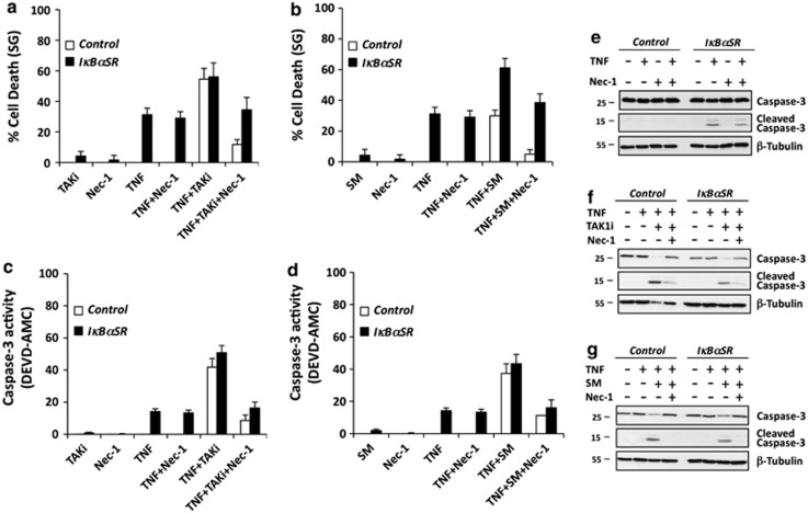
Finally, to further investigate whether the protective role of TAK1 and cIAP1/2 against RIPK1-dependent apoptosis was depending on NF-_κ_B activation, we analyzed the effect of TAK1 inhibition or cIAP1/2 depletion in MEFs unable to mount an NF-κ_B response due to stable expression of an I_κ_B_α mutant resistant to proteasomal degradation (I_κ_B_α_SR).35 As shown in Figure 2, single TNF stimulation induced RIPK1-kinase independent apoptosis in the I_κ_B_α_SR MEFs, but co-stimulation with either TAK1i or SM led to an increase in cell death and caspase-3 activity that could be inhibited by Nec-1.
Figure 2.
TAK1 and cIAP1/2 protect cells against TNF-mediated RIPK1-dependent apoptosis in a NF-_κ_B-independent manner. (a–d) Control MEFs or MEFs overexpressing I_κ_B_α_SR were pretreated with TAK1i (a and c) or SM (b and d) in the presence or absence of Nec-1 and subsequently stimulated with hTNF for 10 h. Cell death (a and b) and caspase-3 activity (c and d) was analyzed at the indicated time using the Fluostar Omega fluorometer. Error bars indicate the standard deviation from triplicate samples. The results are representative of at least two independent experiments. (e–g) Control MEFs or MEFs overexpressing I_κ_B_α_SR were pretreated with media (e), TAK1i (f) or SM (g) in the presence or absence of Nec-1 and subsequently stimulated with hTNF for, respectively, 4 h (e) or 2 h (f and g). Cells were then lysed and immunoblotted as indicated
Together, our results demonstrate that, similar to cIAP1/2 depletion by SM, TAK1 kinase inhibition induces RIPK1 kinase-dependent apoptosis upon TNF stimulation, which could be switched to necroptosis by inhibiting caspase activity. Importantly, the RIPK1 dependency in apoptosis induction originates from the fact that both TAK1 and cIAP1/2 regulate an early NF-_κ_B-independent cell death checkpoint in the TNFR1 pathway, in addition to their reported role in NF-_κ_B activation.
TAK1 kinase inhibition induces complex IIb assembly without affecting RIPK1 ubiquitylation at complex I
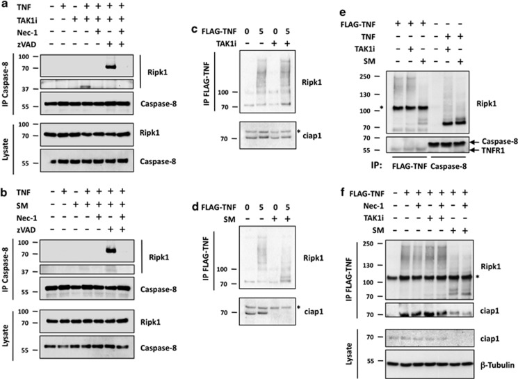
The combination of TNF and SM leads to apoptosis through assembly of a caspase-8-activating complex, referred to as complex IIb, consisting of FADD, procaspase-8 and RIPK1.12, 15, 17 To determine whether TAK1 kinase inhibition similarly induces TNF-mediated RIPK1-dependent apoptosis via complex IIb assembly, we immunoprecipitated caspase-8 in MEFs stimulated either with TNF+TAK1i or TNF+SM, and analyzed RIPK1 recruitment by immunoblotting. These immunoprecipitations were performed in the absence or presence of zVAD-fmk, as previous studies indicated that complex IIb detection requires caspase inhibition to avoid proteolytic cleavage of RIPK1 by capase-8.12, 15, 17, 36 As shown in Figures 3a and b, both TNF+TAK1i and TNF+SM treatments led to complex IIb assembly, which was better detected in the presence of zVAD-fmk. Cleaved fragments of RIPK1 were mostly observed in the absence of zVAD-fmk. Importantly, Nec-1 completely inhibited complex IIb assembly both in the absence or presence of zVAD-fmk, further demonstrating the kinase-dependent role of RIPK1 in TNF+TAK1i and TNF+SM apoptotic settings.
Figure 3.
TAK1 kinase inhibition induces complex IIb assembly without affecting RIPK1 ubiquitylation at complex I. (a and b) Immortalized Ripk1+/+ MEFs were pre-treated for 30 min with the indicated compounds in combination with TAK1i (a) or SM (b), followed by stimulation with hTNF for 2 h. Next, complex II was isolated by immunoprecipitation of caspase-8 and analyzed by immunoblotting. (c and d) Immortalized Ripk1+/+ MEFs were pre-treated for 30 min with TAK1i (c) or SM (d) and subsequently stimulated for 5 min with FLAG-hTNF. Complex I was immunoprecipitated and the ubiquitylation status of TNFR1-bound RIPK1 was analyzed by immunoblotting. The levels of cIAP1 in the lysates are also shown. The asterisk shows a non-specific band recognized by the anti-cIAP1 antibody. (e) Immortalized Ripk1+/+ MEFs were pretreated with TAK1i or SM and stimulated with either FLAG-hTNF for 5 min or hTNF for 2 h. Ubiquitylation status of RIP1 in the different complexes was evaluated by immunoblotting after immunoprecipitation of FLAG-hTNF for complex I or caspase-8 for complex II. (f) Immortalized Ripk1+/+ MEFs were pre-treated for 30 min with TAK1i or SM in the presence or absence of Nec-1 and subsequently stimulated with FLAG-hTNF for 5 min. Complex I was immunoprecipitated and recruitment of RIPK1 to TNFR1 was analyzed by immunoblotting
The recruitment of RIPK1 to complex IIb in cIAP1/2-depleted conditions is thought to originate from defective RIPK1 ubiquitylation at complex I. We therefore compared RIPK1 ubiquitylation status at the receptor complex by immunoprecipitating TNFR1 using FLAG-hTNF in MEFs pre-treated, or not, with either TAK1 inhibitor or SM for 30 min. Although SM pre-treatment greatly affected RIPK1 ubiquitylation, TAK1 kinase inhibition had no effect on RIPK1 ubiquitylation status or on cIAP1 levels (Figures 3c and d). Of note, no difference in RIPK1 ubiquitylation levels was observed at the level complex IIb between TNF+TAK1i and TNF+SM treatments (Figure 3e). Also, contrary to its crucial role in complex IIb assembly, we found that RIPK1 kinase activity was dispensable for the recruitment and ubiquitylation of RIPK1 at complex I (Figure 3f).
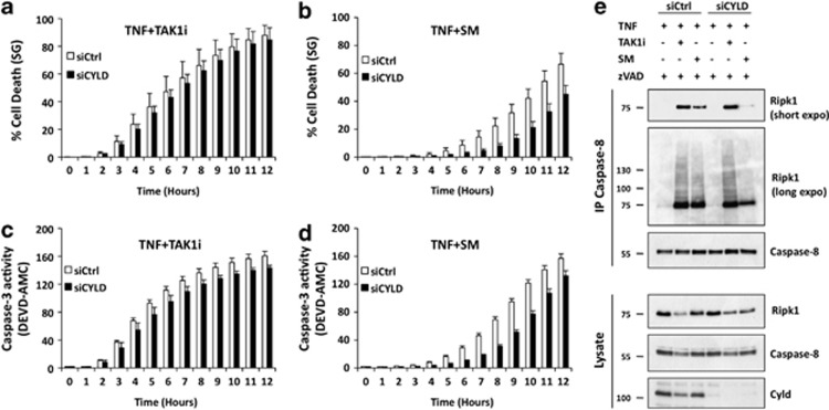
Because the de-ubiquitinase CYLD was suggested to promote complex IIb assembly under SM conditions by dismantling the Ub chains remaining on RIPK1 at complex I,12 we next compared the effect of CYLD repression on apoptotic cell death induced by TNF+TAK1i and TNF+SM. As shown in Figures 4a–d (and Supplementary Figure S6), CYLD repression provided protection to apoptosis induced by TNF+SM, but not by TNF+TAK1i. Those effects, respectively, correlated with defective and unaffected complex IIb formation (Figure 4e).
Figure 4.
CYLD knockdown protects cells against RIPK1-dependent apoptosis induced by TNF and SM, but not by TNF and TAK1i. (a–e) Immortalized Ripk1+/+ MEFs were transfected with a control siRNA or siRNA-targeting CYLD. After 72 h, cells were pretreated with TAK1i (a and c) or SM (b and d) for 30 min followed by stimulation with hTNF. Cell death (a and b) and caspase-3 activity (c and d) were analyzed at the indicated time points using the Fluostar Omega fluorometer. Error bars indicate the standard deviation from triplicate samples. The results are representative of at least two independent experiments. (e) Immortalized Ripk1+/+ MEFs were transfected with a control siRNA or siRNA-targeting CYLD. After 72 h, cells were pretreated with TAK1i or SM in the presence of zVAD for 30 min followed by the stimulation with hTNF for 2 h. Complex II assembly was analyzed by immunoblotting after immunoprecipitation of caspase-8
Our data therefore indicate that it is not per se the Ub chains conjugated to RIPK1 at complex I but instead the recruitment of TAK1 to these Ub chains that, either directly or indirectly, regulates the integration of RIPK1 in complex IIb. The fact that CYLD repression does not provide protection under TAK1 kinase inhibition highlights the role of TAK1 downstream of RIPK1 ubiquitylation in the early NF-_κ_B-independent cell death checkpoint of the TNFR1 pathway.
RIPK3 deficiency affects TNFR1-mediated RIPK1 kinase-dependent apoptosis
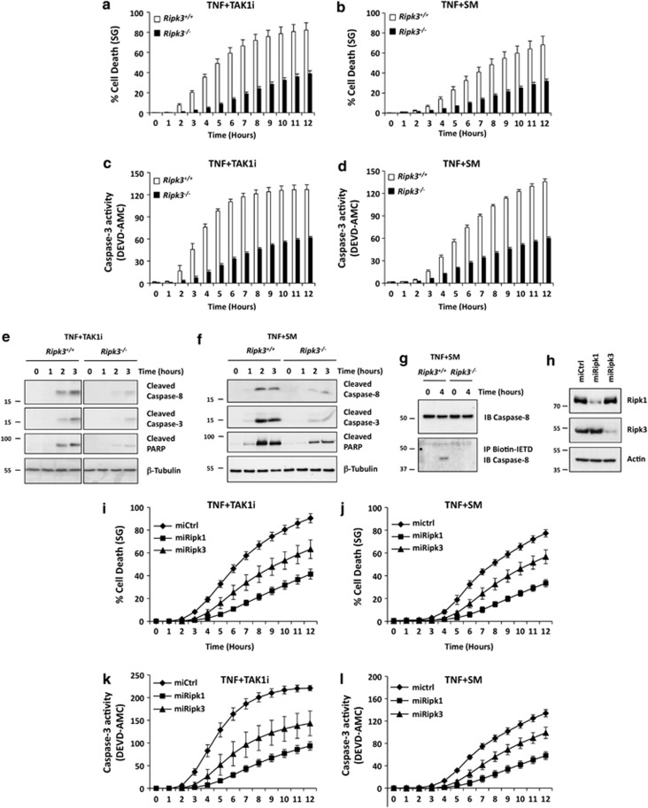
RIPK3 has emerged as an essential kinase in necroptosis induction25, 26, 27 and, as a consequence, RIPK3 repression has become a proof of principle to highlight the contribution of necroptosis in a defined phenotype; although initial reports on RIPK3 indicated that its ectopic expression could also induce apoptosis.37, 38 To investigate whether RIPK3 could contribute to TNF-induced RIPK1-dependent apoptosis, we stimulated immortalized Ripk3+/+ and _Ripk3_−/− MEFs with TNF+SM and TNF+TAK1i. Remarkably, we observed that RIPK3 deficiency provided partial protection to both apoptotic triggers (Figures 5a–d, Supplementary Figure S7). The reduction in RIPK1-dependent apoptosis was accompanied by a reduction in caspase-8, caspase-3 and PARP cleavage (Figures 5e and f), indicating a role for RIPK3 upstream or at the level of caspase-8. To further validate the role of RIPK3 in TNFR1-mediated RIPK1-dependent caspase-8 activation, we immunoprecipitated active caspase-8 using biotin-IETD-fmk in Ripk3+/+ and _Ripk3_−/− primary MEFs stimulated with TNF+SM, and again observed defective caspase-8 activation in the absence of RIPK3 (Figure 5g). The role of RIPK3 in TNF+TAK1i- and TNF+SM-induced apoptosis was also confirmed by repressing RIPK3 in the Ripk1+/+ MEFs using miRNA (Figures 5h–l, Supplementary Figure S8). Interestingly, RIPK3 deficiency did not provide protection to RIPK1-independent apoptosis induced by Fas ligation (Supplementary Figure S9).
Figure 5.
RIPK3 deficiency affects TNFR1-mediated RIPK1-dependent apoptosis. (a–d) Immortalized Ripk3+/+ and _Ripk3_−/− MEFs were pre-treated for 30 min with TAK1i (a and c) or SM (b and d) and subsequently stimulated with hTNF. Cell death (a and b) and caspase-3 activity (c and d) were analyzed at the indicated time points using the Fluostar Omega fluorometer. Error bars indicate the standard deviation from triplicate samples. The results are representative of at least three independent experiments. (e and f) Immortalized Ripk3+/+ and _Ripk3_−/− MEFs were pre-treated for 30 min with SM (e) or TAK1i (f), followed by stimulation with hTNF for the indicated periods of time. Cells were then lysed and immunoblotted as indicated. (g) Primary Ripk3+/+ and _Ripk3_−/− MEFs were pre-treated for 30 min with biotinylated-IETD-fmk and SM and subsequently stimulated with hTNF for 4 h. Cells were then lysed and caspase-8 bound to biotinylated-IETD-fmk was immunoprecipitated. The levels of pro-caspase-8 in the lysates and of cleaved-caspase-8 in the immunoprecipitate were then revealed by immunoblotting. (h–l) Immortalized Ripk1+/+ MEFs were lentivirally transduced with either a control miRNA or a miRNA-targeting RIPK1 or RIPK3. (h) Repression of RIPK1 or RIPK3 protein levels was revealed by immunoblotting. (i–l) miRNA-transduced Ripk1+/+ MEFs were pre-treated for 30 min with TAK1i (i and k) or SM (j and l) and subsequently stimulated with hTNF. Cell death (i and j) or caspase-3 activity (k and l) was analyzed at the indicated time points using the Fluostar Omega fluorometer. Error bars indicate the standard deviation from triplicate samples. The results are representative of at least two independent experiments
Our results therefore demonstrate that RIPK3 specifically contributes to TNFR1-mediated RIPK1-dependent apoptosis, in addition to its well-established role in TNF-induced necroptosis.
RIPK3 differentially regulates TNF-mediated apoptosis and necroptosis
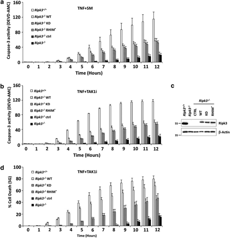
Necroptosis induction relies on both RIPK1 and RIPK3 kinase activities, and on the physical interaction between RIPK1 and RIPK3 via their respective RHIM domains.25, 26 To test whether these domains of RIPK3 are also required for its contribution to TNF-mediated RIPK1-dependent apoptosis, we lentivirally reconstituted _Ripk3_−/− MEFs with an empty vector (ctrl), with wild-type (WT) RIPK3 and with two mutants of RIPK3 reported to inhibit necroptosis induction: a kinase-dead mutant (KD)25 and a RHIM domain mutant (RHIM*).26 Interestingly, we found that reconstitution of _Ripk3_−/− MEFs with all three RIPK3 variants equally rescued caspase-3 activation upon TNF+SM or TNF+TAK1i treatment (Figures 6a and b). The extent of rescue correlated with the relative RIPK3 expression levels between the _Ripk3_−/− reconstituted cells and the Ripk3+/+ MEFs (Figure 6c). Nevertheless, we observed that the cells reconstituted with WT RIPK3 succumbed to the treatment almost as efficiently as the Ripk3+/+ MEFs, whereas the cells reconstituted with KD and RHIM*, RIPK3 only partially recovered the cell death sensitivity (Figure 6d). Because cells reconstituted with the two RIPK3 mutants are unable to rescue any sensitivity to RIPK3-dependent necroptosis (Supplementary Figure S10), the difference in cell death sensitivity between the three RIPK3 variants upon TNF+TAK1i treatment presumably originates from the fact that the ectopic expression of WT RIPK3 in _Ripk3_−/− MEFs induces necroptosis on top of apoptosis, which is impossible for the two RIPK3 mutants.
Figure 6.
The kinase activity of RIPK3 and its intact RHIM domain are dispensable for TNFR1-mediated RIPK1-dependent apoptosis. (a–d) Immortalized _Ripk3_−/− MEFs were lentivirally reconstituted with an empty control vector (CTRL), wild-type Ripk3 (WT), kinase-dead Ripk3 (KD) or RHIM mutated Ripk3 (RHIM*). (a–d) The reconstituted _Ripk3_−/− MEFs were pre-treated for 30 min with SM (a) or TAK1i (b) and subsequently stimulated with hTNF. Caspase-3 activity (a and b) and cell death (d) were analyzed at the indicated time points using the Fluostar Omega fluorometer. Error bars indicate the standard deviation from triplicate samples. The results are representative of at least three independent experiments. (c) RIPK3 expression levels in Ripk3+/+ and reconstituted _Ripk3_−/− MEFs were compared by immunoblotting
Together, our data indicate that RIPK3 contributes to TNF-mediated RIPK1-dependent apoptosis independently of its kinase activity or intact RHIM, and therefore demonstrate that RIPK3 differentially regulates apoptosis and necroptosis downstream of TNFR1.
Reactive oxygen species (ROS) scavenging inhibits TNFR1-mediated RIPK1-dependent apoptosis
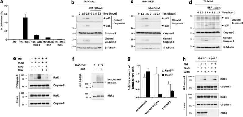
It was recently suggested that, in the context of the inflammasome, RIPK3 induces caspase-8 activation through ROS production via an uncharacterized mechanism.39 To investigate whether the contribution of RIPK3 to caspase-8 activation in our cellular system also involves ROS generation, we first analyzed the effect of ROS scavenging, using butylated hydroxyanisole (BHA) and _n_-acetylcysteine, on RIPK1-dependent apoptosis induced by TNF+TAK1i and TNF+SM. We found that ROS scavenging protected the MEFs from these apoptotic triggers even more potently than Nec-1 (Figure 7a). Importantly, this protection was associated with the inhibition of caspase-8 and caspase-3 processing (Figures 7b–d), implying a role for ROS in the early signaling phase of RIPK1-dependent apoptosis. We therefore analyzed whether ROS scavenging could interfere with complex IIb assembly, and found that 30 min pre-treatment with BHA almost completely inhibited the association of RIPK1 with caspase-8 (Figure 7e). Of note, BHA pre-treatment had no effects on the recruitment of RIPK1 to complex I (Figure 7f), demonstrating a role for ROS in the transition from complex I to complex IIb.
Figure 7.
ROS scavenging inhibits TNFR1-mediated RIPK1-dependent apoptosis. (a) Immortalized Ripk1+/+ MEFs were pre-treated for 30 min with TAK1i in the presence of Nec-1, BHA or NAC and subsequently stimulated with hTNF for 4 h. Cell death was analyzed using the Fluostar Omega fluorescence plate reader. Error bars indicate standard deviation from triplicate samples. Experiments are representative of at least three independent experiments. (b–d) Immortalized Ripk1+/+ MEFs were pre-treated for 30 min with TAK1i (b and c) or SM (d) in the presence or absence of BHA (b and d) or NAC (c). Cells were then stimulated by hTNF for the indicated time and cell lysates were analyzed by immunoblotting. (e) Immortalized Ripk1+/+ MEFs were pre-treated with the indicated compounds for 30 min and then stimulated with hTNF for 2 h. TNFR1 complex IIb was isolated by caspase-8 immunoprecipitation and analyzed by immunoblotting. (f) Immortalized Ripk1+/+ MEFs were pre-treated or not with BHA for 30 min before stimulation with FLAG-hTNF for 5 min. TNFR1 complex I was analyzed by immunoprecipitation using FLAG beads followed by immunoblotting. (g) Immortalized Ripk3+/+ and _Ripk3_−/− MEFs were left unstimulated or pre-treated for 30 min with TAK1i in the presence or absence of zVAD-fmk and subsequently stimulated with hTNF for 3 h. DHR-123 was added for the last 30 min of incubation at 37 °C. Cellular ROS in PI-negative cells was monitored using the BD LSRII flow cytometer. The results are normalized to the percentage of DHR123+/PI- cells detected in unstimulated Ripk3+/+ MEFs. Error bars indicate standard deviation from triplicate samples. The results are representative of two independent experiments. (h) Immortalized Ripk3+/+ and _Ripk3_−/− MEFs were pre-treated with TAK1i in the presence or absence of zVAD-fmk, followed by stimulation with hTNF for 2 h. TNFR1 complex IIb assembly was analyzed by immunoprecipitation of caspase-8
RIPK3 regulates TNF-mediated RIPK1-dependent apoptosis downstream of complex IIb assembly, possibly via ROS generation
Knowing that caspase-8 activation was positively regulated by both RIPK3 and ROS in our TNF+TAK1i and TNF+SM settings, and that a RIPK3-ROS-caspase-8 activation axis had previously been suggested,39 we next analyzed the effect of RIPK3 deficiency on ROS generation upon TNF+TAK1i treatment. As RIPK3 repression is known to affect ROS levels under necroptotic conditions,40 we included TNF+TAK1i+zVAD-fmk treatment as a positive control in our experiments. Remarkably, we found that RIPK3 deficiency affected ROS levels in both necroptotic and apoptotic conditions, whereas no difference in ROS levels was detected in unstimulated conditions (Figure 7g). However, and contrary to ROS scavenging, we found that RIPK3 deficiency did not affect complex IIb assembly (Figure 7h), suggesting a role for RIPK3 in TNF-mediated RIPK1-dependent apoptosis downstream of complex IIb assembly, possibly via ROS generation.
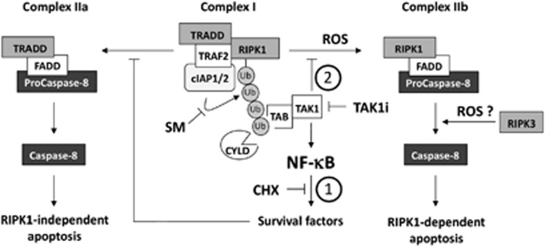
Together, our data therefore demonstrate that ROS production is required upstream and possibly downstream of complex IIb assembly for full caspase-8 activation, and that RIPK3 only contributes to TNF-mediated RIPK1-dependent apoptosis downstream of complex IIb assembly, possibly via ROS generation (Figure 8).
Figure 8.
Model for the two cell death checkpoints in TNF-induced apoptosis. TNF stimulation leads to the assembly of the plasma membrane TNFR1 complex I. Within this complex, RIPK1 is rapidly conjugated with Ub chains. These Ub chains act as scaffolds for the recruitment and activation of the TAB2/3-TAK1 complex and the IKK complex, which subsequently leads to the activation of the canonical NF-_κ_B signaling pathways that drive transcription of genes that prevent cell death — the first cell death checkpoint (1). Accordingly, when the NF-_κ_B response is inhibited, either genetically or by the use of the general translation inhibitor cycloheximide (CHX), TNFR1 ligation switches from a pro-survival to a RIPK1-independent pro-apoptotic response by assembly of the cytoplasmic TNFR1 complex IIa. In addition to its role in NF-_κ_B activation, TAK1 regulates a second cell death checkpoint at the level of complex I that prevents RIPK1 from integrating the death complex (2). Indeed, when TAK1 activity in complex I is affected, either indirectly by avoiding TAK1 recruitment through the use of Smac Mimetics (SM) — which affects RIPK1 ubiquitylation — or directly by inhibiting its kinase activity (TAK1i), TNF stimulation induces RIPK1 kinase-dependent apoptosis by assembly of the cytosolic death complex IIb. Because CYLD acts upstream of TAK1 recruitment, its repression does not protect cells from apoptosis induced by TAK1 kinase inhibition. RIPK3 contributes to full caspase-8 activation by acting downstream of complex IIb, in a kinase and intact RHIM-independent manner. ROS regulates TNF-induced RIPK1-dependent caspase activation by acting upstream of complex IIb assembly, and possibly also downstream of complex IIb via RIPK3
Discussion
Depending on the cellular context, TNF can induce RIPK1-independent or -dependent apoptosis by assembly of two different cytosolic death complexes, respectively, complex IIa or IIb.11 RIPK1-independent apoptosis is classically obtained by the addition of transcriptional or translational inhibitors, whereas RIPK1-dependent apoptosis is mostly observed in conditions of cIAP1/2 depletion, obtained either by the use of SM or more physiologically after stimulation with TWEAK, which induces cIAP1/2 degradation upon binding to its cognate receptor Fn14.12, 15, 17, 21, 41 As both approaches interfere with the canonical NF-_κ_B-mediated pro-survival response, it is believed that the decision to undergo either complex IIa or IIb apoptosis is regulated at a different level. For instance, the Ub chains conjugated to RIPK1 by cIAP1/2 at the receptor complex I are thought to constitute the decisive factor preventing RIPK1 from integrating complex II and limiting its contribution to the apoptotic dismantlement of the cell.23 cIAP1/2-mediated ubiquitylation of RIPK1, and potentially of other proteins within complex I, provides docking sites for the recruitment of the TAB-TAK1 complex.15, 18, 42 Here, we show that inhibition of TAK1 kinase activity triggers TNF-mediated complex IIb-dependent apoptosis independently of NF-_κ_B and without affecting ubiquitylation of RIPK1 at complex I, therefore demonstrating that the recruitment of TAK1 to the Ub chains, and not the Ub chains themselves, regulates the death potential of RIPK1. Consistently, CYLD repression only provided protection to TNF-mediated death in conditions affecting RIPK1 ubiquitylation in complex I, and not upon TAK1 kinase inhibition, supporting a role for TAK1 downstream of RIPK1 ubiquitylation in this early NF-_κ_B-independent cell death checkpoint. Further investigation is needed to understand how TAK1 regulates the death potential of RIPK1, but it is tempting to speculate that TAK1, either directly or indirectly, negatively regulates RIPK1 kinase activity at the level of complex I. Indeed, we found that TNF-mediated RIPK1-dependent apoptosis relies on RIPK1 activity, and previously reported that the sensitization of L929 cells to TNF-induced necroptosis upon TAK1 kinase inhibition was associated with enhanced RIPK1 kinase activity.40 Interestingly, it was recently reported that NEMO also negatively regulates TNF-mediated RIPK1-dependent apoptosis and necroptosis independently of NF-_κ_B.43, 44 Whether TAK1 and NEMO collaborate in the early NF-_κ_B-independent cell death checkpoint downstream of TNFR1 by regulating RIPK1 kinase activity is an interesting possibility that will deserve future attention.
The interest for necroptosis has grown dramatically over the past few years, mainly due to recent studies demonstrating its importance under physiological and pathological conditions.24 Although the molecular mechanisms regulating necroptosis are not yet fully understood, it is well established that the kinase activities of both RIPK1 and RIPK3, and the physical interaction between these two kinases, are crucial steps in mediating this type of cell death. As a consequence, the inhibition of RIPK1 kinase activity by Nec-1 and the repression of RIPK3 levels, either by genetically or by RNA interference, have become commonly used approaches to estimate the contribution of necroptosis both in vitro and in vivo.29, 45 The results presented in this study, however, demonstrate that none of these two approaches is an absolute tool to characterize necroptosis, as both RIPK1 kinase activity and RIPK3 are also required for TNF-mediated apoptosis under TNF+SM or TNF+TAK1i treatment. In line with this, our results demonstrate that ROS generation is not only required for TNF-mediated necroptosis,40 as in this study ROS scavenging also inhibited caspase activation and complex IIb assembly upon TNF stimulation. The fact that RIPK3 deficiency affects the extent of caspase-8 activation and ROS levels without disturbing complex IIb assembly itself suggests the involvement of ROS both upstream and downstream of complex IIb, with RIPK3 only contributing to ROS generation downstream of complex IIb. The idea that RIPK3 contributes to apoptosis independently of RIPK1 binding is supported by the results from our reconstitution experiment, in which both WT RIPK3 and the RHIM mutant similarly rescued caspase activation upon TNF+SM or TNF+TAK1i treatment. In addition, a RIPK1-independent role of RIPK3 in ROS-mediated caspase-8 activation has also recently been reported for inflammasome activation.39 In the context of necroptosis, two recent studies provided a potential link between RIPK3 and mitochondrial ROS production that involves binding of RIPK3, and not RIPK1, to MLKL.46, 47 However, MLKL probably does not fulfill this function under the apoptotic conditions studied here, as the interaction between RIPK3 and MLKL was shown to depend on RIPK3 kinase activity and our results show that the KD mutant of RIPK3 also recapitulate the sensitivity to TNF-induced apoptosis. Our results on the kinase-independent role of RIPK3 in TNF-mediated apoptosis are in line with early work reporting apoptosis induction upon ectopic expression of KD RIPK3 mutants.37, 48 It was also previously reported that RIPK3 could, directly or indirectly, interact with the prodomain of caspase-8 upon ectopic expression.37 It is therefore possible that the contribution of RIPK3 to TNF-mediated apoptosis involves another mechanism than ROS production. Further studies are therefore required to better understand the scaffolding role of RIPK3 in TNF-induced RIPK1-dependent apoptosis and to decipher the link between RIPK3, ROS and caspase-8 under these apoptotic conditions.
Our results demonstrate that, in addition to its well-established role in necroptosis, RIPK3 contributes to TNF-induced RIPK1-dependent apoptosis in a way that does not require its intact RHIM domain and its kinase activity. Our work therefore opens doors for the re-interpretation of the specific necroptotic phenotypes reported in previous studies on RIPK3. This study also highlights the need to identify new molecules, or molecular mechanisms, to specifically interfere with necroptosis induction. Importantly, as RIPK3 kinase activity and the RHIM domain are required for necroptosis but dispensable for apoptosis induced by TNF+SM and TNF+TAKi, the development of specific RIPK3 kinase inhibitors or small molecules that prevent homotypic RHIM domain interactions remain an interesting future challenge to specifically inhibit necroptosis. In line with this, the development of a RIPK3 kinase-dead knock-in mouse line will be of great value to estimate the relative contribution of RIPK3-dependent necroptosis versus apoptosis in physiological and pathological conditions.
Materials and Methods
Plasmids
The sequences encoding WT Ripk3 and the mutated versions of Ripk3 were cloned into pENTR3C using the cloneEZ pcr cloning kit (GenScript, Piscataway, NJ, USA). Next, these sequences were transferred into a modified pLenti6-V5-puromycin destination vector using the LR gateway recombination system (Life Technologies, Carlsbad, CA, USA). The KD contains the K51A substitution. The RHIM domain mutant was generated by mutating the four consecutive amino acids QIGN (449–451) into AAAA.
Cell lines
SV40 large T-immortalized Ripk1+/+, _Ripk1_−/−, Ripk3+/+ and _Ripk3_−/− MEFs have previously been described.49 Primary Ripk3+/+ and _Ripk3_−/− MEFs were isolated from E12.5 littermate embryos following standard protocol. The Tak1+/+ and _Tak1_−/− MEFs were kindly provided by Dr. Takeuchi.30 The MEFs and HEK293T cells were cultured in Dulbecco's modified Eagle's medium supplemented with 10% fetal calf serum, penicillin (100 IU/ml), streptomycin (0.1 mg/ml), L-glutamine (200 _μ_M) and sodium pyruvate (400 _μ_M). Reconstitution of the _Ripk3_−/− MEFs was done by lentiviral transduction. 1 × 10⁶ HEK293T cells were transfected using calcium phosphate with the empty pLenti6-V5-puromycin plasmid or with the plasmids containing the different versions of Ripk3 in combination with pCAGGS-CrmA and the lentiviral packaging vectors pMD2-VSVG and pCMV-ΔR8.91. The medium was changed after 6 h, and sample was collected 48 h post transfection. The virus-containing supernatant was then used to infect the _Ripk3_−/− MEFs. The infected MEFs were then selected for 4 days by adding 2.5 _μ_g/ml puromycin to the medium. Control retroviral pLXSN vector and pLXSN-I_κ_B_α_SR vector were used to infect immortalized wt MEFs. Stable control and I_κ_B_α_SR-expressing cells were obtained through selection with G418 at 0.5 _μ_g/ml for 1 week.
Antibodies, cytokines and reagents
Antibodies were purchased from the following companies:
Anti-RIP1 (BD Biosciences, San Jose, CA, USA, #610459), anti-RIP3 (Sigma-Aldrich, St Louis, MO, USA, #R4277), anti-caspase-8 (Enzo Life Sciences Inc., Farmingdale, NY, USA, #ALX-804-447), anti-caspase-8 (Cell Signaling, Danvers, MA, USA, #9429), anti-caspase 3 (Cell Signaling, #9662), anti-β tubulin (Abcam, Cambridge, UK, #ab6046-200), anti-cleaved PARP (Asp214; Cell Signaling, #9544S). The anti-cIAP1 antibody was a kind gift from Professor Silke (University of Melbourne).34 Recombinant human TNF-α, produced and purified to at least 99% homogeneity in our laboratory, has a specific biological activity of 3 × 10⁷ IU/mg and was used at 600 IU/ml (20 ng/ml) to stimulate MEFs. Recombinant FLAG-hTNF-α was purchased from VIB Protein Service Facility (Ghent, Belgium) and was used at 1.5 _μ_g/ml. The general protein translation inhibitor CHX (Sigma-Aldrich) was used as 0.25 _μ_g/ml (Figure 1c) and 5 _μ_g/ml (Figure 3a). Nec-1 (Calbiochem, Merck KGaA, Darmstadt, Germany) was used at 10 _μ_M. The TAK1 kinase inhibitor, NP-009245 (AnalytiCon Discovery GmbH, Potsdam, Germany), a derivative of (5Z)-7-Oxozeaenol, was used at 1 _μ_M. The caspase peptide inhibitor, zVAD-fmk (Bachem, Bubendorf, Switzerland), was used at 20 _μ_M. Compound A (TetraLogic Pharmaceuticals Inc., Malvern, PA, USA) was used at 500 nM. BHA (Sigma-Aldrich) was used at 100 _μ_M. _N_-Acetyl-L-cysteine (Sigma-Aldrich) was used at 5 mM.
Analysis of cell death and caspase-3 activity using the Fluostar Omega fluorescence plate reader (BMG Labtech GmbH, Ortenberg, Germany)
MEFs were seeded at least in triplicate at 7500 cells per well in a 96-well adherent plate using L15 medium (Leibovitz medium) or DMEM. The next day, the cells were pre-treated with the indicated compounds for 30 min and then stimulated with hTNF (600 IU/ml) in the presence of 5 _μ_M SytoxGreen (Life Technologies) and 20 _μ_M DEVD-AMC (PeptaNova GmbH, Sandhausen, Germany). SytoxGreen and DEVD-AMC fluorescence intensity were measured in function of the time at intervals of 1 h by using a Fluostar Omega fluorescence plate reader, with excitation/emission filters of 485/520 nm for SytoxGreen and 360/460 nm for DEVD-AMC. The gains set at 1100, 20 flashes per well were taken, with orbital averaging with a diameter of 3 mm. Cell death was calculated by subtracting the induced SytoxGreen fluorescence from the background fluorescence and by dividing the obtained result by the maximal fluorescence (minus the background fluorescence) obtained by permeabilization of the cells using Triton X-100 at a final concentration of 0.1%. The caspase-3 activity was calculated by subtracting the induced DEVD-AMC fluorescence from the background fluorescence and by dividing the obtained result with the SytoxGreen fluorescence obtained from the Triton X-100 permeabilized cells, which served to normalized the value to the amount of cells seeded. All experiments were performed at least twice in triplicates. Validation of the system is presented in Supplementary Figure S3.
Intracellular ROS measurement
MEFs were seeded in triplicate at 3 × 10⁵ cells/well (six-well plate). The next day, the cells were pre-treated with TAK1 kinase inhibitor for 30 min and stimulated with hTNF for an additional 3 h. In the last 30 min of incubation at 37 °C, the cells were supplemented with dihydrorodamine 123 (DHR123). Following treatment, the cells were trypsinized and re-suspended in 1 ml of PI-containing medium. Cellular ROS production was determined by measuring the conversion of DHR123 to R123 by flow cytometry on a triple-laser (405, 488 and 635 nm) LSR-II using the FACSDiva software (BD Biosciences). Only viable cells (PI negative) were gated for the analysis of ROS production. ROS measurement was performed twice in triplicate.
Immunoprecipitation
MEFs were seeded at 2 × 10⁶ cells/10 cm plate the day before stimulation. After treatment with the indicated triggers, MEFs were washed with PBS and lysed in NP-40 lysis buffer (150 mM NaCl, 1% NP-40, 10% glycerol, 10 mM Tris-HCl pH 8) supplemented with EDTA-free protease inhibitor cocktail tablets (Roche Diagnostics, Basel, Switzerland, #11873580001) and phosphatase inhibitor cocktail tablets (Roche Diagnostics, #04906837001). For TNFR1 complex I immunoprecipitation (5 min post stimulation), TNFRI bound to human recombinant FLAG-hTNF was immunoprecipitated overnight using anti-FLAG M2 affinity gel (Sigma-Aldrich). For TNFR1 complex II immunoprecipitation (2 h post stimulation), endogenous caspase-8 was immunoprecipitated overnight using anti-caspase-8-coupled protein A beads. For immunoprecipitation of processed caspase-8, MEFs were first pre-treated with biotin-IETD-fmk (MBL International, Woburn, MA, USA, #JM-1121-20C) for 30 min and then stimulated with hTNF and SM for an additional 3.5 h. The cells were then washed with PBS, lysed and processed caspase-8 bound to biotin-IETD-fmk was immunoprecipitated overnight using streptavidin beads (Life Technologies, #S59G). After all immunoprecipitations, the beads were washed three times in NP-40 lysis buffer and the immunoprecipitated proteins were eluted by adding 60 _μ_l of 2 × Laemmli buffer to the beads. Complexes were subsequently analyzed by immunoblotting (20 _μ_l/lane).
RNAi-mediated knockdown
1 × 10⁶ MEF cells were seeded in a 75-cm2 flask and transfected the next day according to the manufacturer's protocol using 25 nM siRNA-targeting CYLD or 25 nM siCONTROL non-targeting siRNA (ON-TARGET_plus_ SMART pool siRNA; Dharmacon, Thermo Fisher Scientific, Waltham, MA, USA). After 48 h, the cells were trypsinized and seeded. The next day, the cells were stimulated with hTNF and cell death or recruitment of RIPK1 to caspase-8 was determined as described above. Knockdown efficiency was tested by immunoblotting. To achieve a stable miRNA-mediated knockdown of RIPK1 and RIPK3 in the immortalized Ripk1+/+ MEFs, oligos against the 3′UTR of RIPK1 and RIPK3 were cloned into BLOCK-iT HiPerform Lentiviral Pol II miR RNAi Expression System with EmGFP (Life Technologies) and subsequently cloned into the pLenti6.2-V5 destination vector (Life Technologies) by the gateway system. These miRNA-containing plasmids were introduced into immortalized Ripk1+/+ MEFs by lentiviral transduction as described above.
Acknowledgments
We thank Dr. Nozomi Takahashi for constructive scientific discussion. We are also grateful to Kim Newton and Vishva Dixit for providing us with the RIPK3 knockout mouse line that was used to generate Ripk3+/+ and _Ripk3_−/− MEFs, and TetraLogic Pharmaceuticals, Inc. for providing us with the SM CmpA. Y.D. is holder of a Ph.D. fellowship from the Agency for Innovation by Science and Technology (IWT). M.J.M.B. has a tenure track position in the Multidisciplinary Research Program of Ghent University (GROUP-ID). Research in his group is supported by grants from the Research Foundation Flanders (FWO G.0172.12N) and Interuniversity Attraction Poles (IAP 7). Research in the Vandenabeele group is supported by the European grants (Euregional PACT II), Belgian grants (Interuniversity Attraction Poles, IAP 7/32), Flemish grants (Research Foundation Flanders – FWO G.0875.11, FWO G.0973.11, FWO G.0A45.12N, Methusalem grant – BOF09/01M00709), Ghent University grants (MRP, GROUP-ID consortium), grant from the Foundation against Cancer, F94 and grants from VIB. Research in the Dejardin group is supported by Belgian grants from the Interuniversity Attraction Poles, (IAP 7/32) and the « Fédération belge Contre le Cancer ».
Glossary
RIPK1
receptor interacting protein kinase 1
cIAP1/2
cellular inhibitor of apoptosis 1 and 2
CYLD
cylindromatosis
TNF
tumor necrosis factor
TNFR1
TNF receptor 1
TAK1
TGF-_β_-activated kinase 1
ROS
reactive oxygen species
TRADD
TNF receptor-associated death domain
TAB2/3
TAK1-binding protein 2/3
NF-_κ_B
nuclear factor kappa B
IKK
inhibitor of nuclear factor kappa B kinase
CHX
cycloheximide
FADD
Fas-associated protein with death domain
SM
Smac Mimetic
Nec-1
necrostatin-1
zVAD-fmk
Z-Val-Ala-DL-Asp-fluoromethylketone
Ub
ubiquitin
BHA
butylated hydroxyanisole
KD
kinase-dead mutant
The authors declare no conflict of interest.
Footnotes
Supplementary Information accompanies this paper on Cell Death and Differentiation website (http://www.nature.com/cdd)
Edited by G Melino
Supplementary Material
Supplementary Information
References
- Varfolomeev EE, Ashkenazi A. Tumor necrosis factor: an apoptosis JuNKie. Cell. 2004;116:491–497. doi: 10.1016/s0092-8674(04)00166-7. [DOI] [PubMed] [Google Scholar]
- Vandenabeele P, Declercq W, Vanhaesebroeck B, Grooten J, Fiers W. Both TNF receptors are required for TNF-mediated induction of apoptosis in PC60 cells. J Immunol. 1995;154:2904–2913. [PubMed] [Google Scholar]
- Wilson NS, Dixit V, Ashkenazi A. Death receptor signal transducers: nodes of coordination in immune signaling networks. Nat Immunol. 2009;10:348–355. doi: 10.1038/ni.1714. [DOI] [PubMed] [Google Scholar]
- Vandenabeele P, Declercq W, Van Herreweghe F, Vanden Berghe T. The role of the kinases RIP1 and RIP3 in TNF-induced necrosis. Sci Signal. 2010;3:re4. doi: 10.1126/scisignal.3115re4. [DOI] [PubMed] [Google Scholar]
- Micheau O, Tschopp J. Induction of TNF receptor I-mediated apoptosis via two sequential signaling complexes. Cell. 2003;114:181–190. doi: 10.1016/s0092-8674(03)00521-x. [DOI] [PubMed] [Google Scholar]
- Walczak H. TNF and ubiquitin at the crossroads of gene activation, cell death, inflammation, and cancer. Immunol Rev. 2011;244:9–28. doi: 10.1111/j.1600-065X.2011.01066.x. [DOI] [PubMed] [Google Scholar]
- Gerlach B, Cordier SM, Schmukle AC, Emmerich CH, Rieser E, Haas TL, et al. Linear ubiquitination prevents inflammation and regulates immune signalling. Nature. 2011;471:591–596. doi: 10.1038/nature09816. [DOI] [PubMed] [Google Scholar]
- Dynek JN, Goncharov T, Dueber EC, Fedorova AV, Izrael-Tomasevic A, Phu L, et al. c-IAP1 and UbcH5 promote K11-linked polyubiquitination of RIP1 in TNF signalling. EMBO J. 2010;29:4198–4209. doi: 10.1038/emboj.2010.300. [DOI] [PMC free article] [PubMed] [Google Scholar]
- Newton K, Matsumoto ML, Wertz IE, Kirkpatrick DS, Lill JR, Tan J, et al. Ubiquitin chain editing revealed by polyubiquitin linkage-specific antibodies. Cell. 2008;134:668–678. doi: 10.1016/j.cell.2008.07.039. [DOI] [PubMed] [Google Scholar]
- Mahoney DJ, Cheung HH, Mrad RL, Plenchette S, Simard C, Enwere E, et al. Both cIAP1 and cIAP2 regulate TNFalpha-mediated NF-kappaB activation. Proc Natl Acad Sci USA. 2008;105:11778–11783. doi: 10.1073/pnas.0711122105. [DOI] [PMC free article] [PubMed] [Google Scholar]
- Declercq W, Vanden Berghe T, Vandenabeele P. RIP kinases at the crossroads of cell death and survival. Cell. 2009;138:229–232. doi: 10.1016/j.cell.2009.07.006. [DOI] [PubMed] [Google Scholar]
- Wang L, Du F, Wang X. TNF-alpha induces two distinct caspase-8 activation pathways. Cell. 2008;133:693–703. doi: 10.1016/j.cell.2008.03.036. [DOI] [PubMed] [Google Scholar]
- Gentle IE, Wong WW, Evans JM, Bankovacki A, Cook WD, Khan NR, et al. In TNF-stimulated cells, RIPK1 promotes cell survival by stabilizing TRAF2 and cIAP1, which limits induction of non-canonical NF-kappaB and activation of caspase-8. J Biol Chem. 2011;286:13282–13291. doi: 10.1074/jbc.M110.216226. [DOI] [PMC free article] [PubMed] [Google Scholar]
- Varfolomeev E, Goncharov T, Fedorova AV, Dynek JN, Zobel K, Deshayes K, et al. c-IAP1 and c-IAP2 are critical mediators of tumor necrosis factor alpha (TNFalpha)-induced NF-kappaB activation. J Biol Chem. 2008;283:24295–24299. doi: 10.1074/jbc.C800128200. [DOI] [PMC free article] [PubMed] [Google Scholar]
- Bertrand MJ, Milutinovic S, Dickson KM, Ho WC, Boudreault A, Durkin J, et al. cIAP1 and cIAP2 facilitate cancer cell survival by functioning as E3 ligases that promote RIP1 ubiquitination. Mol Cell. 2008;30:689–700. doi: 10.1016/j.molcel.2008.05.014. [DOI] [PubMed] [Google Scholar]
- Moulin M, Anderton H, Voss AK, Thomas T, Wong WW, Bankovacki A, et al. IAPs limit activation of RIP kinases by TNF receptor 1 during development. EMBO J. 2012;31:1679–1691. doi: 10.1038/emboj.2012.18. [DOI] [PMC free article] [PubMed] [Google Scholar]
- Petersen SL, Wang L, Yalcin-Chin A, Li L, Peyton M, Minna J, et al. Autocrine TNFalpha signaling renders human cancer cells susceptible to Smac-mimetic-induced apoptosis. Cancer Cell. 2007;12:445–456. doi: 10.1016/j.ccr.2007.08.029. [DOI] [PMC free article] [PubMed] [Google Scholar]
- Wong WW, Gentle IE, Nachbur U, Anderton H, Vaux DL, Silke J. RIPK1 is not essential for TNFR1-induced activation of NF-kappaB. Cell Death Differ. 2010;17:482–487. doi: 10.1038/cdd.2009.178. [DOI] [PubMed] [Google Scholar]
- Park SM, Yoon JB, Lee TH. Receptor interacting protein is ubiquitinated by cellular inhibitor of apoptosis proteins (c-IAP1 and c-IAP2) in vitro. FEBS Lett. 2004;566:151–156. doi: 10.1016/j.febslet.2004.04.021. [DOI] [PubMed] [Google Scholar]
- Haas TL, Emmerich CH, Gerlach B, Schmukle AC, Cordier SM, Rieser E, et al. Recruitment of the linear ubiquitin chain assembly complex stabilizes the TNF-R1 signaling complex and is required for TNF-mediated gene induction. Mol Cell. 2009;36:831–844. doi: 10.1016/j.molcel.2009.10.013. [DOI] [PubMed] [Google Scholar]
- Vince JE, Pantaki D, Feltham R, Mace PD, Cordier SM, Schmukle AC, et al. TRAF2 must bind to cellular inhibitors of apoptosis for tumor necrosis factor (tnf) to efficiently activate nf-{kappa}b and to prevent tnf-induced apoptosis. J Biol Chem. 2009;284:35906–35915. doi: 10.1074/jbc.M109.072256. [DOI] [PMC free article] [PubMed] [Google Scholar]
- O'Donnell MA, Legarda-Addison D, Skountzos P, Yeh WC, Ting AT. Ubiquitination of RIP1 regulates an NF-kappaB-independent cell-death switch in TNF signaling. Curr Biol. 2007;17:418–424. doi: 10.1016/j.cub.2007.01.027. [DOI] [PMC free article] [PubMed] [Google Scholar]
- Vucic D, Dixit VM, Wertz IE. Ubiquitylation in apoptosis: a post-translational modification at the edge of life and death. Nat Rev. Mol Cell Biol. 2011;12:439–452. doi: 10.1038/nrm3143. [DOI] [PubMed] [Google Scholar]
- Vandenabeele P, Galluzzi L, Vanden Berghe T, Kroemer G. Molecular mechanisms of necroptosis: an ordered cellular explosion. Nat Rev. Mol Cell Biol. 2010;11:700–714. doi: 10.1038/nrm2970. [DOI] [PubMed] [Google Scholar]
- He S, Wang L, Miao L, Wang T, Du F, Zhao L, et al. Receptor interacting protein kinase-3 determines cellular necrotic response to TNF-alpha. Cell. 2009;137:1100–1111. doi: 10.1016/j.cell.2009.05.021. [DOI] [PubMed] [Google Scholar]
- Cho YS, Challa S, Moquin D, Genga R, Ray TD, Guildford M, et al. Phosphorylation-driven assembly of the RIP1-RIP3 complex regulates programmed necrosis and virus-induced inflammation. Cell. 2009;137:1112–1123. doi: 10.1016/j.cell.2009.05.037. [DOI] [PMC free article] [PubMed] [Google Scholar]
- Zhang DW, Shao J, Lin J, Zhang N, Lu BJ, Lin SC, et al. RIP3, an energy metabolism regulator that switches TNF-induced cell death from apoptosis to necrosis. Science. 2009;325:332–336. doi: 10.1126/science.1172308. [DOI] [PubMed] [Google Scholar]
- Degterev A, Hitomi J, Germscheid M, Ch'en IL, Korkina O, Teng X, et al. Identification of RIP1 kinase as a specific cellular target of necrostatins. Nat Chem Biol. 2008;4:313–321. doi: 10.1038/nchembio.83. [DOI] [PMC free article] [PubMed] [Google Scholar]
- Galluzzi L, Vitale I, Abrams JM, Alnemri ES, Baehrecke EH, Blagosklonny MV, et al. Molecular definitions of cell death subroutines: recommendations of the Nomenclature Committee on Cell Death 2012. Cell Death Differ. 2012;19:107–120. doi: 10.1038/cdd.2011.96. [DOI] [PMC free article] [PubMed] [Google Scholar]
- Sato S, Sanjo H, Takeda K, Ninomiya-Tsuji J, Yamamoto M, Kawai T, et al. Essential function for the kinase TAK1 in innate and adaptive immune responses. Nat Immunol. 2005;6:1087–1095. doi: 10.1038/ni1255. [DOI] [PubMed] [Google Scholar]
- Shim JH, Xiao C, Paschal AE, Bailey ST, Rao P, Hayden MS, et al. TAK1, but not TAB1 or TAB2, plays an essential role in multiple signaling pathways in vivo. Genes Dev. 2005;19:2668–2681. doi: 10.1101/gad.1360605. [DOI] [PMC free article] [PubMed] [Google Scholar]
- Thiefes A, Wolter S, Mushinski JF, Hoffmann E, Dittrich-Breiholz O, Graue N, et al. Simultaneous blockade of NFkappaB, JNK, and p38 MAPK by a kinase-inactive mutant of the protein kinase TAK1 sensitizes cells to apoptosis and affects a distinct spectrum of tumor necrosis factor [corrected] target genes. J Biol Chem. 2005;280:27728–27741. doi: 10.1074/jbc.M411657200. [DOI] [PubMed] [Google Scholar]
- Arslan SC, Scheidereit C. The prevalence of TNFalpha-induced necrosis over apoptosis is determined by TAK1-RIP1 interplay. PloS One. 2011;6:e26069. doi: 10.1371/journal.pone.0026069. [DOI] [PMC free article] [PubMed] [Google Scholar]
- Vince JE, Wong WW, Khan N, Feltham R, Chau D, Ahmed AU, et al. IAP antagonists target cIAP1 to induce TNFalpha-dependent apoptosis. Cell. 2007;131:682–693. doi: 10.1016/j.cell.2007.10.037. [DOI] [PubMed] [Google Scholar]
- Brown K, Gerstberger S, Carlson L, Franzoso G, Siebenlist U. Control of I kappa B-alpha proteolysis by site-specific, signal-induced phosphorylation. Science. 1995;267:1485–1488. doi: 10.1126/science.7878466. [DOI] [PubMed] [Google Scholar]
- Geserick P, Hupe M, Moulin M, Wong WW, Feoktistova M, Kellert B, et al. Cellular IAPs inhibit a cryptic CD95-induced cell death by limiting RIP1 kinase recruitment. J Cell Biol. 2009;187:1037–1054. doi: 10.1083/jcb.200904158. [DOI] [PMC free article] [PubMed] [Google Scholar]
- Sun X, Lee J, Navas T, Baldwin DT, Stewart TA, Dixit VM. RIP3, a novel apoptosis-inducing kinase. J Biol Chem. 1999;274:16871–16875. doi: 10.1074/jbc.274.24.16871. [DOI] [PubMed] [Google Scholar]
- Yu PW, Huang BC, Shen M, Quast J, Chan E, Xu X, et al. Identification of RIP3, a RIP-like kinase that activates apoptosis and NFkappaB. Curr Biol. 1999;9:539–542. doi: 10.1016/s0960-9822(99)80239-5. [DOI] [PubMed] [Google Scholar]
- Vince JE, Wong WW, Gentle I, Lawlor KE, Allam R, O'Reilly L, et al. Inhibitor of apoptosis proteins limit RIP3 kinase-dependent interleukin-1 activation. Immunity. 2012;36:215–227. doi: 10.1016/j.immuni.2012.01.012. [DOI] [PubMed] [Google Scholar]
- Vanlangenakker N, Vanden Berghe T, Bogaert P, Laukens B, Zobel K, Deshayes K, et al. cIAP1 and TAK1 protect cells from TNF-induced necrosis by preventing RIP1/RIP3-dependent reactive oxygen species production. Cell Death Differ. 2011;18:656–665. doi: 10.1038/cdd.2010.138. [DOI] [PMC free article] [PubMed] [Google Scholar]
- Ikner A, Ashkenazi A. TWEAK induces apoptosis through a death-signaling complex comprising receptor-interacting protein 1 (RIP1), Fas-associated death domain (FADD), and caspase-8. J Biol Chem. 2011;286:21546–21554. doi: 10.1074/jbc.M110.203745. [DOI] [PMC free article] [PubMed] [Google Scholar]
- Ea CK, Deng L, Xia ZP, Pineda G, Chen ZJ. Activation of IKK by TNFalpha requires site-specific ubiquitination of RIP1 and polyubiquitin binding by NEMO. Mol Cell. 2006;22:245–257. doi: 10.1016/j.molcel.2006.03.026. [DOI] [PubMed] [Google Scholar]
- O'Donnell MA, Hase H, Legarda D, Ting AT. NEMO inhibits programmed necrosis in an NFkappaB-independent manner by restraining RIP1. PLoS One. 2012;7:e41238. doi: 10.1371/journal.pone.0041238. [DOI] [PMC free article] [PubMed] [Google Scholar]
- Legarda-Addison D, Hase H, O'Donnell MA, Ting AT. NEMO/IKKgamma regulates an early NF-kappaB-independent cell-death checkpoint during TNF signaling. Cell Death Differ. 2009;16:1279–1288. doi: 10.1038/cdd.2009.41. [DOI] [PMC free article] [PubMed] [Google Scholar]
- Duprez L, Takahashi N, Van Hauwermeiren F, Vandendriessche B, Goossens V, Vanden Berghe T, et al. RIP kinase-dependent necrosis drives lethal systemic inflammatory response syndrome. Immunity. 2011;35:908–918. doi: 10.1016/j.immuni.2011.09.020. [DOI] [PubMed] [Google Scholar]
- Sun L, Wang H, Wang Z, He S, Chen S, Liao D, et al. Mixed lineage kinase domain-like protein mediates necrosis signaling downstream of RIP3 kinase. Cell. 2012;148:213–227. doi: 10.1016/j.cell.2011.11.031. [DOI] [PubMed] [Google Scholar]
- Zhao J, Jitkaew S, Cai Z, Choksi S, Li Q, Luo J, et al. Mixed lineage kinase domain-like is a key receptor interacting protein 3 downstream component of TNF-induced necrosis. Proc Natl Acad Sci USA. 2012;109:5322–5327. doi: 10.1073/pnas.1200012109. [DOI] [PMC free article] [PubMed] [Google Scholar]
- Kasof GM, Prosser JC, Liu D, Lorenzi MV, Gomes BC. The RIP-like kinase, RIP3, induces apoptosis and NF-kappaB nuclear translocation and localizes to mitochondria. FEBS Lett. 2000;473:285–291. doi: 10.1016/s0014-5793(00)01473-3. [DOI] [PubMed] [Google Scholar]
- Jouan-Lanhouet S, Arshad MI, Piquet-Pellorce C, Martin-Chouly C, Le Moigne-Muller G, Van Herreweghe F, et al. TRAIL induces necroptosis involving RIPK1/RIPK3-dependent PARP-1 activation. Cell Death Differ. 2012;19:2003–2014. doi: 10.1038/cdd.2012.90. [DOI] [PMC free article] [PubMed] [Google Scholar]
Associated Data
This section collects any data citations, data availability statements, or supplementary materials included in this article.
Supplementary Materials
Supplementary Information
