Enhanced translation of a chloroplast-expressed RbcS gene restores small subunit levels and photosynthesis in nuclear RbcS antisense plants (original) (raw)
Abstract
Ribulose-1,5-bisphosphate carboxylase/oxygenase (Rubisco) is a key enzyme that converts atmospheric carbon to food and supports life on this planet. Its low catalytic activity and specificity for oxygen leads to photorespiration, severely limiting photosynthesis and crop productivity. Consequently, Rubisco is a primary target for genetic engineering. Separate localization of the genes in the nuclear and chloroplast genomes and a complex assembly process resulting in a very low catalytic activity of hybrid Rubisco enzymes have rendered several earlier attempts of Rubisco engineering unsuccessful. Here we demonstrate that the _Rbc_S gene, when integrated at a transcriptionally active spacer region of the chloroplast genome, in a nuclear _Rbc_S antisense line and expressed under the regulation of heterologous (gene 10) or native (_psb_A) UTRs, results in the assembly of a functional holoenzyme and normal plant growth under ambient CO2 conditions, fully shortcircuiting nuclear control of gene regulation. There was ≈150-fold more _Rbc_S transcript in chloroplast transgenic lines when compared with the nuclear _Rbc_S antisense line, whereas the wild type has 7-fold more transcript. The small subunit protein levels in the gene 10/_Rbc_S and _psb_A/_Rbc_S plants were 60% and 106%, respectively, of the wild type. Photosynthesis of gene 10/_Rbc_S plants was approximately double that of the antisense plants, whereas that of _psb_A/_Rbc_S plants was restored almost completely to the wild-type rates. These results have opened an avenue for using chloroplast engineering for the evaluation of foreign Rubisco genes in planta that eventually can result in achieving efficient photosynthesis and increased crop productivity.
Ribulose-1,5-bisphosphate carboxylase/oxygenase (Rubisco) is the most prevalent enzyme on this planet, accounting for 30–50% of total soluble protein in the chloroplast; it fixes carbon dioxide, but oxygenase activity severely limits photosynthesis and crop productivity (1, 2). Rubisco consists of eight large subunits (LSUs) and eight small subunits (SSUs). The SSU is imported from the cytosol, and both subunits undergo several posttranslational modifications before assembly into functional holoenzyme (3).
Genetic manipulation of Rubisco to improve its function has involved both nuclear and plastid genetic engineering, mostly in tobacco. When the plastid _rbc_L gene encoding the LSU was deleted and expressed via the nuclear genome, transgenic plants exhibited a severe Rubisco deficiency (4). When the _rbc_L gene from Chromatium vinosum was expressed via the nuclear genome of Rubisco-deficient plants, it was poorly transcribed, and no foreign LSU was detected (5).
In yet another approach, a mutated _rbc_L gene, engineered into the chloroplast genome of tobacco, resulted in a Rubisco with a specificity factor and carboxylation rate of only 25% of the wild type (6). Also, when the tobacco _rbc_L gene was replaced with a heterologous gene from cyanobacteria or Helianthus annus, Rubisco-deficient plants were obtained (7). Engineering the nuclear genome of Arabidopsis with a cDNA encoding a pea SSU also resulted in a hybrid Rubisco that was compromised in catalysis (8). Engineering the chloroplast genome with the _rbc_L-_rbc_S operon from a nongreen alga resulted in lack of Rubisco assembly (9). Engineering the L2 form of Rubisco from Rhodospirillum rubrum resulted in transgenic plants that were unable to survive at ambient CO2 levels (10).
Yet another approach for Rubisco manipulation was to engineer the _Rbc_S gene into the chloroplast genome. Whitney and Andrews (11) expressed two copies of a His-tagged _Rbc_S gene in transgenic chloroplasts and obtained less than ≈1% accumulation of the SSU. When a nuclear _Rbc_S antisense tobacco mutant (12–14) was used for engineering the _Rbc_S gene via the chloroplast genome, it resulted in decreased photosynthetic activity and retarded growth (15). This mutant line is an ideal target for Rubisco engineering, because any successful Rubisco assembly would be easily discernible. The _Rbc_S gene was expressed under the regulation of a chloroplast ribosome binding site (RBS) (GGAGG); the chloroplast transgenic plants accumulated abundant plastid _Rbc_S transcript, but it was not translated (15). The failure of several attempts to introduce into the chloroplast genome an _Rbc_S gene that can be expressed properly to produce a fully functional Rubisco cast doubt on the usefulness of this approach for Rubisco engineering.
It is evident that a completely functional eukaryotic Rubisco has never been assembled in any foreign host. Therefore, in this study, _Rbc_S cDNA was engineered into a transcriptionally active spacer region of the chloroplast genome of nuclear _Rbc_S antisense tobacco plant. In addition, two different 5′ UTRs were used to facilitate enhanced translation of the _Rbc_S cDNA. This report demonstrates successful Rubisco expression and assembly from plastid-derived SSUs and LSUs of Rubisco to achieve normal plant growth under ambient CO2 conditions and photosynthesis.
Materials and Methods
Construction of Chloroplast Transformation Vectors. The _Rbc_S cDNA was amplified by using PCR from the vector pTSA (a kind gift from Xing-Hai Zhang, University of Illinois at Urbana–Champaign, Urbana) incorporating an _Nco_I restriction enzyme site at the 5′ end to facilitate its cloning downstream to gene 10 and _psb_A 5′ UTRs. The gene 10 5′-UTR/_Rbc_S cDNA and _psb_A promoter/5′-UTR/_Rbc_S cDNA cassettes were cloned into the multiple cloning site of the basic chloroplast transformation vector pLD-CtV (16, 17) to yield pLDADg10_Rbc_S and pLDAD_psb_A_Rbc_S, respectively.
Chloroplast Transformation and Regeneration of Transgenic Plants. Seeds from nuclear _Rbc_S antisense tobacco plants (12) were surface-sterilized and germinated on Murashige and Skoog salt mixture without organic components (MSO)-kanamycin (50 mg/liter) media (18). All the germinating seedlings were resistant to kanamycin, indicating homozygous status of the transgene. Seedlings were transferred to small jars and maintained at 26°C under a 16-h light/8-h dark photoperiod. Leaves obtained from the nuclear _Rbc_S antisense plants were used for particle bombardment, and these original mother plants were maintained in tissue culture or in soil to be used as controls for subsequent analysis.
Particle bombardment with the chloroplast transformation vectors pLDADg10_Rbc_S and pLDAD_psb_A_Rbc_S was carried out as described (18). Putative chloroplast transgenic shoots were screened for site-specific integration of the transgene cassette by using PCR (detailed below). Positive shoots were subjected to another round of selection, and regenerating shoots were transferred to small jars containing Murashige and Skoog salt mixture without organic components (MSO)-spectinomycin (500 mg/liter) medium. After 3–4 weeks, plants were transferred to soil and maintained at 26°C under a 16-h light/8-h dark photoperiod in a growth chamber.
Continuous Illumination. Leaf samples of comparable age and morphology, grown under a normal photoperiod of 16-h light/8-h dark were harvested and frozen in liquid nitrogen for subsequent Northern and Western analyses [zero days (0D)]. Light intensity was maintained at ≈200 μE·m-2·s-1 [1 Einstein (E) is equal to 1 mol of photons] with fluorescent lights. Afterward, plants were kept under continuous light, and leaf tissue was harvested after 1, 3, or 5 days for Northern and Western analyses. The nuclear _Rbc_S antisense line used for this study was the original mother plant from which the chloroplast transgenic plants were obtained.
PCR Screening and Southern Analysis. Total cellular DNA was isolated from 100 mg of leaf tissue by using DNeasy Plant Mini Kit (Qiagen, Valencia, CA). To investigate homoplasmy, _Sma_I-digested total plant DNA was probed with the radiolabeled DNA fragment P1 (Fig. 1_A_). Prehybridization and hybridization were carried out per manufacturer protocol (Stratagene). The radio-labeled probe was prepared by using Ready-To-Go DNA labeling beads per manufacturer protocol (Amersham Pharmacia Biosciences). To confirm the presence of chloroplast-localized _Rbc_S, total plant DNA from Nt-pLDADg10_Rbc_S was digested with _Xba_I and that from Nt-pLDAD_psb_A_Rbc_S with _Spe_I. A radiolabeled 0.5-kb _Rbc_S cDNA fragment was used as the probe to detect plastid-localized _Rbc_S cDNA.
Fig. 1.
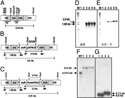
(A) Schematic representation of the 16S-23S region of wild-type tobacco chloroplast genome. The _Bam_HI/_Bgl_II restriction fragment, P1, was used as a probe to investigate homoplasmy. The _Apa_I restriction fragment, P2, was used as a probe to detect 16S rRNA transcript levels in Northern analysis. Also shown are transgenic chloroplast genomes harboring pLDADg10_Rbc_S(B) or pLDAD_psb_A_Rbc_S (C) as well as the annealing positions of primers 3P/3M and 5P/2M and expected amplicon sizes. (D) PCR analysis with primer pair 3P/3M. Lane M, 1-kb DNA ladder; lanes 1 and 2, untransformed; lanes 3 and 4, Nt-pLDADg10_Rbc_S; lanes 5 and 6, Nt-LDAD_psb_A_Rbc_S. (E) PCR analysis with primer pair 5P/2M. Lane M, 1-kb DNA ladder; lanes 1 and 2, untransformed; lanes 3 and 4, Nt-pLDADg10_Rbc_S; lanes 5 and 6, Nt-pLDAD_psb_A_Rbc_S. (F) Southern blot analysis. Total DNA was digested with _Sma_I and probed with a radiolabeled _Bgl_II/_Bam_HI fragment, P1. WT, untransformed; lanes 1 and 2, Nt-pLDADg10_Rbc_S; lanes 3 and 4, Nt-pLDAD_psb_A_Rbc_S. (G) Southern blot analysis. Total DNA was probed with _Rbc_S. Nt-pLDADg10_Rbc_S was digested with _Xba_I (lanes 1 and 2) and Nt-pLDAD_psb_A_Rbc_S was probed with _Spe_I (lanes 3 and 4).
Northern Analysis. Total cellular RNA was isolated from leaves of similar developmental age by using RNeasy Plant Mini Kit (Qiagen). Northern blot analysis (5 μg of RNA per sample) was carried out as described (19). Northern blots were probed with a radiolabeled 0.5-kb _Rbc_S cDNA fragment to detect relative _Rbc_S transcript levels and a radiolabeled 0.9 _Apa_I-digested fragment (P2) (Fig. 1 A) representing 16S rRNA-encoding DNA. Relative transcript levels were measured by using spot densitometry (Alphaimager 3300, Alpha Innotech, San Leandro, CA). For the densitometric measurement of chloroplast transcripts, lane 1 (Fig. 2 A and C) was used, and for determining the antisense and wild-type transcript ratio, lane 3 (Fig. 2 A and C) was used. The measured values were plotted as a bar chart (Fig. 2 B and D).
Fig. 2.
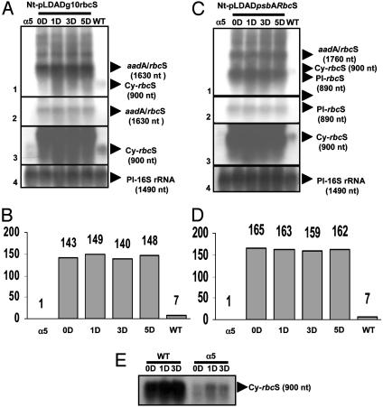
Analysis of transcript levels under normal photoperiod (0D) or continuous light for 1, 3, and 5 days. (A) Nt-pLDADg10_Rbc_S. (B) Densitometry analysis of the Northern blot shown in A. (C) Nt-pLDAD_psb_A_Rbc_S. (D) Densitometry analysis of the Northern blot shown in C. Lanes 1–3 represent the Northern blot exposed for different durations. Cy-_Rbc_S, cytosolic _Rbc_S mRNA (900 nt), plastid (Pl) 16S rRNA (1,490 nt) was used as a control for equal loading (lane 4). (E) Relative _Rbc_S transcript levels in wild-type and _Rbc_S antisense plant (α5). An equal amount of RNA (5 μg) was loaded per sample.
Protein Extraction and Western Analysis. Total soluble protein was extracted from 100 mg (fresh weight) of leaf tissue by using 2× Laemmli buffer. Protein was quantified in three replicates by using the Bio-Rad RC-DC protein quantification kit per manufacturer protocol. An equal volume (10 μl) of the protein extract (qualitative analysis) or equal quantity (10 μg, quantitative analysis) of total soluble protein was subjected to 0.1% SDS/14% PAGE, and Western blot analysis was performed by using antibody against tobacco Rubisco (20). Alkaline phosphatase-linked secondary antibody was used to detect the Rubisco LSUs and SSUs by using Lumi-Phos WB chemiluminescent reagent (Pierce). The SDS/PAGE gels were stained by using GelCode blue stain (Pierce). Relative SSU levels were measured by using spot densitometry (Alphaimager 3300, Alphaimager Corp.). The measured values were plotted as a bar chart (Figs. 3 B and D and 4 B and D).
Fig. 3.
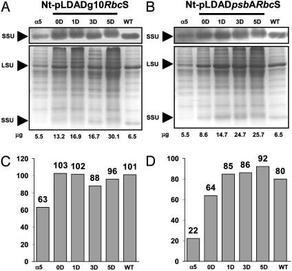
Qualitative comparison of SSU protein levels in 100 mg (fresh weight) of leaf tissue and an equal volume (10 μl of each) examined on a 14% SDS/PAGE gel. (A and C, Upper) Immunoblot probed with anti-Rubisco antibody. (A and C, Lower) GelCode blue-stained SDS/PAGE gel. Numbers below the stained gel indicate the amount of protein loaded (in μg). Shown are Nt-pLDADg10_Rbc_S (A) or Nt-pLDAD_psb_A_Rbc_S (C) grown under normal photoperiod (0D) or under continuous light for 1, 3, and 5 days. (B and D) Densitometry analysis of the immunoblots shown in A and C, respectively.
Fig. 4.
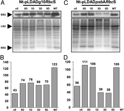
Quantitative comparison of SSU protein level in 10 μg of total protein examined on a 14% SDS/PAGE gel. (A and C, Upper) Immunoblot probed with anti-Rubisco antibody. (A and C, Lower) GelCode blue-stained SDS/PAGE gel. Shown are the Nt-pLDADg10_Rbc_S line (A) or Nt-pLDAD_psb_A_Rbc_S (C) grown under normal photoperiod (0D) or exposed to continuous light for 1, 3, and 5 days. (B and D) Densitometry analysis of the immunoblots shown in A and C, respectively.
Measurement of Photosynthetic CO2 Assimilation Rate. The photosynthetic CO2 assimilation rates were measured with an LI-6400 instrument (Li-Cor, Lincoln, NE). The following parameters were used: 28°C leaf temperature, 21% O2, 1,200 μmol·m-2·s-1 light, 40% relative humidity, and CO2 concentration ranging from 0 to 1,200 μbar (1 bar = 100 kPa). Two leaves per plant of similar developmental age (≈50 days old) were assayed.
Results and Discussion
Chloroplast Transformation Vectors. The tobacco chloroplast transformation vector pLD-CtV targets the transgene cassette to the 16S-_trn_I/_trn_A-23S region of the chloroplast genome (Fig. 1 A; see refs. 16 and 17 for details). To achieve enhanced translation, the tobacco _Rbc_S cDNA along with its transit peptide-coding sequence was cloned into the multiple cloning site of pLD-CtV downstream of two different 5′ UTRs. A _psb_A3′ UTR is already present in pLD-CtV, which aids in transcript stability (21). The transit peptide sequence was retained for chloroplast expression in both the vectors, because it is probably involved in positively affecting _Rbc_S mRNA abundance and transcript stability in chloroplasts (11, 15).
In the chloroplast transformation vector pLDADg10_Rbc_S, expression of the _Rbc_S gene is regulated by a gene 10 5′ UTR (Fig. 1_B_). The gene 10 5′ UTR is derived from bacteriophage and is known to enhance translation in chloroplasts considerably when fused to endogenous promoters (22). In addition, it is free from developmental or light regulation, because it is derived from a foreign source. In this expression cassette, _Rbc_S cDNA should be transcribed as a dicistron in the chloroplasts, because the upstream gene, _aad_A, is devoid of a 3′ UTR and the gene 10 5′ UTR is promoterless. In pLDAD_psb_A_Rbc_S, expression of the _Rbc_S gene is regulated by the _psb_A promoter/5′ UTR (Fig. 1_C_). Because of the presence of a separate promoter, this expression cassette should produce an _Rbc_S monocistron in addition to an _aad_A-_Rbc_S dicistron.
Confirmation of Transgene Integration and Determination of Homoplasmy. Both chloroplast transformation vectors were introduced into the chloroplasts of nuclear _Rbc_S antisense leaves via particle bombardment. The presence of an origin of replication in the chloroplast transformation vector allows the vector to replicate independently and provide more copies for integration, thus expediting the process of achieving homoplasmy even in the first round of selection (16, 17). After 4–5 weeks, resistant shoots were obtained on RMOP-spectinomycin (500 mg/liter) medium. Putative chloroplast transgenic clones were screened initially for site-specific transgene integration by using primer pair 3P and 3M. Primer 3P anneals to the DNA sequence upstream of the flanking region used in the chloroplast transformation vector, and primer 3M anneals within the _aad_A coding sequence (Fig. 1_B_). True chloroplast transgenic shoots should produce an amplicon of 1.65 kb that should be absent in nuclear transgenic plants or mutants. PCR data from two clones, each of which has incorporated the transgene cassette, harboring pLDADg10_Rbc_S and pLDAD_psb_A_Rbc_S are shown (Fig. 1_D_). The presence of a 1.65-kb PCR product in the chloroplast transgenic plants confirms site-specific integration of the transgene cassette. To verify the presence of the complete transgene cassette, primers 5P and 2M were used. Primer 5P anneals within the _aad_A coding region, and 2M anneals to the right flanking region of the chloroplast transformation vector (Fig. 1 B and C). Shoots that have incorporated the pLDADg10_Rbc_S cassette produced a 2.15-kb amplicon, whereas shoots successfully transformed with the pLDAD_psb_A_Rbc_S vector produced a 2.3-kb amplicon (Fig. 1_E_). These PCR-positive shoots were divided into small pieces and placed on RMOP-spectinomycin medium for a second round of selection to achieve homoplasmy.
Homoplasmy was investigated by Southern blot analysis. Restriction digestion of total plant DNA with _Sma_I should produce a 4.01-kb fragment in the wild-type plastome when probed with a _Bam_HI/_Bg_lII radiolabeled fragment P1 (Fig. 1 A). Plants successfully transformed with the pLDADg10_Rbc_S and pLDAD_psb_A_Rbc_S should produce a fragment of 6.0 and 6.15 kb, respectively (Fig. 1 B and C). Southern blot analysis confirmed the presence of a transformed plastid genome in the selected lines. The wild-type sample generated the predicted fragment of 4.01 kb, whereas samples from the Nt-pLDADg10_Rbc_S and Nt-pLDAD_psb_A_Rbc_S generated the predicted fragment of 6.0 and 6.15 kb, respectively (Fig. 1_F_). Absence of a wild-type fragment of 4.01 kb in the chloroplast transgenic plants confirmed the homoplasmy in these plants; i.e., all the chloroplast genomes were uniformly transformed with _Rbc_S cDNA. To confirm the presence of _Rbc_S cDNA, total plant DNA from Nt-pLDADg10_Rbc_S plants was digested with _Xba_I, and that from Nt-pLDAD_psb_A_Rbc_S was digested with _Spe_I. The presence of 0.60-kb (Nt-pLDADg10_Rbc_S) and 0.73-kb (Nt-pLDAD_psb_A_Rbc_S) fragments in the Southern blots probed with a radio-labeled _Rbc_S cDNA fragment confirmed the presence of _Rbc_S cDNA (Fig. 1_G_).
Transcript Analysis of RbcS in Chloroplast Transgenic Plants. The chloroplast-expressed _Rbc_S cDNA is composed of the transit peptide sequence along with the coding sequence for the SSU of Rubisco. The nuclear _Rbc_S transcript is ≈900 nt long, and it encodes the SSU precursor (12). In Nt-pLDADg10_Rbc_S plants, _Rbc_S cDNA is expressed under the regulation of a promoterless gene 10 5′ UTR. The _Rbc_S cDNA thus should be transcribed as a dicistron of ≈1,630 nt. Total cellular RNA isolated from a nuclear _Rbc_S antisense plant, an Nt-pLDADg10_Rbc_S plant grown under normal photoperiod (0D) or exposed to continuous light for 1, 3, or 5 days, and a wild-type tobacco plant were probed with radiolabeled _Rbc_S cDNA. From the autoradiogram, it is evident that the _aad_A/_Rbc_S dicistron (1,630 nt) is highly abundant, whereas the nuclear transcript (900 nt) is barely detectable (Fig. 2 A, lane 2). The blot had to be exposed for a longer duration to perform densitometry analysis (Fig. 2 A, lanes 1 and 3). Densitometry analysis revealed that at 0D, the plastid-derived _Rbc_S transcript was 143-fold more than the transcript levels in the nuclear _Rbc_S antisense plant (Fig. 2_B_), which is in sharp contrast to an only 20-fold increase in plastid-derived _Rbc_S transcript level observed in an earlier study in which the transgene was integrated into the single-copy region of the tobacco chloroplast genome (15). The _Rbc_S transcript levels in the wild type were only 6.8-fold that of the antisense, similar to that reported previously (12, 15). As expected, there was no increase in _Rbc_S transcription during continuous exposure to light in the chloroplast-engineered plant. The Northern blot had to be overexposed to observe the nuclear transcript in the nuclear _Rbc_S antisense and wild-type plants.
In Nt-pLDAD_psb_A_Rbc_S line, _Rbc_S is expected to be transcribed as a monocistron as well as an _aad_A-_Rbc_S dicistron. The monocistron and the dicistron should be 890 and 1,760 nt long, respectively. (Fig. 2_C_). In this case also, the nuclear transcripts were undetectable after normal exposure of the blot (Fig. 2_C_, lane 2). Therefore, the blot was exposed for a longer duration to perform densitometry analysis. Based on densitometry analysis, the chloroplast-derived _Rbc_S transcripts were 165-fold more than the transcript levels in the nuclear _Rbc_S antisense plants (Fig. 2_D_). In an earlier study, _Rbc_S was engineered into a transcriptionally silent region of the chloroplast genome, but the transcript abundance was only 5-fold more than the nuclear-derived wild-type _Rbc_S transcript levels (6); no _Rbc_S antisense plants were used in this study, and a direct comparison could not be made. However, we report here a 25-fold increase in _Rbc_S transcript abundance over the wild-type nuclear transcript level. As expected, there was no discernible effect of continuous light exposure on transcription of _Rbc_S cDNA integrated into plastid genomes (Fig. 2_C_) in these plants as well. Engineering of another transgene coding for the human serum albumin at the same integration site, under the regulation of _psb_A promoter, resulted in similar transcript abundance under light and dark, again confirming that light does not influence transcription (19). The native _psb_A transcript composed of the 5′ and 3′ UTRs has been shown to be stable for ≈40 h (23). Therefore, transcript stability should result in similar _Rbc_S transcript levels even under continuous light. For controls, transcript analysis also was performed on a wild-type and a nuclear _Rbc_S antisense plant maintained under normal photoperiod (0D) or exposed to continuous light for 1 and 3 days (Fig. 2_E_). Continued light exposure initially had only a slight enhancing effect on the steady-state transcript levels in the nuclear antisense plants, and the levels decreased on the third day (Fig. 2_E_). As expected, there was a 2-fold increase in _Rbc_S transcript levels in the wild type after continued light exposure, because the _Rbc_S transcription is known to be light-regulated (24).
The large increase (20- to 25-fold) in steady-state _Rbc_S transcript levels in chloroplast transgenic plants as compared with the wild type can be attributed partly to the high copy number (≈20,000) of the chloroplast-engineered gene when integrated into the inverted repeat region. All the earlier efforts of manipulating the chloroplast genome for Rubisco engineering integrated the transgene into transcriptionally silent regions. The site of integration used here is the intergenic region between the _trn_I-trn_A genes in the rrn operon present in the inverted repeat regions of the tobacco chloroplast genome. This site is transcriptionally active because of the read-through transcription from the upstream 16S promoter (P_rrn), which is responsible for transcribing six native genes. This is responsible for the high accumulation of the foreign transcripts when transgenes are integrated at this site; chloroplast foreign transcripts were observed to be 169-fold higher than the best nuclear transgenic lines even in the absence of a promoter for this transgene (25). To rule out any loading artifact, the blots were probed with 16S rRNA-encoding DNA fragment. An equal amount of transcripts corresponding to 16S rRNA (1,490 nt) was detected in all the samples, confirming equal loading of total RNA (Fig. 2 A and C, lane 4).
Qualitative Analysis of SSU. Two different 5′ UTRs were used in this study to enhance the translation of _Rbc_S cDNA. The T7 gene 10 5′ UTR is independent of any cellular control in plants and should facilitate constitutive translation of the transgene. The second chloroplast transformation vector has the _psb_A promoter/5′ UTR regulating the expression of _Rbc_S cDNA. All plants were analyzed further for total protein and the level of SSUs and LSUs (Fig. 3 A and C). On an equal fresh-weight basis, both transformed lines Nt-pLDADg10_Rbc_S and Nt-pLDAD_psb_A_Rbc_S contained a higher level of total soluble protein when compared with nuclear antisense and the wild-type plants. This result is in contrast to observations made earlier in which _Rbc_S cDNA was expressed under the regulation of the Shine–Dalgarno (GGAGG) sequence in the chloroplast genome of nuclear _Rbc_S antisense plants. In that study, the total soluble protein on an equal leaf-area basis in the chloroplast transgenic plant was similar to the nuclear _Rbc_S antisense plants and one third of wild type (15). In the present study, an equal volume (10 μl) of protein sample derived from equal fresh weight of leaf tissue (100 mg) was subjected to SDS/PAGE. The total protein content in leaves of Nt-pLDADg10_Rbc_S under a normal photoperiod (0D) was 2.4- and 2.0-fold more when compared with the nuclear antisense and wild type, respectively (Fig. 3_A_). Furthermore, there was a 2.3-fold increase in the total protein content in these transgenic plants by the fifth day of continuous light exposure (Fig. 3_A_). The higher level of protein under a normal photoperiod (0D) was accompanied by an increased level of SSU, which was 102% when compared with the wild type by Western blot analysis (Fig. 3_B_). A corresponding increase in the level of LSU is clearly visible in the stained protein gel.
Similarly, the total soluble protein in leaves of Nt-pLDAD_psb_A_Rbc_S under a normal photoperiod (0D) was 1.6- and 1.3-fold more when compared with nuclear antisense and wild type, respectively (Fig. 3_C_). There was a 3.0-fold increase in the total soluble protein in these transformed plants on the fifth day of continuous light exposure. Western blot analysis revealed that SSU levels in Nt-pLDAD_psb_A_Rbc_S plant exposed to continuous light for 3 days were 107% of wild-type levels (Fig. 3_D_).
Quantitative Analysis of SSU. SDS gel electrophoresis and Western blots were carried out also by using an equal quantity of protein (i.e., 10 μg per sample). In the Nt-pLDADg10_Rbc_S plant, the percentage SSU level was 58% greater under normal photoperiod (0D) than the nuclear antisense plant, but it was still only 60% of the wild type (Fig. 4 A and B). On the other hand, in the Nt-pLDAD_psb_A_Rbc_S plant, SSU levels under a normal photoperiod (0D) were 106% of wild-type level (Fig. 4 C and D). In both transgenic lines, the levels decreased after 3 and 5 days of continuous light exposure (Fig. 4 A and C). The decrease was particularly prominent in the Nt-pLDAD_psb_A_Rbc_S plant (Fig. 4_C_). Similar observations were made in three different independent experiments. The dynamics of chloroplast-derived _Rbc_S expression in these plants are not understood completely at this point, although it is known that chloroplast proteases are activated under stress caused by prolonged light exposure (26).
From the protein analysis it is apparent that the use of 5′ UTRs for chloroplast-specific expression of _Rbc_S results in enhanced levels of the SSU. The increased levels of SSU are an index of the increased amount of Rubisco, which is also apparent in the GelCode blue-stained SDS/PAGE gels. The gene 10 5′ UTR enhanced the SSU level to 60% of the wild-type level. The Nt-pLDAD_psb_A_Rbc_S line accumulated 106% of SSU when compared with the wild type under normal photoperiod (0D). The _psb_A 5′ UTR has been shown to result in overexpression of several foreign proteins (19, 27), and it was found to be the best among the UTRs investigated in enhancing light-mediated translation (28). However, it is intriguing that in the present study, overexpression of SSU far beyond the wild-type levels was not observed. Continuous light had no enhancing effect on SSU levels after 1 day of exposure, and the levels actually dropped on the third and fifth days. Although the transcript levels remained very high, protein levels were not increased further. Lack of correlation between transcript levels and translation was evident also in an earlier study in which similar transcript levels of chloroplast-engineered human serum albumin resulted in a higher protein level during light but lower protein level under dark (19). Additionally, continued light exposure results in the activation of proteases in the chloroplast that may have contributed to the diminished amounts of SSU (26). It has been shown that the expression of _Rbc_S and _rbc_L is coordinated by adjustments of subunit stoichiometries in response to the abundance of unassembled subunits (13, 29). The question that arises then is: Do the _rbc_L transcripts (10,000 copies in the large single-copy region) and LSU levels become limiting for SSU assembly?
The presence of abundant SSU in the chloroplast transgenic lines indicates that posttranslational cleavage of its transit peptide and assembly into the holoenzyme was achieved successfully inside the chloroplast. In light of this observation, most of the arguments put forth in an earlier, unsuccessful study regarding inefficient posttranslational modification of chloroplastsynthesized SSU (11) have been overcome in our investigations. Expression of foreign proteins from the transcriptionally active _trn_I-_trn_A intergenic region has resulted in the highest level of foreign protein accumulation ever reported in plants, i.e., 46.1% (30). Also, proteins ranging from 11 to 83 kDa in size have been expressed successfully (31, 32) at very high levels. The _trn_I-_trn_A intergenic region has also allowed for single-step multigene engineering for insect resistance and phytoremediation (30, 33, 34). Successful engineering of 11 biopharmaceutical proteins, vaccine antigens, and 6 agronomic traits (for herbicide, insect, disease resistance, drought/salt tolerance, phytoremediation, etc) at this intergenic site make it a preferred site for chloroplast genetic engineering in general (18, 35).
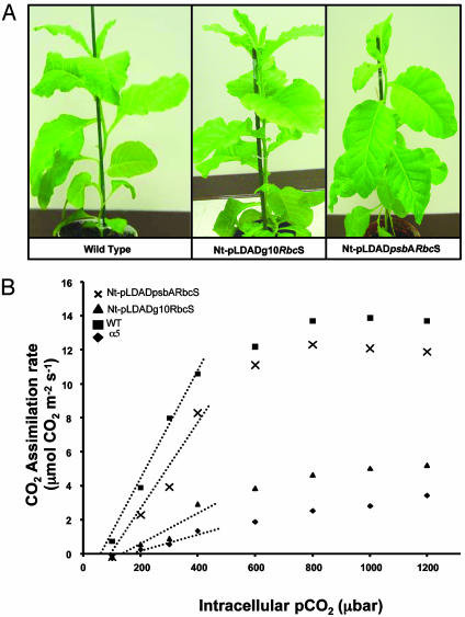
Plant Growth and Photosynthetic CO2 Assimilation Rate. The nuclear _Rbc_S antisense plants are easily distinguishable from wild-type plants, because they have retarded growth and delayed flowering (14). Expression of a promoterless gene 10 5′-UTR-regulated _Rbc_S cDNA in the chloroplasts of nuclear _Rbc_S antisense plant (Nt-pLDADg10_Rbc_S line) resulted in a phenotype that was similar to the wild-type plants under the growth conditions used. However, these plants started flowering ≈27–35 days earlier than the wild-type plants (data not shown) for reasons that remain to be determined. A similar observation was made for five different Nt-pLDADg10_Rbc_S transgenic lines. Comparison between the 29-day-old (after transfer to soil) Nt-pLDADg10_Rbc_S line and wild-type plant is shown in Fig. 5_A_. Expression of _psb_A 5′-UTR-regulated _Rbc_S cDNA in the Nt-pLDAD-_psb_A_Rbc_S plants also resulted in the restoration of normal growth and flowering time.
Fig. 5.
(A) Comparative growth of wild-type, Nt-pLDADg10_Rbc_S, and Nt-pLDAD_psb_A_Rbc_S plants 29 days after transfer to soil in pots. (B) Photosynthetic CO2 assimilation rates under 21% O2. The calculated (dotted lines) initial slopes of CO2 assimilation (μmol CO2 m-2 s-1 μbar-1) were: α5, 0.0051; Nt-pLDADg10_Rbc_S, 0.0096; Nt-pLDAD_psb_A_Rbc_S, 0.0271; and wild type, 0.0336. These data were determined from linear regressions of the data from 100–400 μbar CO2.
Young, fully expanded leaves were subjected to gas-exchange measurements for determining photosynthetic CO2 assimilation. Photosynthesis of the Nt-pLDAD_psb_A_Rbc_S plants was nearly restored to the wild-type level (Fig. 5_B_), indicating increased Rubisco activity in the chloroplast transgenic line derived from nuclear Rbc_S antisense plant. The initial slope of the CO_2 response curve, which is determined by the amount and activity of Rubisco, in the Nt-pLDAD_psb_A_Rbc_S line, is comparable with the wild type. Photosynthesis of the Nt-pLDADg10_Rbc_S line was not restored completely, but it was ≈2-fold greater than that of the nuclear _Rbc_S antisense plant as determined by comparison of the initial slope of CO2 assimilation.
Conclusions
Successful complementation of the SSU deficiency in a nuclear _Rbc_S antisense line by chloroplast-derived SSU opens avenues for additional research in this field, which has been seriously limited by our inability to engineer _Rbc_S genes via the chloroplast genome. Genetic engineering of Rubisco has been complicated by the separate location of its genes, mechanisms that integrate gene expression in the chloroplast and nucleus, and the low catalytic ability of hybrid Rubisco proteins obtained previously with chloroplast or nuclear transformation. Existing eukaryotic Rubiscos with kinetic properties that can result in greater photosynthesis if introduced into C3 crop plants are being identified by biochemical analysis and improved modeling of photosynthesis, which takes into account the crop canopy structure (36). However, demonstrating that a foreign Rubisco will undergo possibly essential posttranslational modifications and whether it can be properly assembled and expressed at a high level in a foreign host has not been feasible yet. The ability to express the SSU gene successfully from the chloroplast genome, as we have demonstrated here, will facilitate the expression and analysis of foreign Rubisco genes, because only chloroplast transformation will be required and both genes can be engineered simultaneously in a single compartment. Then, the ability of a selected foreign Rubisco to increase photosynthesis and crop productivity can be evaluated further in planta. Use of a nuclear _rbc_S antisense line to hyperexpress mutant or foreign _Rbc_S genes in the chloroplast genome also can serve as a model system to study the dynamics of Rubisco assembly. For example, continued study of the transformed plants created here should provide additional insight into the mechanisms controlling Rubisco expression, because nuclear control via the regulation of SSU expression has been short-circuited. Specifically, consequences of the physical relocation of the _Rbc_S gene into its preevolutionary state on the processing of SSU and effect of light and developmental cues on the expression of _Rbc_S gene can be elucidated. It will be vital to determine whether nuclear control of Rubisco expression is necessary for a proper response to external signals in the environment and thus maximal growth. Consequently, the approach and methods developed here should greatly facilitate future Rubisco engineering to achieve the long-cherished goal of increasing plant productivity.
Acknowledgments
This article is dedicated to the late Professor Lawrence Bogorad. We thank Dr. Steve Rodermel for the nuclear _Rbc_S antisense seeds and Dr. Xing-Hai Zhang for the pTSA clone, harboring the _Rbc_S cDNA. We are extremely grateful to Dr. P. V. V. Prasad for help with the photosynthesis measurements. We also thank Dr. Maria Gallo-Meagher and Dr. Mukesh Jain for generous help. This work was supported by funds from U.S. Department of Agriculture Grant 3611-21000-017-00D (to H.D.).
Abbreviations: Rubisco, ribulose-1,5-bisphosphate carboxylase/oxygenase; LSU, large subunit; SSU, small subunit; 0D, zero days.
References
- 1.Ogren, W. L. (2003) Photosynth. Res. 76**,** 53-63. [DOI] [PubMed] [Google Scholar]
- 2.Spreitzer, R. J. & Salvucci, M. E. (2002) Annu. Rev. Plant Biol. 53**,** 449-475. [DOI] [PubMed] [Google Scholar]
- 3.Houtz, R. L. & Portis, A. R. (2003) Arch. Biochem. Biophys. 414**,** 150-158. [DOI] [PubMed] [Google Scholar]
- 4.Kanevski, I. & Maliga, P. (1994) Proc. Natl. Acad. Sci. USA 91**,** 1969-1973. [DOI] [PMC free article] [PubMed] [Google Scholar]
- 5.Madgwick, P. J., Colliver, S. P., Banks, F. M., Habash, D. Z., Dulieu, H., Parry, M. A. J. & Paul, M. J. (2002) Ann. Appl. Biol. 140**,** 13-19. [Google Scholar]
- 6.Whitney, S. M., Von Caemmerer, S., Hudson, G. S. & Andrews, T. J. (1999) Plant Physiol. 121**,** 579-588. [DOI] [PMC free article] [PubMed] [Google Scholar]
- 7.Kanevski, I., Maliga, P., Rhoades, D. F. & Gutteridge, S. (1999) Plant Physiol. 119**,** 133-141. [DOI] [PMC free article] [PubMed] [Google Scholar]
- 8.Getzoff, T. P., Zhu, G. H., Bohnert, H. J. & Jensen, R. G. (1998) Plant Physiol. 116**,** 695-702. [DOI] [PMC free article] [PubMed] [Google Scholar]
- 9.Whitney, S. M., Baldett, P., Hudson, G. S. & Andrews, T. J. (2001) Plant J. 26**,** 535-547. [DOI] [PubMed] [Google Scholar]
- 10.Whitney, S. M. & Andrews, T. J. (2001) Proc. Natl. Acad. Sci. USA 98**,** 14738-14743. [DOI] [PMC free article] [PubMed] [Google Scholar]
- 11.Whitney, S. M. & Andrews, T. J. (2001) Plant Cell 13**,** 193-205. [DOI] [PMC free article] [PubMed] [Google Scholar]
- 12.Rodermel, S. R., Abbott, M. S. & Bogorad, L. (1988) Cell 55**,** 673-681. [DOI] [PubMed] [Google Scholar]
- 13.Jiang, C. Z. & Rodermel, S. R. (1995) Plant Physiol. 107**,** 215-224. [DOI] [PMC free article] [PubMed] [Google Scholar]
- 14.Tsai, C. H., Miller, A., Spalding, M. & Rodermel, S. (1997) Plant Physiol. 115**,** 907-914. [DOI] [PMC free article] [PubMed] [Google Scholar]
- 15.Zhang, X. H., Ewy, R. G., Widholm, J. M. & Portis, A. R. (2002) Plant Cell Physiol. 43**,** 1302-1313. [DOI] [PubMed] [Google Scholar]
- 16.Guda, C., Lee, S. B. & Daniell, H. (2000) Plant Cell Rep. 19**,** 257-262. [DOI] [PubMed] [Google Scholar]
- 17.Daniell, H., Datta, R., Varma, S., Gray, S. & Lee, S. B. (1998) Nat. Biotechnol. 16**,** 345-348. [DOI] [PMC free article] [PubMed] [Google Scholar]
- 18.Daniell, H., Ruiz, O. N. & Dhingra, A. (2004) Methods Mol. Biol. 286**,** 111-137. [DOI] [PubMed] [Google Scholar]
- 19.Fernández-San Millán, A., Mingo-Castel, A., Miller, M. & Daniell, H. (2003) Plant Biotechnol. J. 1**,** 71-79. [DOI] [PMC free article] [PubMed] [Google Scholar]
- 20.Eckardt, N. A., Snyder, G. W., Portis, A. R. & Ogren, W. L. (1997) Plant Physiol. 113**,** 575-586. [DOI] [PMC free article] [PubMed] [Google Scholar]
- 21.Stern, D. B., Higgs, D. C. & Yang, J. J. (1997) Trends Plant Sci. 2**,** 308-315. [Google Scholar]
- 22.Staub, J. M., Garcia, B., Graves, J., Hajdukiewicz, P. T. J., Hunter, P., Nehra, N., Paradkar, V., Schlittler, M., Carroll, J. A., Spatola, L., et al. (2000) Nat. Biotechnol. 18**,** 333-338. [DOI] [PubMed] [Google Scholar]
- 23.Kim, J., Christopher, D. A. & Mullet, J. E. (1993) Plant Mol. Biol. 22**,** 447-463. [DOI] [PubMed] [Google Scholar]
- 24.Tobin, E. M. & Silverthorne, J. (1985) Annu. Rev. Plant Physiol. 36**,** 569-593. [Google Scholar]
- 25.Lee, S. B., Kwon, H. B., Kwon, S. J., Park, S. C., Jeong, M. J., Han, S. E., Byun, M. O. & Daniell, H. (2003) Mol. Breed. 11**,** 1-13. [Google Scholar]
- 26.Zheng, B., Halperin, T., Hruskova-Heidingsfeldova, O., Adam, Z. & Clarke, A. K. (2002) Physiol. Plant. 114**,** 92-101. [DOI] [PubMed] [Google Scholar]
- 27.Staub, J. M. & Maliga, P. (1994) Plant J. 6**,** 547-553. [DOI] [PubMed] [Google Scholar]
- 28.Eibl, C., Zou, Z. R., Beck, A., Kim, M., Mullet, J. & Koop, H. U. (1999) Plant J. 19**,** 333-345. [DOI] [PubMed] [Google Scholar]
- 29.Rodermel, S. (1999) Photosynth. Res. 59**,** 105-123. [Google Scholar]
- 30.DeCosa, B., Moar, W., Lee, S. B., Miller, M. & Daniell, H. (2001) Nat. Biotechnol. 19**,** 71-74. [DOI] [PMC free article] [PubMed] [Google Scholar]
- 31.Daniell, H., Lee, S. B., Panchal, T. & Wiebe, P. O. (2001) J. Mol. Biol. 311**,** 1001-1009. [DOI] [PMC free article] [PubMed] [Google Scholar]
- 32.Daniell, H., Watson, J., Koya, V. & Leppla, S. H. (2004) Vaccine, in press. [DOI] [PMC free article] [PubMed]
- 33.Ruiz, O., Hussein, H., Terry, N. & Daniell, H. (2003) Plant Physiol. 132**,** 1344-1352. [DOI] [PMC free article] [PubMed] [Google Scholar]
- 34.Daniell, H. & Dhingra, A. (2002) Curr. Opin. Biotechnol. 13**,** 136-141. [DOI] [PMC free article] [PubMed] [Google Scholar]
- 35.Devine, A. L. & Daniell, H. (2004) in Plastids, Annual Plant Reviews, ed. Moller, S. (Blackwell, Oxford), Vol. 13, Chap. 10, in press.
- 36.Zhu, X.-G., Portis, A. R., Jr., & Long, S. P. (2004) Plant Cell Environ. 27**,** 155-165. [Google Scholar]
