Nitric oxide negatively regulates abscisic acid signaling in guard cells by S-nitrosylation of OST1 (original) (raw)
Significance
Drought stress induces the accumulation of the plant stress hormone abscisic acid (ABA). ABA then quickly activates the protein kinase OST1/SnRK2.6 to phosphorylate a number of proteins in guard cells, resulting in stomatal closure to reduce transpirational water loss. How SnRK2.6 is deactivated and how ABA signaling may be desensitized are unclear. This study found that nitric oxide (NO) resulting from ABA signaling causes S-nitrosylation of SnRK2.6 at a cysteine residue close to the kinase catalytic site, which blocks the kinase activity. Dysfunction of S-nitrosoglutathione (GSNO) reductase causes GSNO overaccumulation in guard cells and ABA insensitivity in stomatal regulation. This work thus reveals how ABA-induced NO functions in guard cells to inactivate SnRK2.6 to negatively feedback regulate ABA signaling.
Keywords: NO, ABA, drought, GSNOR, stomata
Abstract
The phytohormone abscisic acid (ABA) plays important roles in plant development and adaptation to environmental stress. ABA induces the production of nitric oxide (NO) in guard cells, but how NO regulates ABA signaling is not understood. Here, we show that NO negatively regulates ABA signaling in guard cells by inhibiting open stomata 1 (OST1)/sucrose nonfermenting 1 (SNF1)-related protein kinase 2.6 (SnRK2.6) through S-nitrosylation. We found that SnRK2.6 is S-nitrosylated at cysteine 137, a residue adjacent to the kinase catalytic site. Dysfunction in the S-nitrosoglutathione (GSNO) reductase (GSNOR) gene in the gsnor1-3 mutant causes NO overaccumulation in guard cells, constitutive S-nitrosylation of SnRK2.6, and impairment of ABA-induced stomatal closure. Introduction of the Cys137 to Ser mutated SnRK2.6 into the gsnor1-3/ost1-3 double-mutant partially suppressed the effect of gsnor1-3 on ABA-induced stomatal closure. A cysteine residue corresponding to Cys137 of SnRK2.6 is present in several yeast and human protein kinases and can be S-nitrosylated, suggesting that the S-nitrosylation may be an evolutionarily conserved mechanism for protein kinase regulation.
Abscisic acid (ABA) plays critical roles in seed dormancy and germination, plant growth, and adaptation to environmental challenges (1, 2). Stresses, such as drought and high salt conditions, increase ABA concentration in plants as a result of ABA biosynthesis or ABA release from its inactive, conjugated forms (3). In the presence of ABA, the ABA receptors in the PYR1 (Pyrabactin Resistance 1)/PYL (PYR1-Like)/RCAR (Regulatory Component of ABA receptor) protein family bind to and inhibit the activity of clade A protein phosphatase 2Cs (PP2Cs), which are considered as coreceptors and negative regulators of ABA signaling (4–6). This process then results in the release of sucrose nonfermenting 1 (SNF1)-related protein kinase 2s (SnRK2s) from suppression by the PP2Cs. As central components of the ABA signaling pathway, the activated SnRK2s phosphorylate dozens of downstream effectors to regulate various physiological processes, including stomatal closure, root growth and development, seed dormancy, seed germination, and flowering (7).
As the gateway for photosynthetic CO2 uptake and transpirational water loss, stomata are critical for plant growth and physiology (8). ABA regulates stomatal movement and mutations in ABA biosynthesis genes (9), or in the PYL or SnRK2.6 (also known as OST1) genes cause open-stomata phenotypes (10). On the other hand, dysfunction of the PP2Cs or overexpression of RCAR1/PYL9 causes stomatal closure (5). Among the three SnRK2s, SnRK2.2, -2.3, and -2.6, which are most important for ABA signaling, SnRK2.6 is preferentially expressed in guard cells and plays a critical role in stomatal regulation, whereas SnRK2.2 and -2.3 are mainly expressed in seeds and young seedlings and are thus more important for seed germination and seedling growth (4, 11). SnRK2.6 phosphorylates the slow (S-type) anion channel associated 1 and inward potassium channel KAT1 (K+ channel in Arabidopsis thaliana 1) to cause stomatal closure (12, 13).
ABA also triggers the generation of several second-messenger molecules, such as calcium, inositol phospholipids, and nitric oxide (NO) and reactive oxygen species (ROS) (14–17). These second messengers are involved in ABA regulation of stomatal closure and other physiological processes (15, 18–20). Among these second messengers, NO has been well documented to have important roles in ABA signal transduction. ABA induces NO generation in roots and guard cells (15, 21). Exogenous application of NO can trigger stomatal closure, whereas application of the NO scavenger 2-(4-carboxyphenyl)-4,4,5,5-tetramethylimidazoline-1-oxyl-3-oxide (c-PTIO) inhibits stomatal closure (22), suggesting a positive role of exogenous NO in stomatal closure. Several studies have suggested that exogenous NO may affect stomatal responses to ABA by regulating an inward K+ channel (20), an anion channel (23), and the generation of nitrated cGMP (24), although the direct target of NO in ABA signaling remains unclear.
In plants, NO is produced by the nitrite-dependent nitrate reductase pathway (15, 25) and a pathway dependent on the nitric oxide associated 1 (NOA1) protein (21), although NOA1 is not an NO synthase (26). NO regulates many physiological processes in plants, including responses to phytohormones, such as ABA, cytokinin, auxin, gibberellins, and salicylic acid, immunity against pathogens, senescence, and flowering (15, 27–31). The deficiency in NO generation in the nia1nia2noa1 triple mutant results in ABA-hypersensitive stomatal closure (21), suggesting a negative role of endogenous NO in ABA signaling. NO overaccumulates in Arabidopsis gsnor1(S-nitrosoglutathione reductase 1)/hot5 (sensitive to hot temperatures 5)/par2 (paraquat resistant 2) mutant plants that are impaired in the GSNO reductase gene (32–34). The gsnor1-3 mutant is hypersensitive to heat stress (33) and bacterial pathogen (28, 31), but is more resistant to oxidative stress (34). Characterization of gsnor1 mutant plants suggested that GSNOR regulates multiple developmental and metabolic programs in Arabidopsis (35). In cytokinin signaling, NO causes S-nitrosylation and inhibition of the histidine phosphotransfer protein AHP1 (Arabidopsis histidine phosphotransfer protein 1) (27). In salicylic acid signaling, the S-nitrosylation of the salicylic acid receptor NPR1 facilitates its oligomerization (31). In addition, NO regulates cell death in plant immunity by S-nitrosylation of the NADPH oxidase AtRBOHD (28).
Here, we show that GSNO and Cys-NO (S-nitrosocysteine) can inhibit SnRK2.6 by S-nitrosylation. The S-nitrosylation of SnRK2.6 occurs at Cys137, which is adjacent to the catalytic loop of the kinase. A Cys137 to Ser mutation causes the kinase to be resistant to inhibition by GSNO in vitro, whereas a Cys137 to Trp mutation results in an inactive kinase in vitro and in vivo. Introduction of the Cys137 to Ser mutated form of SnRK2.6 into gsnor1-3/ost1-3 (open stomata 1–3, a null allele of snrk2.6) partially suppresses the defect in stomatal closure caused by overaccumulation of SNOs because of the gsnor1-3 mutation. Our results suggest that ABA induced S-nitrosylation of SnRK2.6 functions to negatively feedback regulate ABA signaling in plants. Moreover, the S-nitrosylation of cysteine137 is evolutionarily conserved in some AMPK/SNF1-related kinases and glycogen synthase kinase 3/SHAGGY-like kinases (SKs) in plants, yeast, and mammals, suggesting that S-nitrosylation–mediated inhibition may be a general regulatory mechanism for these eukaryotic protein kinases.
Results
GSNO Inhibits SnRK2.6 by S-Nitrosylation of Cys-137 in Vitro.
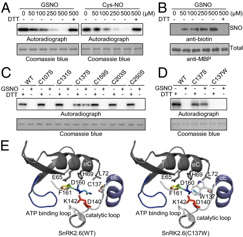
To test whether NO may regulate the activity of components in the core ABA signaling pathway, we tested the effects of the NO donors, GSNO, Cys-NO, and SNAP (S-Nitroso-N-acetylpenicillamine), on the activity of SnRK2.6 in vitro because SnRK2.6 is a key player in the ABA regulation of guard cells and its activity can be readily assayed in vitro and in vivo. As shown in Fig. 1_A_ and Fig. S1, GSNO, Cys-NO, and SNAP inhibited the activity of SnRK2.6 in a dose-dependent manner, but such inhibitory effects were reversed by DTT, indicating that the effects of NO donors rely on the thiol-based redox status. To determine whether SnRK2.6 is S-nitrosylated in the presence of GSNO, we used a biotin-switch method in which the S-nitrosylated cysteine is labeled with biotin and subsequently detected by antibiotin immunoblot analysis (27, 31). As shown in Fig. 1_B_, GSNO induced S-nitrosylation of SnRK2.6 in a dose-dependent manner, and application of DTT abolished the GSNO induced S-nitrosylation. Six cysteine residues in SnRK2.6 are putative target sites of S-nitrosylation. To determine which cysteine is required for GSNO-mediated inhibition, we mutated each cysteine (C) to serine (S) separately and tested the effect of GSNO on the mutated SnRK2.6. Of the six mutations, only the Cys137Ser (C137S) mutation eliminated the inhibitory effect of GNSO (Fig. 1_C_). The C137S mutation also caused a slight reduction in SnRK2.6 kinase activity (Fig. 1 C and D). The SnRK2.6C137W with a Trp substitution at Cys137 that may mimic S-nitrosylation (27, 36) did not show any detectable kinase activity (Fig. 1_D_). Based on the crystal structure of SnRK2.6 resolved by a recent study (37), the side-chain of C137 is exposed on the surface and has only 8 Å distance from D140 in the catalytic loop and D160 in the Mg-binding loop in the catalytic cleft of SnRK2.6 (Fig. 1_E_, Left). The C137W mutation likely interferes directly with the kinase catalytic activity.
Fig. 1.
S-nitrosylation at Cys-137 inhibits the activity of SnRK2.6. (A) Nitric oxide donors GSNO and Cys-NO inhibit the activity of SnRK2.6 in a dose-dependent manner. MBP–SnRK2.6 incubated with indicated concentration of GSNO (Left) and Cys-NO (Right) for 10 min and then [γ-32P]ATP was added to determine the autophosphorylation of SnRK2.6. In the rightmost lane (DTT+), 1 mM DTT was added into the reaction before adding [γ-32P]ATP. (B) GSNO causes S-nitrosylation of SnRK2.6 as detected by the biotin-switch assay. (C) Effects of C-to-S site-directed mutation of the six cysteines on SnRK2.6 activity upon GSNO (50 μM) or DTT treatment. (D) Effects of C137S and C137W mutations on the kinase activity of SnRK2.6. (E) Structure of SnRK2.6 showing the position of Cys-137 (Left) and Trp-137 (Right). Residues E65, H69, L72, K142, D160, F161, and C137 (W137) are shown by sticks.
To further verify the S-nitrosylation of SnRK2.6 at Cys137, we subjected freshly purified SnRK2.6 to a biotin-switch assay, where free cysteines are blocked with S-methymethane thiosulfonate (MMTS) and nitrosylated cysteines are labeled with biotin. Mass spectrometry was performed immediately after the labeling of biotin. The assay detected the S-nitrosylation of Cys137 in SnRK2.6 but none of the other cysteine was S-nitrosylated in the presence of the blocking reagent MMTS (Fig. S2). In the absence of MMTS, Cys131, Cys159, Cys203, and Cys250 were also labeled by biotin (Fig. S3). The peptide containing Cys107 was too short to be detected by mass spectrometry after trypsin digestion. These results suggested that S-nitrosylation occurred specifically at Cys137 after GSNO treatment. We also detected the dehydrogenation of Cys137 and Cys131 in the tryptic fragments after the biotin-switch assay (Fig. S4), suggesting that a disulfide bond may form between Cys137 and Cys131 in some SnRK2.6 molecules. Interestingly, the C131S mutation in SnRK2.6 (C131S) appeared to increase the level of detectable S-nitrosylation relative to the wild-type SnRK2.6 (WT) (Fig. S5). When Cys137 was mutated, virtually no S-nitrosylated SnRK2.6 was detected in the presence of GSNO (Fig. S5). Collectively, these results show that SnRK2.6 is S-nitrosylated at Cys137 in the presence of NO donors and that the S-nitrosylation of SnRK2.6 blocks its kinase activity in vitro.
ABA Enhances the S-Nitrosylation of SnRK2.6 in Vivo.
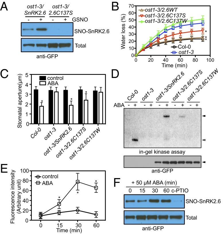
To further confirm that the Cys137 is modified by S-nitrosylation in vivo, we subjected total protein extracts from ost1-3 mutant plants complemented with native promoter driven GFP-tagged wild-type SnRK2.6 (ost1-3/SnRK2.6 WT -GFP) to biotin-switch analysis. Biotin-labeled SnRK2.6-GFP was purified with streptavidin beads and detected by anti-GFP immunoblot analysis. As shown in Fig. 2_A_, a low level of S-nitrosylation of SnRK2.6 could be detected under control conditions and exogenous GSNO treatment on the seedlings increased the level of S-nitrosylated SnRK2.6 in ost1-3/SnRK2.6 WT -GFP transgenic plants. In ost1-3/SnRK2.6 C137S -GFP plants, in which the Cys137 is mutated to Serine, S-nitrosylated SnRK2.6 was undetectable. These results suggest that S-nitrosylation of SnRK2.6 at Cys137 occurs in vivo.
Fig. 2.
ABA up-regulates the S-nitrosylation of Cys-137 of SnRK2.6 in vivo. (A) GSNO-induced S-nitrosylation of wild-type but not Cys173Ser mutated form of SnRK2.6-GFP from plants. Twelve-day-old seedlings were treated with or without 250 μM GSNO for 30 min. The biotin-switch and immunoblot assays were performed as described in SI Materials and Methods. (B and C) Analysis of water loss (B) and stomata responses to 10 μM ABA (C) in the wild-type, ost1-3 mutant, and ost1-3 transgenic lines expressing SnRK2.6WT-GFP, SnRK2.6C137S-GFP, and SnRK2.6C137W-GFP. Treatment with water was used as control. Error bars indicate SD. n = 3–5 independent experiments. More than 20 stomata were measured for each treatment in each experiment in C. Student’s t test, *P < 0.05 (significantly different from _ost1-3_). (_D_) In-gel kinase assay showing SnRK2.6 activity before (−) and after (+) 50 μM ABA treatment in the wild-type, the _ost1-3_ mutant, and _ost1-3_ transgenic lines expressing SnRK2.6WT-GFP, SnRK2.6C137S-GFP, and SnRK2.6C137W-GFP. The positions of SnRK2.6 and GFP-fused SnRK2.6 are indicated by open and closed triangles, respectively. (_E_) Average FDA-FM fluorescence intensity indicating NO levels in guard cells upon ABA (10 μM) treatment in Col wild-type. Treatment with water was used as control. Error bars indicate SD (_n_ > 6 from 3 independent experiments). Student’s t test, *P < 0.01. (F) ABA regulation of S-nitrosylation of SnRK2.6 in ost1-3/SnRK2.6 WT -GFP transgenic plants. Twelve-day-old seedlings were treated with or without 50 μM ABA for the indicated times. The seedlings treated with 200 μM c-PTIO were used as a control. The biotin-switch and immunoblot assays were performed as described in SI Materials and Methods.
We compared the water loss and ABA-induced stomatal closure in Col, ost1-3, and transgenic plants expressing wild-type and mutated forms of SnRK2.6. Only SnRK2.6 WT -GFP complemented the water loss (Fig. 2_B_) and stomatal closure (Fig. 2_C_) phenotypes of ost1-3. The transgenic plant ost1-3/SnRK2.6 C137W -GFP, which contains the S-nitrosylation–mimicking mutation at Cys137, was insensitive to ABA-induced stomatal closure (Fig. 2_C_) and had a higher rate of water loss than the wild-type (Fig. 2_B_). SnRK2.6 C137S -GFP partially complemented the phenotypes of ost1-3 (Fig. 2 B and C). Consistent with these phenotypes, in-gel kinase assays showed that SnRK2.6C137S-GFP had a reduced kinase activity and that SnRK2.6C137W-GFP is a “dead” kinase in vivo (Fig. 2_D_). Taken together, our in vitro and in vivo data suggest that S-nitrosylation at Cys137 of SnRK2.6 abolishes its kinase activity.
Consistent with a previous report (15), we found that ABA increased NO generation in guard cells (Fig. 2_E_). To test whether ABA affects the S-nitrosylation of SnRK2.6, we examined the S-nitrosylation of SnRK2.6-GFP upon ABA treatment. Treatment with 50 µM ABA for 15 min did not increase the S-nitrosylation of SnRK2.6. However, ABA treatment for 30 or 60 min increased the level of SnRK2.6 S-nitrosylation (Fig. 2_F_). As expected, preincubation with the NO-scavenger c-PTIO blocked S-nitrosylation of SnRK2.6 (Fig. 2_F_). These results indicate that prolonged treatment with ABA enhances the S-nitrosylation of SnRK2.6.
The NO Overaccumulation Mutant gsnor1-3 Is Insensitive to ABA-Induced Stomatal Closure, a Phenotype That Is Partially Suppressed by the Cys137S Mutation in SnRK2.6.
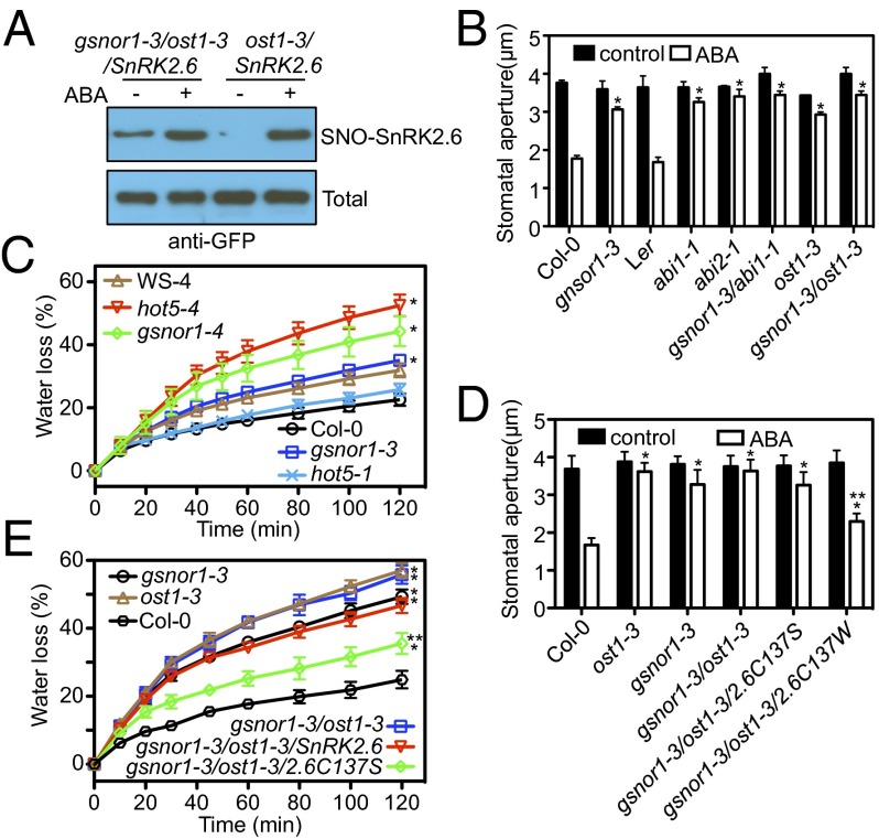
We hypothesized that NO production inside guard cells may negatively regulate ABA signaling through SnRK2.6 S-nitrosylation. To test this hypothesis, we investigated ABA responses in gsnor1-3 mutant plants, which overproduce SNOs because of a deficiency of the GSNO reductase in Arabidopsis (32). S-nitrosylation of proteins such as AHP1 (27) and SABP3 (salicylic acid-binding protein 3) (38), is increased in gsnor1-3 mutant plants. In gsnor1-3 leaves, NO overaccumulated in guard cells, where the GSNOR1 is expressed (Fig. S6 A and B). Without ABA treatment, the level of S-nitrosylation of SnRK2.6 was higher in gsnor1-3/ost1-3/SnRK2.6 than ost1-3/SnRK2.6 plants (Fig. 3_A_). After ABA treatment, the S-nitrosylation levels were similar. In the gsnor1-3 mutant, ABA was substantially less effective in inducing stomatal closure than in the Col-0 wild-type plants (Fig. 3_B_). However, exogenous H2O2, but not SNP or GSNO, could still induce stomatal closure in gsnor1-3 (Fig. S7_A_). The stomatal insensitivity of gnsor1-3 to ABA is similar to that of the ost1-3, abi1-1, and abi2-1 mutants (Fig. 3_B_) (39, 40). Like these single mutants, gsnor1-3/abi1-1 and gsnor1-3/ost1-3 double-mutant plants are equally insensitive to ABA in stomatal closure (Fig. 3_B_). Consistent with the impaired ABA response in guard cells, the excised leaves of gsnor1-3 showed a higher water-loss rate than wild-type leaves (Fig. 3_C_). Two other T-DNA insertion mutants, gsnor1-4 and hot5-4 in the WS (Wassilewskija) background, also exhibited higher water loss than their wild-type control plants (Fig. 3_C_). However, hot5-1, which is another missense allele of GSNOR1 in the Col-0 background (33), showed a similar water loss rate as the Col-0 wild-type (Fig. 3_C_). Immunoblot and GSNOR activity assays revealed that gsnor1-3, gsnor1-4, and hot5-4 were virtually null alleles (Fig. S7 D and E) (33), whereas hot5-1 plants contained a similar amount of GSNOR protein as the wild-type and retained about half of the GSNOR activity (Fig. S7 D and E) (33). Therefore, the transpirational water-loss phenotypes are consistent with the GSNOR protein and activity levels. Consistent with its higher transpirational water loss, the leaf surface temperature of gsnor1-3 was significantly lower than that of the Col-0 wild-type, especially under drought stress (Fig. S8). Our results suggest that increased NO accumulation in the gsnor1 mutants causes elevated S-nitrosylation of SnRK2.6, resulting in ABA insensitivity in guard cells and higher transpirational water loss.
Fig. 3.
Overaccumulation of SNOs in the gnsor1 mutant impairs the stomatal response to ABA and partial suppression of the stomatal defect by C137S mutated form of SnRK2.6. (A) ABA enhances S-nitrosylation of SnRK2.6 in gsnor1-3/ost1-3/SnRK2.6-GFP and ost1-3/SnRK2.6-GFP plants as revealed by a biotin-switch assay. Twelve-day-old seedlings were treated with or without 50 μM ABA for 30 min. The biotin-switch and immunoblot assays were performed as described in SI Materials and Methods. (B) Stomatal responses to exogenous ABA in wild-type and mutant plants. Stomatal apertures were measured in epidermal strips peeled from rosette leaves of 4- to 6-wk-old seedlings of Col-0 wild-type and the indicated mutants after the strips were incubated for 2 h in a buffer without (control) or with 10 μM ABA. n = 3–5 independent experiments. More than 20 stomata were measured for each treatment in each experiment. Error bars represent ± SD. Student’s t test, *P < 0.05 (significantly different from wild-type). (C) Water loss (percentage of initial fresh weight) in the detached rosette leaves of gsnor1 mutant and wild-type plants. Error bars indicate SD. n = 3–5 independent experiments. Student’s t test, *P < 0.05. (D) Stomatal responses to exogenous ABA (10 μM) in wild-type and mutant plants. n = 3 independent experiments. More than 20 stomata were used for each treatment in each experiment, Error bars represent ± SD. Note that stomatal apertures at the control condition were similar for all genotypes because the epidermal strips were placed under light and high humidity conditions and incubated in stomata-opening solution to maximize stomatal opening before ABA treatment. (E) Water loss (percentage of initial fresh weight) in the detached leaves of Col-0 wild-type and mutant plants. Error bars indicate SD. n = 3–5 independent experiments. For D and E, Student’s t tests show significant difference from Col-0 (*P < 0.05), and between gsnor1-3/ost1-3 and gsnor1-3/ost1-3 (**P < 0.05).
We examined the kinase activity of SnRK2.6 in gsnor1 mutant plants using an in-gel kinase assay (11, 41), which involves separating plant proteins in SDS-polyacrylamide gels containing ABF2 fragments as a kinase substrate and renaturation of the proteins with DTT after removal of SDS from the gels before detecting protein bands with kinase activities by incubation with [λ-32P]ATP. The assay revealed higher SnRK2.6 kinase activities in hot5-4 and gsnor1-4 mutant plants (Fig. S7_F_). Because the DTT used for protein renaturation in the assay would reverse S-nitrosylation and possibly some other modifications on Cys residues of SnRK2.6, the results suggest that S-nitrosylated and other Cys-modified SnRK2.6 in the gsnor1 mutants are not further subjected to any irreversible and inhibitory modifications.
To confirm that the ABA insensitivity in stomatal closure in gsnor1 mutant plants is a result of SnRK2.6 S-nitrosylation, we introduced native promoter-driven SnRK2.6 WT and SnRK2.6 C137S into the gsnor1-3 mutant by crossing with ost1-3 mutant plants expressing the SnRK2.6 constructs, and analyzed the water loss and stomatal responses in the resulting gsnor1-3/ost1-3/SnRK2.6 WT and gsnor1-3/ost1-3/SnRK2.6 C137S plants. The SnRK2.6 C173S mutant but not the wild-type SnRK2.6 partially restored ABA-induced stomatal closure in the gsnor1-3 mutant (Fig. 3_D_). The water-loss defect of the gsnor1-3/ost1-3 double-mutant was also partially rescued by introduction of SnRK2.6 C173S but not the wild-type SnRK2.6 (Fig. 3_E_).
The S-Nitrosylated Cysteine Is Evolutionarily Conserved in Eukaryotes.
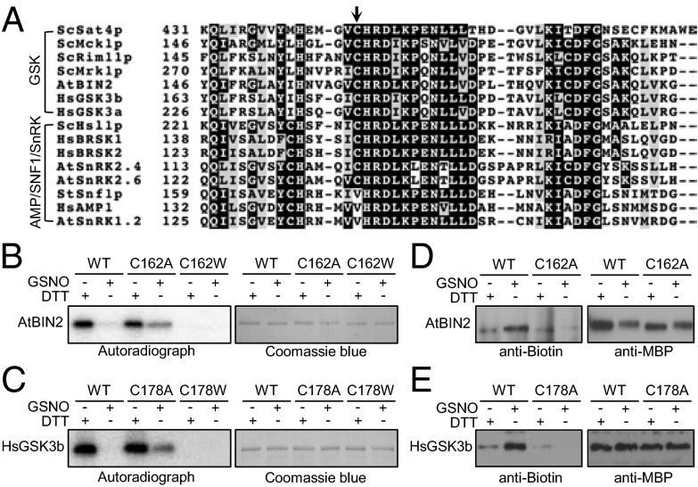
SNF1/AMPKs are conserved in all eukaryotes and play fundamental roles in cellular responses to metabolic stress (42). The 48 SnRKs in Arabidopsis are divided into three subfamilies. All 10 members in the SnRK2 subfamily and SnRK3.1 in the SnRK3 subfamily have the conserved cysteine (cystein-137 in SnRK2.6) that can be potentially S-nitrosylated. The conserved cysteine is also present in SnRK2 orthologs from other plants, including various crop plants (Fig. S9_A_). None of the other 24 members in the SnRK3 subfamily and none of the three members in the SnRK1 subfamily in Arabidopsis has the conserved cysteine (Fig. 4_A_ and Fig. S9_B_). The proteins with the highest sequence homologies to the Arabidopsis SnRK2s, AMPKs in mammals and SNF1 in yeasts have a hydrophobic amino acid rather than cysteine at the corresponding position. Interestingly, two AMPK-related protein kinases in mammals, brain-specific kinases (BRSK) 1 and 2, which are required for neuronal polarization (43), have the corresponding cysteine near their catalytic sites, similar to the Arabidopsis SnRK2s. The yeast SNF1-related kinases Hsl1 and Hal4, but not the SNF1 itself, also have the corresponding cysteine (Fig. 4_A_). Another family of protein kinases that contains the conserved cysteine is the glycogen synthase kinase 3(GSK)/SKs. The conserved cysteine residue is present in 9 of 10 GSKs in Arabidopsis, in the human GSK3α/β, and in the yeast Mck1, Rim11, and Mrk1 (Fig. 4_A_). To test whether the conserved cysteine residues in the AMPK1/SNF1-related kinases and SKs may also be modified by S-nitrosylation, we examined the effects of GSNO on their kinase activities. Of these protein kinases, we were able to express the following four in Escherichia coli and the purified recombinant proteins had detectable autophosphorylation activities: HsBRSK1, AtBIN2, HsGSK3b, and ScHsl1p (Fig. S10). All four kinases were inhibited by GSNO in a dose-dependent manner (Fig. S10). We mutated the conserved cysteines, Cys162 in AtBIN2 and Cys178 in HsGSK3b, to alanine (A) or tryptophan (W). GSNO abolished the kinase activities of wild-type BIN2 (brassinosteroid insensitive 2) and GSK3b (Fig. 4 B and C, lanes 1 and 2). The C-to-A mutated forms of BIN2 and GSK3b still retained some kinase activities after the GSNO treatment (Fig. 4 B and C, lanes 3 and 4). The C-to-W mutated forms of BIN2 and GSK3b totally lost their kinase activities (Fig. 4 B and C, lanes 5 and 6). As was the case with SnRK2.6, the S-nitrosylation of these kinases was induced by GSNO, and the mutation of the conserved cysteine blocked the S-nitrosylation of the proteins (Fig. 4 D and E). Our finding of the regulation of BIN2 by S-nitrosylation is consistent with the observation that brassinosteroid treatment induces NO accumulation in maize leaves (44). The conservation of the cysteine and the S-nitrosylation–dependent regulation of the activities of the kinases from diverse organisms suggest that S-nitrosylation–mediated inhibition is a general regulatory mechanism for these eukaryotic protein kinases.
Fig. 4.
S-nitrosylation of Cys137 is evolutionarily conserved across kingdoms. (A) Cys137 of SnRK2.6 is evolutionarily conserved in some AMPK/SNF1-related kinases and GSKs in Arabidopsis, yeast, and mammals. The conserved cysteine is indicated by the arrow. GSNO inhibits the kinase activity of Arabidopsis BIN2 (B) and human GSK3b (C). Recombinant wild-type and mutated MBP-AtBIN2 and MBP-HsGSK3b were incubated with DTT or GSNO (50 μM) for 10 min and then [γ-32P]ATP was added to determine the autophosphorylation of recombinant kinases. GSNO induces the S-nitrosylation of recombinant Arabidopsis MBP-BIN2 (D) and human MBP-GSK3b (E), as determined by the biotin-switch assay. All experiments were repeated at least twice with similar results.
Discussion
Our study has revealed a novel mechanism by which NO regulates ABA signaling in Arabidopsis. We provided evidence that ABA enhances SnRK2.6 S-nitrosylation and the S-nitrosylation feedback inhibits ABA signaling. The evidence includes: (i) exogenous NO donors caused S-nitrosylation of SnRK2.6 and inhibited its activity in vitro, and an S-nitrosylation-mimicking mutation abolished its kinase activity; (ii) ABA enhanced the level of nitrosylated SnRK2.6 in planta; and (iii) NO-overproducing mutant plants were less sensitive to ABA in stomatal closure, but the defect was partially suppressed by ectopic expression of C137S mutated form of SnRK2.6. Our study revealed SnRK2.6 as a new S-nitrosylation target that bridges NO-mediated redox signaling and ABA signaling.
In the absence of ABA, PP2Cs inhibit SnRK2.6 activity by both dephosphorylation at Ser175 and direct binding (45). In the presence of ABA, however, the binding of PYLs to PP2Cs releases SnRK2.6 from inhibition. SnRK2.6 is activated very quickly by ABA because 2 min of ABA treatment is enough to cause a strong activation of SnRK2.6 (41). In contrast to the early activation of SnRK2.6, NO-mediated inhibition of SnRK2.6 is expected to happen later during ABA treatment because the level of S-nitrosylated SnRK2.6 is not increased after 15 min of ABA application (Fig. 2_F_). NO accumulation did not reach peak levels until 30 min after ABA application (Fig. 2_E_) (46). The notion that NO accumulation and SnRK2.6 S-nitrosylation are slow or late events during ABA treatment is consistent with the observation that ABA-induced NO generation is dependent on the quick burst of ROS (47). The accumulation of endogenous NO may function as one of the negative feedback mechanisms to prevent overactivation of ABA signaling in guard cells. This feedback regulation is achieved by S-nitrosylation of SnRK2.6. ABA-activated SnRK2.6 phosphorylates NADPH oxidases to cause ROS production in guard cells (18, 48). The S-nitrosylation of the NADPH oxidase AtRBOHD has been shown to cause inhibition of the NADPH oxidase (28), and thus may also contribute to the NO-mediated negative feedback regulation of ABA signaling. Although detailed dynamic changes in the levels of activated SnRK2.6, NO, and S-nitrosylated SnRK2.6 in guard cells are not yet known, available evidence suggests the following scenario: ABA treatment causes very fast and strong activation of SnRK2.6 in guard cells. The activated SnRK2.6 phosphorylates many downstream effector proteins to cause stomatal closure, and it also phosphorylates the NADPH oxidases AtRBOHD and AtRBOHF to cause a burst of ROS, which in turn causes NO accumulation in the guard cells. When NO accumulates to high levels, it causes S-nitrosylation and inhibition of SnRK2.6 and the NADPH oxidases. This inhibition serves to desensitize ABA signaling. The phenomenon of desensitization of ABA signaling was observed 20 y ago, when water deficit stress was shown to reduce the stomatal sensitivity to ABA (49).
In gsnor1 mutant plants, overaccumulated SNOs cause constitutive S-nitrosylation of SnRK2.6 such that the kinase is inactivated, and thus the stomata are insensitive to ABA (Fig. 3). In support of our conclusion that endogenous NO has a negative role in ABA signaling in guard cells, a recent study demonstrated that the NO-deficient mutant nia1nia2noa1 is hypersensitive to ABA in stomatal closure (21). The kinase activity of S-nitrosylated SnRK2.6 may be resumed quickly, depending on cellular redox homeostasis. Protein S-nitrosylation depends on the proximity of the protein to NO sources, and on factors for transnitrosylation and denitrosylation, such as thioredoxin and glutathione levels (50). The reversible inhibition of SnRK2 by S-nitrosylation may fine-tune the strength or duration of SnRK2.6 activation in response to ABA. Such dynamic control may be important for plants to balance stress resistance with growth that cannot happen if stomata are fully closed as a result of continued activation of SnRK2.6. Because NO originates from nitrogen sources, NO-mediated stomatal regulation through SnRK2.6 S-nitrosylation may also help coordinate nitrogen supply with photosynthetic carbon availability.
The negative effect of endogenous NO on ABA signaling in guard cells is supported by genetic analysis using both NO deficient and overaccumulation mutants (Fig. 3) (21). On the other hand, several studies have found a positive role of exogenous application of NO in ABA-induced stomatal closure (15, 20, 22, 24). Exogenous NO might promote stomatal closure by regulating ion channels (20, 23), or by generating nitrated cGMP (24). Alternatively, the observed positive role of exogenous NO on ABA responses in guard cells might be caused by secondary effects, such as induction of ROS or other second messengers (24, 47).
The activities of protein kinases are tightly controlled by multiple mechanisms. In most cases, dephosphorylation at the phosphorylated sites in the “activation segment” causes kinase deactivation (51). For SnRK2.6, the phosphorylation of Ser175 is necessary for its full activity (37). The negative regulators of ABA signaling, PP2Cs, inhibit SnRK2.6 partly by dephosphorylation at Ser175. Similarly, in brassinosteroid signaling, AtBIN2 is dephosphorylated and inactivated by BSU1 (bri1 SUPPRESSOR 1), a Kelch-repeat domain-containing protein phosphatase (52). In contrast to the phosphorylation-mediated activation, the phosphorylation of GSK3 at serine 9 by Akt kinase inhibits its activity (53), suggesting a “pseudo-substrate” mechanism to reduce the kinase activity (51). Our finding that GSNO-mediated S-nitrosylation of HsBRSK1, ScHsl1p, GSKs, and AtSnRK2s suggests a third way to deactivate these protein kinases. The cysteine in the catalytic cleft exists only in certain members of the SnRK/GSK family. In most protein kinases, the conserved cysteine is substituted by a hydrophobic amino acid. Other cysteines not in the position near the catalytic loop could still be modified by S-nitrosylation, leading to the inhibition of some kinases (54–56). The S-nitrosylation of cysteine in the catalytic cleft reported here represents a unique mechanism in the regulation of some of the AMPK/SNF1-related and GSK family of protein kinases.
Materials and Methods
Plant Materials and Growth Condition.
The Arabidopsis wild-type Columbia-0 (Col-0), Wassilewskija-4 (WS-4), and Landsberg (Ler) plants were used in this study. The gsnor1-3 (GABI_315D11), hot5-1 (CS66011), hot5-4 (FLAG_298F11), gsnor1-4 (FLAG_220G07), and ost1-3 (Salk_008068) mutant seeds were ordered from the Arabidopsis Biological Resource Center. Seeds were germinated on half-strength MS agar plates containing 1.5% sucrose. For the measurement of water loss or stomatal apertures, 10-d-old seedlings on MS plates were transferred to soil and grown under short-day conditions (12 h light, 100–120 μmol·m−2·s−1) at 22 °C.
Epidermal Strip Bioassay and Water Loss Measurement.
Stomatal bioassay experiments were performed as described previously (15). Epidermal strips were peeled from the rosette leaves of 4- to 6-wk-old seedlings that were placed under the light and high humidity for 12 h and incubated in stomata-opening solution containing 50 mM KCl, 10 mM MES, pH 6.15 in a growth chamber for 1.5 h to maximize stomatal opening before ABA was added. Stomatal apertures were measured 2 h after 10 μM ABA was added. The apertures of about 60 stomata were measured in three independent experiments.
For the measurement of water loss, detached rosette leaves of 4-wk-old plants were placed in weighing dishes and left on the laboratory bench with light (30–40 μmol·m−2·s−1). Fresh weight was monitored at the indicated times. Water loss was expressed as a percentage of initial fresh weight.
In Vitro Kinase Assay and In-Gel Kinase Assay.
The in vitro kinase assay and in-gel kinase assay were performed as described previously (11); details are provided in SI Materials and Methods.
In Vitro and in Vivo S-Nitrosylation Assays.
In vitro and in vivo S-nitrosylation assays were performed as described (28, 31); details are provided in SI Materials and Methods.
Other Methods.
Details for other methods are provided in SI Materials and Methods, including recombinant protein expression and site-directed mutagenesis, structure information of SnRK2.6, histochemical detection of GUS activity, infrared thermography imaging, NO detection by confocal microscopy, and homology search and sequence alignment. The primers used in this study are listed in Table S1.
Supplementary Material
Supplementary File
Acknowledgments
This work was supported by NIH Grant R01GM059138 (to J.-K.Z.) and National Natural Science Foundation of China Grants 91017001 and 31171363 (to P.W.).
Footnotes
The authors declare no conflict of interest.
References
- 1.Cutler SR, Rodriguez PL, Finkelstein RR, Abrams SR. Abscisic acid: Emergence of a core signaling network. Annu Rev Plant Biol. 2010;61:651–679. doi: 10.1146/annurev-arplant-042809-112122. [DOI] [PubMed] [Google Scholar]
- 2.Klingler JP, Batelli G, Zhu JK. ABA receptors: The START of a new paradigm in phytohormone signalling. J Exp Bot. 2010;61(12):3199–3210. doi: 10.1093/jxb/erq151. [DOI] [PMC free article] [PubMed] [Google Scholar]
- 3.Lee KH, et al. Activation of glucosidase via stress-induced polymerization rapidly increases active pools of abscisic acid. Cell. 2006;126(6):1109–1120. doi: 10.1016/j.cell.2006.07.034. [DOI] [PubMed] [Google Scholar]
- 4.Fujii H, et al. In vitro reconstitution of an abscisic acid signalling pathway. Nature. 2009;462(7273):660–664. doi: 10.1038/nature08599. [DOI] [PMC free article] [PubMed] [Google Scholar]
- 5.Ma Y, et al. Regulators of PP2C phosphatase activity function as abscisic acid sensors. Science. 2009;324(5930):1064–1068. doi: 10.1126/science.1172408. [DOI] [PubMed] [Google Scholar]
- 6.Park S-Y, et al. Abscisic acid inhibits type 2C protein phosphatases via the PYR/PYL family of START proteins. Science. 2009;324(5930):1068–1071. doi: 10.1126/science.1173041. [DOI] [PMC free article] [PubMed] [Google Scholar]
- 7.Wang P, et al. Quantitative phosphoproteomics identifies SnRK2 protein kinase substrates and reveals the effectors of abscisic acid action. Proc Natl Acad Sci USA. 2013;110(27):11205–11210. doi: 10.1073/pnas.1308974110. [DOI] [PMC free article] [PubMed] [Google Scholar]
- 8.Kim TH, Böhmer M, Hu H, Nishimura N, Schroeder JI. Guard cell signal transduction network: advances in understanding abscisic acid, CO2, and Ca2+ signaling. Annu Rev Plant Biol. 2010;61:561–591. doi: 10.1146/annurev-arplant-042809-112226. [DOI] [PMC free article] [PubMed] [Google Scholar]
- 9.Léon-Kloosterziel KM, et al. Isolation and characterization of abscisic acid-deficient Arabidopsis mutants at two new loci. Plant J. 1996;10(4):655–661. doi: 10.1046/j.1365-313x.1996.10040655.x. [DOI] [PubMed] [Google Scholar]
- 10.Gonzalez-Guzman M, et al. Arabidopsis PYR/PYL/RCAR receptors play a major role in quantitative regulation of stomatal aperture and transcriptional response to abscisic acid. Plant Cell. 2012;24(6):2483–2496. doi: 10.1105/tpc.112.098574. [DOI] [PMC free article] [PubMed] [Google Scholar]
- 11.Fujii H, Verslues PE, Zhu J-K. Identification of two protein kinases required for abscisic acid regulation of seed germination, root growth, and gene expression in Arabidopsis. Plant Cell. 2007;19(2):485–494. doi: 10.1105/tpc.106.048538. [DOI] [PMC free article] [PubMed] [Google Scholar]
- 12.Geiger D, et al. Activity of guard cell anion channel SLAC1 is controlled by drought-stress signaling kinase-phosphatase pair. Proc Natl Acad Sci USA. 2009;106(50):21425–21430. doi: 10.1073/pnas.0912021106. [DOI] [PMC free article] [PubMed] [Google Scholar]
- 13.Sato A, et al. Threonine at position 306 of the KAT1 potassium channel is essential for channel activity and is a target site for ABA-activated SnRK2/OST1/SnRK2.6 protein kinase. Biochem J. 2009;424(3):439–448. doi: 10.1042/BJ20091221. [DOI] [PubMed] [Google Scholar]
- 14.Allen GJ, et al. A defined range of guard cell calcium oscillation parameters encodes stomatal movements. Nature. 2001;411(6841):1053–1057. doi: 10.1038/35082575. [DOI] [PubMed] [Google Scholar]
- 15.Desikan R, Griffiths R, Hancock J, Neill S. A new role for an old enzyme: Nitrate reductase-mediated nitric oxide generation is required for abscisic acid-induced stomatal closure in Arabidopsis thaliana. Proc Natl Acad Sci USA. 2002;99(25):16314–16318. doi: 10.1073/pnas.252461999. [DOI] [PMC free article] [PubMed] [Google Scholar]
- 16.Lee Y, et al. Abscisic acid-induced phosphoinositide turnover in guard cell protoplasts of Vicia faba. Plant Physiol. 1996;110(3):987–996. doi: 10.1104/pp.110.3.987. [DOI] [PMC free article] [PubMed] [Google Scholar]
- 17.McAinsh MR, Brownlee C, Hetherington AM. Abscisic acid-induced elevation of guard cell cytosolic Ca2+ precedes stomatal closure. Nature. 1990;343:186–188. [Google Scholar]
- 18.Pei Z-M, et al. Calcium channels activated by hydrogen peroxide mediate abscisic acid signalling in guard cells. Nature. 2000;406(6797):731–734. doi: 10.1038/35021067. [DOI] [PubMed] [Google Scholar]
- 19.Kwak JM, et al. NADPH oxidase AtrbohD and AtrbohF genes function in ROS-dependent ABA signaling in Arabidopsis. EMBO J. 2003;22(11):2623–2633. doi: 10.1093/emboj/cdg277. [DOI] [PMC free article] [PubMed] [Google Scholar]
- 20.Garcia-Mata C, et al. Nitric oxide regulates K+ and Cl− channels in guard cells through a subset of abscisic acid-evoked signaling pathways. Proc Natl Acad Sci USA. 2003;100(19):11116–11121. doi: 10.1073/pnas.1434381100. [DOI] [PMC free article] [PubMed] [Google Scholar]
- 21.Lozano-Juste J, León J. Enhanced abscisic acid-mediated responses in nia1nia2noa1-2 triple mutant impaired in NIA/NR- and AtNOA1-dependent nitric oxide biosynthesis in Arabidopsis. Plant Physiol. 2010;152(2):891–903. doi: 10.1104/pp.109.148023. [DOI] [PMC free article] [PubMed] [Google Scholar]
- 22.Neill SJ, Desikan R, Clarke A, Hancock JT. Nitric oxide is a novel component of abscisic acid signaling in stomatal guard cells. Plant Physiol. 2002;128(1):13–16. [PMC free article] [PubMed] [Google Scholar]
- 23.Vahisalu T, et al. SLAC1 is required for plant guard cell S-type anion channel function in stomatal signalling. Nature. 2008;452(7186):487–491. doi: 10.1038/nature06608. [DOI] [PMC free article] [PubMed] [Google Scholar]
- 24.Joudoi T, et al. Nitrated cyclic GMP modulates guard cell signaling in Arabidopsis. Plant Cell. 2013;25(2):558–571. doi: 10.1105/tpc.112.105049. [DOI] [PMC free article] [PubMed] [Google Scholar]
- 25.Wang P, Du Y, Li Y, Ren D, Song C-P. Hydrogen peroxide-mediated activation of MAP kinase 6 modulates nitric oxide biosynthesis and signal transduction in Arabidopsis. Plant Cell. 2010;22(9):2981–2998. doi: 10.1105/tpc.109.072959. [DOI] [PMC free article] [PubMed] [Google Scholar]
- 26.Moreau M, Lee GI, Wang Y, Crane BR, Klessig DF. AtNOS/AtNOA1 is a functional Arabidopsis thaliana cGTPase and not a nitric-oxide synthase. J Biol Chem. 2008;283(47):32957–32967. doi: 10.1074/jbc.M804838200. [DOI] [PMC free article] [PubMed] [Google Scholar]
- 27.Feng J, et al. S-nitrosylation of phosphotransfer proteins represses cytokinin signaling. Nat Commun. 2013;4:1529. doi: 10.1038/ncomms2541. [DOI] [PubMed] [Google Scholar]
- 28.Yun B-W, et al. S-nitrosylation of NADPH oxidase regulates cell death in plant immunity. Nature. 2011;478(7368):264–268. doi: 10.1038/nature10427. [DOI] [PubMed] [Google Scholar]
- 29.Terrile MC, et al. Nitric oxide influences auxin signaling through S-nitrosylation of the Arabidopsis TRANSPORT INHIBITOR RESPONSE 1 auxin receptor. Plant J. 2012;70(3):492–500. doi: 10.1111/j.1365-313X.2011.04885.x. [DOI] [PMC free article] [PubMed] [Google Scholar]
- 30.He Y, et al. Nitric oxide represses the Arabidopsis floral transition. Science. 2004;305(5692):1968–1971. doi: 10.1126/science.1098837. [DOI] [PubMed] [Google Scholar]
- 31.Tada Y, et al. Plant immunity requires conformational changes [corrected] of NPR1 via S-nitrosylation and thioredoxins. Science. 2008;321(5891):952–956. doi: 10.1126/science.1156970. [DOI] [PMC free article] [PubMed] [Google Scholar]
- 32.Feechan A, et al. A central role for S-nitrosothiols in plant disease resistance. Proc Natl Acad Sci USA. 2005;102(22):8054–8059. doi: 10.1073/pnas.0501456102. [DOI] [PMC free article] [PubMed] [Google Scholar]
- 33.Lee U, Wie C, Fernandez BO, Feelisch M, Vierling E. Modulation of nitrosative stress by S-nitrosoglutathione reductase is critical for thermotolerance and plant growth in Arabidopsis. Plant Cell. 2008;20(3):786–802. doi: 10.1105/tpc.107.052647. [DOI] [PMC free article] [PubMed] [Google Scholar]
- 34.Chen R, et al. The Arabidopsis PARAQUAT RESISTANT2 gene encodes an S-nitrosoglutathione reductase that is a key regulator of cell death. Cell Res. 2009;19(12):1377–1387. doi: 10.1038/cr.2009.117. [DOI] [PubMed] [Google Scholar]
- 35.Xu S, Guerra D, Lee U, Vierling E. S-nitrosoglutathione reductases are low-copy number, cysteine-rich proteins in plants that control multiple developmental and defense responses in Arabidopsis. Front Plant Sci. 2013;4:430. doi: 10.3389/fpls.2013.00430. [DOI] [PMC free article] [PubMed] [Google Scholar]
- 36.Yonashiro R, et al. Mitochondrial ubiquitin ligase MITOL blocks S-nitrosylated MAP1B-light chain 1-mediated mitochondrial dysfunction and neuronal cell death. Proc Natl Acad Sci USA. 2012;109(7):2382–2387. doi: 10.1073/pnas.1114985109. [DOI] [PMC free article] [PubMed] [Google Scholar]
- 37.Ng LM, et al. Structural basis for basal activity and autoactivation of abscisic acid (ABA) signaling SnRK2 kinases. Proc Natl Acad Sci USA. 2011;108(52):21259–21264. doi: 10.1073/pnas.1118651109. [DOI] [PMC free article] [PubMed] [Google Scholar]
- 38.Wang Y-Q, et al. S-nitrosylation of AtSABP3 antagonizes the expression of plant immunity. J Biol Chem. 2009;284(4):2131–2137. doi: 10.1074/jbc.M806782200. [DOI] [PubMed] [Google Scholar]
- 39.Merlot S, Gosti F, Guerrier D, Vavasseur A, Giraudat J. The ABI1 and ABI2 protein phosphatases 2C act in a negative feedback regulatory loop of the abscisic acid signalling pathway. Plant J. 2001;25(3):295–303. doi: 10.1046/j.1365-313x.2001.00965.x. [DOI] [PubMed] [Google Scholar]
- 40.Murata Y, Pei ZM, Mori IC, Schroeder J. Abscisic acid activation of plasma membrane Ca2+ channels in guard cells requires cytosolic NAD(P)H and is differentially disrupted upstream and downstream of reactive oxygen species production in abi1-1 and abi2-1 protein phosphatase 2C mutants. Plant Cell. 2001;13(11):2513–2523. doi: 10.1105/tpc.010210. [DOI] [PMC free article] [PubMed] [Google Scholar]
- 41.Yoshida R, et al. ABA-activated SnRK2 protein kinase is required for dehydration stress signaling in Arabidopsis. Plant Cell Physiol. 2002;43(12):1473–1483. doi: 10.1093/pcp/pcf188. [DOI] [PubMed] [Google Scholar]
- 42.Hardie DG. AMP-activated/SNF1 protein kinases: Conserved guardians of cellular energy. Nat Rev Mol Cell Biol. 2007;8(10):774–785. doi: 10.1038/nrm2249. [DOI] [PubMed] [Google Scholar]
- 43.Kishi M, Pan YA, Crump JG, Sanes JR. Mammalian SAD kinases are required for neuronal polarization. Science. 2005;307(5711):929–932. doi: 10.1126/science.1107403. [DOI] [PubMed] [Google Scholar]
- 44.Zhang A, et al. Nitric oxide mediates brassinosteroid-induced ABA biosynthesis involved in oxidative stress tolerance in maize leaves. Plant Cell Physiol. 2011;52(1):181–192. doi: 10.1093/pcp/pcq187. [DOI] [PubMed] [Google Scholar]
- 45.Soon F-F, et al. Molecular mimicry regulates ABA signaling by SnRK2 kinases and PP2C phosphatases. Science. 2012;335(6064):85–88. doi: 10.1126/science.1215106. [DOI] [PMC free article] [PubMed] [Google Scholar]
- 46.Zhang A, et al. Nitric oxide induced by hydrogen peroxide mediates abscisic acid-induced activation of the mitogen-activated protein kinase cascade involved in antioxidant defense in maize leaves. New Phytol. 2007;175(1):36–50. doi: 10.1111/j.1469-8137.2007.02071.x. [DOI] [PubMed] [Google Scholar]
- 47.Bright J, Desikan R, Hancock JT, Weir IS, Neill SJ. ABA-induced NO generation and stomatal closure in Arabidopsis are dependent on H2O2 synthesis. Plant J. 2006;45(1):113–122. doi: 10.1111/j.1365-313X.2005.02615.x. [DOI] [PubMed] [Google Scholar]
- 48.Sirichandra C, et al. Phosphorylation of the Arabidopsis AtrbohF NADPH oxidase by OST1 protein kinase. FEBS Lett. 2009;583(18):2982–2986. doi: 10.1016/j.febslet.2009.08.033. [DOI] [PubMed] [Google Scholar]
- 49.Peng Z-Y, Weyers JDB. Stomatal sensitivity to abscisic acid following water deficit stress. J Exp Bot. 1994;45(6):835–845. [Google Scholar]
- 50.Martínez-Ruiz A, et al. Specificity in S-nitrosylation: A short-range mechanism for NO signaling? Antioxid Redox Signal. 2013;19(11):1220–1235. doi: 10.1089/ars.2012.5066. [DOI] [PMC free article] [PubMed] [Google Scholar]
- 51.Harwood AJ. Regulation of GSK-3: A cellular multiprocessor. Cell. 2001;105(7):821–824. doi: 10.1016/s0092-8674(01)00412-3. [DOI] [PubMed] [Google Scholar]
- 52.Kim T-W, et al. Brassinosteroid signal transduction from cell-surface receptor kinases to nuclear transcription factors. Nat Cell Biol. 2009;11(10):1254–1260. doi: 10.1038/ncb1970. [DOI] [PMC free article] [PubMed] [Google Scholar]
- 53.Cross DAE, Alessi DR, Cohen P, Andjelkovich M, Hemmings BA. Inhibition of glycogen synthase kinase-3 by insulin mediated by protein kinase B. Nature. 1995;378(6559):785–789. doi: 10.1038/378785a0. [DOI] [PubMed] [Google Scholar]
- 54.Reynaert NL, et al. Nitric oxide represses inhibitory kappaB kinase through S-nitrosylation. Proc Natl Acad Sci USA. 2004;101(24):8945–8950. doi: 10.1073/pnas.0400588101. [DOI] [PMC free article] [PubMed] [Google Scholar]
- 55.Park H-S, Huh S-H, Kim M-S, Lee SH, Choi E-J. Nitric oxide negatively regulates c-Jun N-terminal kinase/stress-activated protein kinase by means of S-nitrosylation. Proc Natl Acad Sci USA. 2000;97(26):14382–14387. doi: 10.1073/pnas.97.26.14382. [DOI] [PMC free article] [PubMed] [Google Scholar]
- 56.Whalen EJ, et al. Regulation of β-adrenergic receptor signaling by S-nitrosylation of G-protein-coupled receptor kinase 2. Cell. 2007;129(3):511–522. doi: 10.1016/j.cell.2007.02.046. [DOI] [PubMed] [Google Scholar]
Associated Data
This section collects any data citations, data availability statements, or supplementary materials included in this article.
Supplementary Materials
Supplementary File


