Gene Recruitment of the Activated INO1 Locus to the Nuclear Membrane (original) (raw)
Abstract
The spatial arrangement of chromatin within the nucleus can affect reactions that occur on the DNA and is likely to be regulated. Here we show that activation of INO1 occurs at the nuclear membrane and requires the integral membrane protein Scs2. Scs2 antagonizes the action of the transcriptional repressor Opi1 under conditions that induce the unfolded protein response (UPR) and, in turn, activate INO1. Whereas repressed INO1 localizes throughout the nucleoplasm, the gene is recruited to the nuclear periphery upon transcriptional activation. Recruitment requires the transcriptional activator Hac1, which is produced upon induction of the UPR, and is constitutive in a strain lacking Opi1. Artificial recruitment of INO1 to the nuclear membrane permits activation in the absence of Scs2, indicating that the intranuclear localization of a gene can profoundly influence its mechanism of activation. Gene recruitment to the nuclear periphery, therefore, is a dynamic process and appears to play an important regulatory role.
A study of the yeast gene INO1 indicates that the recruitment of the gene to the nuclear membrane appears to play an important part in its regulation
Introduction
For over a hundred years, it has been recognized that chromatin is distributed non-randomly within the interphase nucleus (Rabl 1885; Boveri 1909). More recently, three-dimensional fluorescence microscopy studies have established that chromosomes are organized into distinct, evolutionarily conserved subnuclear territories (reviewed by Cockell and Gasser [1999]; Isogai and Tjian [2003]). However, DNA is mobile and can move between these domains (reviewed in Gasser [2002]). Recent studies suggest that the subnuclear localization of genes can have dramatic effects on their chromatin state, rate of recombination, and transcription (Cockell and Gasser 1999; Isogai and Tjian 2003; Bressan et al. 2004). Heterochromatin, for example, is generally found concentrated in close proximity to the nuclear envelope. Several genes conditionally colocalize with heterochromatin under conditions in which they are repressed. The transcriptional regulator Ikaros, for example, interacts both with regulatory sequences upstream of target genes and with repeats enriched at centromeric heterochromatin. When repressed, these genes become colocalized with heterochromatin, suggesting that Ikaros promotes repression by directly recruiting target genes into close proximity with heterochromatin (Brown et al. 1997, 1999; Cobb et al. 2000). Consistent with this view, euchromatic sequences that become colocalized with heterochromatin are transcriptionally silenced (Csink and Henikoff 1996; Dernburg et al. 1996).
In Saccharomyces cerevisiae, genes localized in proximity to telomeres are similarly transcriptionally silenced (Gottschling et al. 1990). Silencing is due to Rap1-dependent recruitment of Sir proteins to telomeres (Gotta et al. 1996), which promotes local histone deacetylation and changes in chromatin structure (reviewed in Rusche et al. [2003]). Physical tethering of telomeres at the nuclear periphery through interactions with the nuclear pore is required for silencing (Gotta et al. 1996; Laroche et al. 1998; Galy et al. 2000; Feuerbach et al. 2002). When a reporter gene flanked by silencer motifs was relocated more than 200 kb away from a telomere, silencing was lost (Maillet et al. 1996). Silencing was restored to this gene by overexpression of SIR genes. Therefore it is thought that tethering serves to promote efficient recruitment of Sir proteins, which are enriched at the nuclear periphery and limiting elsewhere (Maillet et al. 1996). Another example of gene silencing at the nuclear periphery comes from experiments in which defects in the silencer of the HMR locus could be suppressed by artificially tethering this locus to the nuclear membrane (Andrulis et al. 1998). Thus, localization of chromatin to the nuclear periphery has been proposed to play a major role in transcriptional repression.
By contrast, we report here that dynamic recruitment of genes to the nuclear membrane can have profound effects on their activation. The gene under study here is INO1, a target gene of the unfolded protein response (UPR), which encodes inositol 1-phosphate synthase. The UPR is an intracellular signaling pathway that is activated by the accumulation of unfolded proteins in the endoplasmic reticulum (ER), which can be stimulated by treatment with drugs that block protein folding or modification or, in yeast, by starvation for inositol (Cox et al. 1997). These conditions activate Ire1, a transmembrane ER kinase/endoribonuclease (Cox et al. 1993; Mori et al. 1993), which, through its endonuclease activity, initiates nonconventional splicing of the mRNA encoding the transcription activator Hac1 (Cox and Walter 1996; Shamu and Walter 1996; Kawahara et al. 1997; Sidrauski and Walter 1997). Only spliced HAC1 mRNA is translated to produce the transcription factor; the Ire1-mediated splicing reaction, therefore, constitutes the key switch step in the UPR (Sidrauski et al. 1996; Ruegsegger et al. 2001).
Hac1 is a basic-leucine zipper transcription factor that binds directly to unfolded protein response elements (UPREs) in the promoters of most target genes to promote transcriptional activation (Cox and Walter 1996; Travers et al. 2000; Patil et al. 2004). However, a subset of UPR target genes uses a different mode of activation. Transcriptional activation of these genes, including INO1, depends on Hac1 and Ire1. These target genes contain an upstream activating sequence that is regulated by the availability of inositol, the UASINO element, in their promoters that is repressed by Opi1 under non-UPR conditions (Greenberg et al. 1982; Cox et al. 1997). Opi1 repression is relieved in a Hac1-dependent manner upon induction of the UPR (Cox et al. 1997). Positively acting transcription factors Ino2 and Ino4 then promote transcription from UASINO-containing promoters (Loewy and Henry 1984; Ambroziak and Henry 1994; Schwank et al. 1995). Our previous work established that the production of Hac1 by UPR induction functions upstream of Opi1, suggesting that the role of the UPR is to counteract Opi1-mediated repression (Cox et al. 1997).
To understand the regulation of UASINO-controlled genes by the UPR, we have examined the molecular events leading to the activation of INO1. We find that Scs2, an integral protein of the nuclear and ER membrane that was recently shown to play a role in telomeric silencing (Craven and Petes 2001; Cuperus and Shore 2002), is required to activate INO1. We observe dynamic INO1 recruitment to the nuclear membrane under activating conditions. Importantly, we find that recruitment requires Hac1 and is opposed by Opi1. Furthermore, we show that artificial recruitment of INO1 to the nuclear membrane can bypass the requirement for Scs2. Gene recruitment to the nuclear membrane therefore plays an instrumental role in INO1 activation.
Results
Abundance and Localization of the Transcriptional Regulators Ino2, Ino4, and Opi1 Are Unaffected by UPR Induction
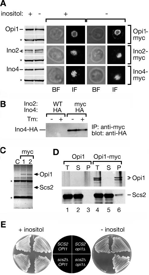
To characterize the molecular basis of transcriptional activation of INO1, we first asked whether the steady-state levels of the known transcriptional regulators—the activators Ino2 and Ino4 and the repressor Opi1—were affected by induction of the UPR. To this end, we monitored the levels of _myc_-tagged proteins by Western blotting after UPR induction by inositol starvation (Figure 1A). Induction of the UPR did not result in a significant change of the abundance of any of the proteins. Thus, in contrast to what has been suggested in previous studies (Ashburner and Lopes 1995a, 1995b; Cox et al. 1997; Schwank et al. 1997; Wagner et al. 1999), INO1 transcription is not regulated through adjustment of the abundance of these regulators.
Figure 1. Scs2 Regulates the Function of Opi1 on the Nuclear Membrane.
(A) Steady state protein levels and localization of Opi1, Ino2, and Ino4 under repressing and activating conditions. Strains expressing _myc_-tagged Opi1, Ino2, or Ino4 (Longtine et al. 1998) were grown in the presence (INO1 repressing condition) or absence (INO1 activating condition) of _myo_-inositol for 4.5 h. Tagged proteins were analyzed by Western blotting (size-fractionated blots on the left, designated Opi1, Ino2, and Ino4) and indirect immunofluorescence (photomicrographs on the right). For Western blot analysis, 25 μg of crude lysates were immunoblotted using monoclonal antibodies against either the myc epitope (top bands in each set) or, as a loading control, Pgk1 (bottom bands in each set; indicated with an asterisk). Immunofluorescence experiments were carried out using anti-myc antibodies and anti-mouse Alexafluor 488. Bright-field (BF) and indirect fluorescent (IF) images for a single z slice through the center of the cell were collected by confocal microscopy.
(B) Ino2 and Ino4 heterodimerize under both repressing and activating conditions. Cells expressing either HA-tagged Ino4 (negative control) or HA-tagged Ino4 and _myc_-tagged Ino2 were grown in the presence or absence of 1 μg/ml tunicamycin (Tm; an inhibitor of protein glycosylation that induces protein misfolding in the ER) for 4.5 h and lysed. Proteins were immunoprecipitated using the anti-myc monoclonal antibody. Immunoprecipitates were size-fractionated by SDS-PAGE and immunoblotted using the anti-HA monoclonal antibody. (Continued on next page)
(C) Coimmunoprecipitation of Scs2 with Opi1. Detergent-solubilized microsomal membranes from either an untagged control strain (lane C) or duplicate preparations from the Opi1-myc tagged strain (myc lanes, 1 and 2) were subjected to immunoprecipitation using monoclonal anti-myc agarose. Immunoprecipitated proteins were size-fractionated by SDS-PAGE and stained with colloidal blue. Opi1-myc and the band that was excised and identified by mass spectrometry as Scs2 are indicated. IgG heavy and light chain bands are indicated with an asterisk.
(D) Coimmunoprecipitation with tagged proteins. Immunoprecipitation analysis was carried out on strains expressing either Scs2-HA alone (lanes 1–3) or Scs2-HA together with Opi1p-myc (lanes 4–6). Equal fractions of the total (T), supernatant (S), and bound (B) fractions were size-fractionated by SDS-PAGE and immunoblotted using anti-myc or anti-HA monoclonal antibodies.
(E) Epistasis analysis. Haploid progeny from an _OPI1/opi1_Δ_SCS2/scs2_Δ double heterozygous diploid strain having the indicated genotypes were streaked onto minimal medium with (+ inositol) or without (– inositol) 100 μg/ml _myo_-inositol and incubated for 2 d at 37 °C.
Next, we tested whether the subcellular localization of these regulators is modulated. We examined the localization of _myc_-tagged Opi1, Ino2, and Ino4 by indirect immunofluorescence (Figure 1A). Again, we observed no significant change upon UPR induction: Ino2 and Ino4 localized to the nucleus under both repressing and activating conditions. Localization of Opi1 also showed no change. Like Ino2 and Ino4, Opi1 localized to the nucleus under both conditions. However, in agreement with recent data by Loewen et al. (2003), we found that Opi1 was concentrated at the nuclear membrane and diffusely distributed throughout the nucleoplasm (Figure 1A). Furthermore, coimmunoprecipitation experiments showed that Ino2 and Ino4 heterodimerize under both conditions, suggesting that this interaction is not regulated (Figure 1B). Taken together, these observations therefore pose an interesting puzzle: How is regulation achieved when the localization and abundance of all three regulators is unchanged between activating and repressing conditions?
Opi1 Is Regulated by an Integral ER/Nuclear Membrane Protein
To begin to explore a possible functional significance of Opi1's unusual localization pattern at the nuclear membrane, we sought to identify binding partners that might tether Opi1 to the membrane. To this end, we immunoprecipitated _myc_-tagged Opi1 under nondenaturing conditions from mildly detergent-solubilized microsomal membranes. Bands that were enriched in the immunoprecipitated fraction from the _myc_-tagged strain were identified by matrix-assisted laser desorption ionization mass spectrometry (Figure 1C). This procedure identified Scs2, a bona fide integral membrane protein known to reside in nuclear membranes and ER (Nikawa et al. 1995; Kagiwada et al. 1998; Kagiwada and Zen 2003). To confirm that Scs2 and Opi1 interact, we performed coimmunoprecipitation analysis from extracts of strains expressing _myc_-tagged Opi1 and hemagglutinin (HA)-tagged Scs2. We observed specific recovery of Scs2-HA in Opi1-myc immunoprecipitates (Figure 1D). Recent results from a genome-wide immunoprecipitation study (Gavin et al. 2002) and in vitro peptide binding studies (Loewen et al. 2003) corroborate the interaction between Opi1 and Scs2.
In contrast to Opi1, the transcriptional repressor Scs2 has been implicated in the activation of INO1 transcription: Overexpression of SCS2 suppresses the Ino– growth phenotype in cells that cannot activate the UPR (Nikawa et al. 1995), and loss of Scs2 impairs activation of INO1 (Kagiwada et al. 1998; Kagiwada and Zen 2003). Therefore, either Scs2 is the downstream target of Opi1-mediated repression, or Scs2 functions upstream to relieve Opi1-mediated repression. To distinguish between these possibilities, we analyzed the growth of the double mutant in the absence of inositol. As shown in Figure 1E, _opi1_Δ cells grew in absence of inositol because INO1 is constitutively expressed. In contrast, _scs2_Δ cells did not grow under these conditions. Double mutant _opi1_Δ _scs2_Δ cells grew in the absence of inositol, indicating that Scs2 functions to regulate Opi1 and is dispensable in the absence of Opi1. Given that Scs2 is an integral membrane protein, these data suggest that regulation of Opi1 occurs at the nuclear membrane.
Ino2 and Ino4 Bind to the INO1 Promoter Constitutively
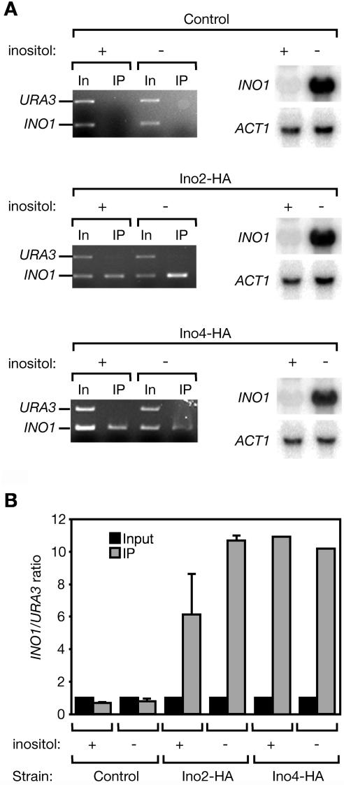
Ino2 and Ino4 have been shown by gel-shift analysis of yeast extracts to bind directly to the UASINO in the INO1 promoter (Lopes and Henry 1991; Ambroziak and Henry 1994; Bachhawat et al. 1995; Schwank et al. 1995). Binding was observed in extracts from cells grown under repressing or activating conditions, and was increased in the absence of Opi1 (Wagner et al. 1999). To monitor the interaction of Ino2 and Ino4 with the INO1 promoter in vivo, we used chromatin immunoprecipitation (ChIP) (Solomon et al. 1988; Dedon et al. 1991). Consistent with the gel-shift experiments, we found that Ino2-HA and Ino4-HA bound to the INO1 promoter under both repressing and activating conditions (Figure 2A). Real-time quantitative PCR analysis of immunoprecipitated DNA confirmed that both Ino2 and Ino4 associated with the INO1 promoter constitutively (Figure 2B). Although we observed an increase in the association of Ino2 with the INO1 promoter under inducing conditions compared with repressing conditions, these results argue that occupancy of the promoter by Ino2/Ino4 is not sufficient for activation but that it must be a subsequent step in the activation process that is regulated by the UPR.
Figure 2. Ino2/Ino4 Bind to the INO1 Promoter Constitutively.
(A) Untagged control cells (upper images), or cells in which the endogenous copies of INO2 and INO4 were replaced with HA-tagged Ino2 (center images) or HA-tagged Ino4 (lower images) were harvested in mid-logarithmic phase and washed into medium with or without _myo_-inositol. After 4.5 h, about 1.5 × 10⁸ cells were harvested and processed for Northern blot analysis (light images with dark bands, right). Northern blots were probed against both INO1 and ACT1 (loading control) mRNA. The remaining cells were fixed with formaldehyde and lysed. Chromatin was sheared by sonication and then subjected to immunoprecipitation with anti-HA agarose. Input DNA (In) and immunoprecipitated DNA (IP) were analyzed by PCR using primers to amplify the INO1 promoter and the URA3 gene. Amplified DNA was size-fractionated by electrophoresis on ethidium bromide-stained agarose gels (dark images with light bands, left).
(B) Quantitative PCR analysis. Input and IP fractions were analyzed by real-time quantitative PCR. The ratio of INO1 promoter to URA3 template in the reaction is shown. Error bars represent the standard error of the mean (SEM) between experiments.
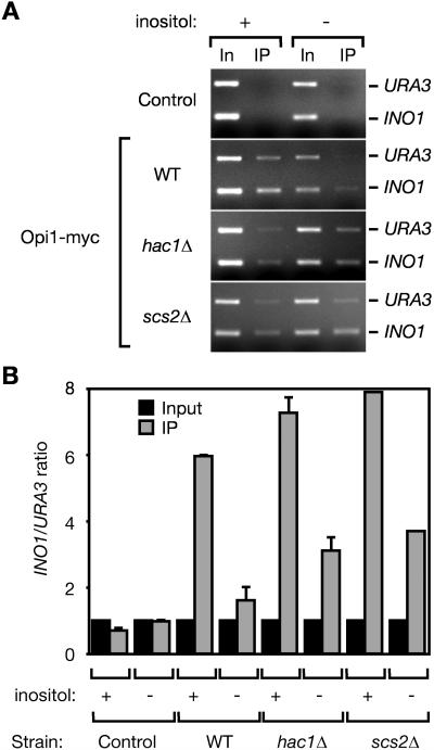
The molecular mechanism by which Opi1 represses transcription is not understood. In particular, it is not clear whether Opi1 binds to the INO1 promoter directly. Early gel-shift experiments using yeast lysates suggested that Opi1 might interact with DNA (Lopes and Henry 1991). However, this association has not been confirmed, and its significance is unknown. We used ChIP analysis and real-time quantitative PCR to assess the interaction of Opi1 with the INO1 promoter in vivo. We observed specific enrichment of the INO1 promoter by immunoprecipitation of Opi1 from cells grown in the presence of inositol (repressing condition) but no significant enrichment of the INO1 promoter by immunoprecipitation of Opi1 from cells starved for inositol (activating condition; Figure 3). By contrast, when we performed the immunoprecipitations from either _hac1_Δ or _scs2_Δ strains, we observed greater enrichment of the INO1 promoter sequences from cells grown under both activating and repressing conditions. These results are consistent with the notion that Opi1 binds to chromatin at the INO1 promoter and that the function of Hac1 and Scs2 is to promote Opi1 dissociation.
Figure 3. UPR-Dependent Dissociation of Opi1 from Chromatin.
(A) Chromatin-associated Opi1 dissociates upon activation of the UPR. Cells of the indicated genotypes were harvested after growth for 4.5 h with or without _myo_-inositol, fixed, and processed as in Figure 2. The _scs2_Δ mutant was transformed with pRS315-Opi1-myc, a CEN ARS plasmid that expresses Opi1-myc at endogenous levels. Input DNA (In) and immunoprecipitated DNA (IP) were analyzed by PCR using primers to amplify the INO1 promoter and the URA3 gene. Amplified DNA was separated by electrophoresis on ethidium bromide–stained agarose gels.
(B) Quantitative PCR analysis. Input and IP fractions were analyzed by real-time quantitative PCR. The ratio of INO1 promoter to URA3 template in the reaction is shown. Error bars represent the SEM between experiments.
In contrast to immunoprecipitation of Ino2 and Ino4, which specifically recovered the INO1 promoter and not the control URA3 sequences (see Figure 2), immunoprecipitates of Opi1 recovered significant amounts of URA3 sequences as well (Figure 3A, upper bands). It is clear from the quantitative PCR analysis that Opi1 binding to the INO1 promoter is specific (Figure 3B). The different conditions used in the qualitative gel analysis (measuring PCR products after many cycles) and the quantitative PCR (measuring PCR products in the linear range of amplification) are likely to account for this difference.
The INO1 Gene Relocalizes within the Nucleus upon UPR Activation
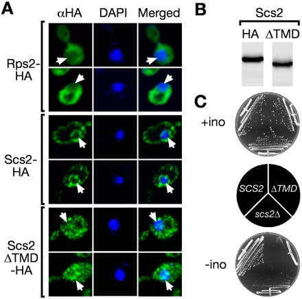
Since Opi1 dissociation from the INO1 promoter correlates with activation and requires Hac1 and Scs2, an integral nuclear membrane protein, we wondered whether activation might occur at the nuclear periphery and thus might be dependent on the subnuclear positioning of the gene. Consistent with this hypothesis, we found that a form of Scs2 (Scs2ΔTMD) lacking the transmembrane domain, which was localized throughout the cell and was not excluded from the nucleus (Figure 4A, compare cytosolic protein Rps2 to Scs2ΔTMD for colocalization with 4′,6′-diamidino-2-phenylindole), and was nonfunctional, rendering cells inositol auxotrophs, despite being expressed at levels comparable to full length Scs2 (Figure 4B and 4C).
Figure 4. Membrane Association Is Essential for Scs2 Function.
The carboxyl-terminal transmembrane domain of Scs2 was removed by replacement with three copies of the HA epitope (Scs2ΔTMD-HA; Longtine et al. [1998]).
(A) Scs2ΔTMD localization. Ribosomal protein S2 (Rps2-HA), Scs2-HA, and Scs2ΔTMD-HA were localized by immunofluorescence against the HA epitope. DNA was stained with 4′,6′-diamidino-2-phenylindole. Images were collected in a single z-plane (≤ 0.7 μm thick) by confocal microscopy. Unlike Rps2-HA, which was excluded from the nucleus (indicated with white arrows), Scs2ΔTMD-HA staining was uniform and evident in the nucleoplasm.
(B) Scs2ΔTMD steady-state levels. Equal amounts of whole cell extract from cells expressing either Scs2-HA or Scs2ΔTMD-HA were analyzed by immunoblotting.
(C) Scs2ΔTMD is nonfunctional. Strains expressing the indicated forms of Scs2 were streaked onto medium with or without _myo_-inositol and incubated for 2 d at 37 °C.
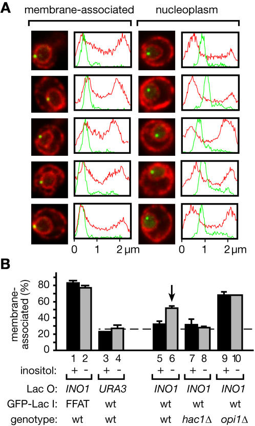
If INO1 were regulated at the nuclear periphery, then the INO1 locus should colocalize with the nuclear membrane under activating conditions. To test this idea, we constructed a strain in which an array of Lac operator (Lac O in Figure 5) binding sites was integrated adjacent to the INO1 locus (Robinett et al. 1996). The strain also expressed a green fluorescent protein (GFP)-Lac repressor fusion protein (GFP-Lac I in Figures 5 and 6) that binds to the Lac operator array to allow localization of the INO1 gene. In a control strain, we integrated the same Lac operator array adjacent to the URA3 locus. Cells were fixed and GFP was visualized by indirect immunofluorescence. Most cells showed a single intranuclear spot localizing the tagged gene; the remaining cells showed two spots due to their post-replication state in the cell cycle. In both the tagged INO1 and the tagged URA3 strains, we simultaneously visualized the ER and nuclear membrane by indirect immunofluorescence against Sec63-myc using a different fluorophore (Figure 5A).
Figure 5. The INO1 Gene Is Recruited to the Nuclear Membrane upon Activation.
An array of Lac operator repeats was integrated at INO1 or URA3 in strains expressing GFP-Lac repressor and _myc_-tagged Sec63. GFP-Lac repressor and Sec63-myc were localized in fixed cells by indirect immunofluorescence. Data were collected from single z sections representing the maximal, most focused signal from the Lac repressor.
(A) Two classes of subnuclear localization. Shown are five representative examples of localization patterns that were scored as membrane-associated (photomicrographs and plots on left) or nucleoplasmic (right). For each image, the fluorescence intensity was plotted for each channel along a line that intersects both the Lac repressor spot and the center of the nucleus.
(B) INO1 is recruited to the nuclear membrane upon activation. The fraction of cells that scored as membrane-associated is plotted for each strain grown in the presence (+) or absence (–) of inositol. The site of integration of the Lac operator (Lac O), the version of the GFP-Lac repressor (GFP-Lac I; either wild-type or having the FFAT membrane-targeting signal) expressed, and the relevant genotype of each strain is indicated. The dashed line represents the mean membrane association of the URA3 gene. The vertical arrow indicates the frequency of membrane association in the wild-type strain under activating conditions. Error bars represent the SEM between separate experiments. Each experiment scored at least 30 cells. The total number of cells (and experiments) scored for each column were: bar 1, 70 (2); bar 2, 66 (2); bar 3, 39 (1); bar 4, 71 (2); bar 5, 140 (4); bar 6, 88 (2); bar 7, 88 (2); bar 8, 92 (3); bar 9, 74 (2); and bar 10, 38 (1).
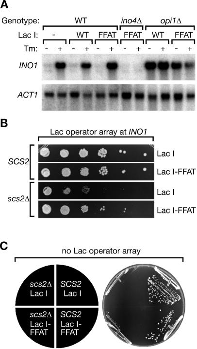
Figure 6. Artificial Relocalization of INO1 Bypasses the Requirement for Scs2.
(A) Northern blot analysis of membrane-targeted INO1. Strains of the indicated genotypes having the Lac operator array integrated at INO1 and expressing either the wild-type GFP-Lac repressor or GFP-FFAT-Lac repressor were grown in the presence or absence of 1 μg/ml tunicamycin (Tm) for 4.5 h, harvested, and analyzed by Northern blot. Blots were probed for either INO1 or ACT1 (as a loading control) mRNA. The wild-type strain CRY1, lacking both the Lac operator array and the Lac repressor, was included in the first two lanes for comparison.
(B) Wild-type or _scs2_Δ mutant strains in which the Lac operator had been integrated at INO1 were transformed with either GFP-Lac repressor or GFP-FFAT-Lac repressor. The resulting transformants were serially diluted (tenfold between wells) and spotted onto medium lacking inositol, uracil, and histidine, and incubated for 2 d at 37 °C.
(C) Wild-type and _scs2_Δ mutant strains transformed with either GFP-Lac repressor or GFP-FFAT-Lac repressor, but lacking the Lac operator, were streaked onto medium lacking inositol and histidine and incubated for 2 d at 37 °C.
To ask whether INO1 associates with the nuclear membrane, we developed stringent criteria for scoring INO1 localization (Figure 5A). Using confocal microscopy, we collected a single z slice through each cell that captured the brightest, most focused point of the GFP-visualized Lac operator array. Images in which this slice traversed the nucleus (i.e., cells that showed a clear nuclear membrane ring staining with a "hole" of nucleoplasm), were binned into two groups: Cells in which the peak of the spot corresponding to the tagged gene coincided with nuclear membrane staining were scored as membrane-associated, and cells in which the peak of the spot corresponding to the tagged gene was offset from nuclear membrane staining were scored as nucleoplasmic. This procedure allowed us to determine the fraction of cells in a given population in which the tagged gene colocalized with the membrane, thus providing a quantitative measure for membrane association. Five examples of each group, with fluorescence intensity plotted along a line bisecting the nucleus and the spot, are shown in Figure 5A.
To confirm that our scoring criterion would identify nuclear membrane association in a meaningful way, we applied it to two controls. As a control for membrane association, we localized INO1 in a strain expressing GFP-Lac repressor fused to a peptide motif from Opi1 containing two phenylalanines in an acidic tract (FFAT motif), which serves as a nuclear membrane–targeting signal (Loewen et al. 2003). This motif was shown to bind to Scs2 and to be required for Opi1 targeting to the nuclear envelope (Loewen et al. 2003). Importantly, targeting of Opi1 to the nuclear membrane still occurred in the absence of Scs2 in an FFAT-dependent manner (Loewen et al. 2003), indicating that, in addition to Scs2, there must exist another, yet-unidentified receptor for FFAT in the nuclear membrane. As shown in Figure 5B, the localization of INO1 scored as 85% membrane-associated (Figure 5B, bar 1), confirming both our scoring criteria and the previous result that FFAT indeed promotes nuclear membrane targeting.
As a control for random distribution, we localized URA3 in a strain expressing GFP-Lac repressor without the FFAT targeting signal. URA3 scored as 23% membrane-associated (Figure 5B, bar 3). Induction of the UPR after depletion of inositol had no effect on the localization of either FFAT-tagged INO1 or URA3 in these strains (Figure 5B, bars 2 and 4). Given that 25% of the volume of the nucleus is contained in the outer shell represented by only 10% of the radius, this level of background is consistent with a random distribution of the URA3 gene throughout the nuclear volume. Based on the spatial resolution of our data (Figure 5A), a spot only 10% of the radius distant from the membrane signal would have been scored as membrane-associated. We therefore defined the mean frequency of membrane-association of the URA3 control between these two conditions (25% ± 3%) as the baseline for subsequent comparisons (Figure 5B, dashed line).
We next compared the membrane association of INO1 under repressing and activating conditions. Under repressing conditions, the membrane association of INO1 was only slightly greater than the baseline (32% ± 3%; Figure 5B, bar 5). In striking contrast, when INO1 was activated, the frequency of membrane association of INO1 increased significantly over baseline (52% ± 3%; Figure 5B, bar 6). Thus, we conclude that, in a significant portion of cells, the INO1 gene became associated with the nuclear membrane under UPR-inducing conditions.
To confirm that the observed recruitment was indeed due to UPR induction, we compared the membrane association of INO1 under repressing or activating conditions in the _hac1_Δ mutant. Because Hac1 is required for activation of INO1, we predicted that membrane association would be prevented in this mutant. Indeed, INO1 failed to become membrane associated in _hac1_Δ mutants starved for inositol (Figure 5B, bars 7 and 8). Our earlier experiments suggested that Hac1 functions to promote dissociation of Opi1 from the INO1 promoter. We therefore tested next whether the presence of Opi1 prevents membrane association. To this end, we determined INO1 localization in the _opi1_Δ strain, in which INO1 is constitutively transcribed (Cox et al. 1997). Indeed, we observed a high degree of membrane association, both in the presence and absence of inositol (68% ± 5%; Figure 5B, bars 9 and 10).
Artificial Recruitment of INO1 Suppresses the _scs2_Δ Ino– Phenotype
The experiments described above indicate that there is a correlation between membrane association of INO1 and its transcriptional activation. To establish causality, we examined the effect of artificially targeting INO1 to the nuclear membrane. In an otherwise wild-type background, artificial targeting of INO1 to the nuclear membrane via FFAT-Lac repressor binding (same strain as in Figure 5B, bars 1 and 2) had no effect on INO1 expression as assessed by Northern blot analysis (Figure 6A) or on the growth of the wild-type strain in the absence of inositol (Figure 6B; compare top two panels). This result suggests that membrane targeting per se is not sufficient to cause activation. In contrast, in the _scs2_Δ mutant we observed that the inositol-requiring growth phenotype of the strain was suppressed by expression of the membrane-targeted FFAT-Lac repressor (Figure 6B; compare bottom two panels). This effect was strictly dependent on having the Lac operator array integrated at the INO1 locus; expressing GFP-FFAT-Lac repressor in the absence of the array (Figure 6C)—or if the array was integrated at the URA3 locus (unpublished data)—did not improve the growth of the _scs2_Δ mutant in the absence of inositol. Consistent with the previous report that FFAT does not require Scs2 to promote nuclear membrane targeting, we observed approximately 50% membrane association of INO1 in the strain expressing the FFAT-Lac repressor (78 cells counted, unpublished data). Thus, the defect in transcription of INO1 in the _scs2_Δ mutant could be rescued, at least partially, through artificial targeting of INO1 to the nuclear membrane. This result demonstrates that nuclear membrane association is functionally important for achieving INO1 transcriptional activation.
Discussion
It is becoming increasingly clear that the spatial arrangement of chromosomes within the nucleus is important for controlling the reactions that occur on DNA and might be regulated (reviewed in Cockell and Gasser [1999]; Isogai and Tjian [2003]). Here we have shown that activation of INO1 occurs at the nuclear membrane and requires the integral membrane protein Scs2. Moreover, artificial recruitment of INO1 to the nuclear membrane permits activation in the absence of Scs2, indicating that the precise intranuclear localization of a gene can profoundly influence its activation. Most importantly, we have shown that the localization of INO1 depends on its activation state; gene recruitment therefore is a dynamic process and appears to play an important regulatory role.
Regulation of Gene Localization
The nucleoplasm is bounded by the inner nuclear membrane, which provides a template that is likely to play a major role in organizing the genome. It is clear from numerous microscopic and biochemical studies that chromatin interacts with nuclear membrane proteins, associated proteins such as filamentous lamins, and nuclear pore complexes (DuPraw, 1965; Murray and Davies, 1979; Paddy, 1990; Worman et al., 1990; Belmont et al., 1993; Glass et al., 1993; Foisner and Gerace, 1993; Sukegawa and Blobel, 1993; Luderus et al., 1994; Marshall et al., 1996). Indeed, several transcriptionally regulated genes have been shown to colocalize with heterochromatin at the nuclear periphery when repressed (Csink and Henikoff 1996; Dernburg et al. 1996; Brown et al. 1997, 1999). Likewise, silencing of genes near telomeres requires physical tethering of telomeres to nuclear pore complexes at the nuclear periphery (Gotta et al. 1996; Maillet et al. 1996; Andrulis et al. 1998; Laroche et al. 1998; Galy et al. 2000; Andrulis et al. 2002; Feuerbach et al. 2002).
Thus, the nuclear periphery has been generally regarded as a transcriptionally repressive environment (Gotta et al. 1996; Maillet et al. 1996; Andrulis et al. 1998; Laroche et al. 1998; Galy et al. 2000; Andrulis et al. 2002; Feuerbach et al. 2002). In contrast, the work presented here shows that gene recruitment to the nuclear periphery can be important for transcriptional activation. This conclusion is supported by a recent study published while this manuscript was in preparation (Casolari et al. 2004). These authors found that a subset of actively transcribed genes associates with components of nuclear pore complexes and that activation of GAL genes correlates with their recruitment from the nucleoplasm to the nuclear periphery and pore-complex protein association (Casolari et al. 2004). The results presented here argue that recruitment of genes to the nuclear periphery is controlled by transcriptional regulators and is important for achieving transcriptional activation. Thus, together, the work by Casolari et al. (2004) and the work presented here demonstrate that gene recruitment to the nuclear periphery can have a general role in activating transcription.
This notion is consistent with the “gene gating hypothesis” put forward by Blobel (1985). As proposed in this hypothesis, transcription of certain genes may be obligatorily coupled to mRNA export through a particular nuclear pore complex. It remains to be shown for INO1, however, whether gene recruitment to the nuclear periphery involves interaction with nuclear pore complex components. Several other scenarios could explain why INO1 activation might require gene recruitment to the nuclear periphery. First, INO1 transcriptional activation requires the SAGA histone acetylase, and both the SWI/SNF and INO80 chromatin remodeling complexes (Kodaki et al. 1995; Pollard and Peterson 1997; Ebbert et al. 1999; Shen et al. 2000; Dietz et al. 2003). Conversely, repression requires the Sin3/Rpd3 histone deacetylase and the ISW chromatin remodeling complex (Hudak et al. 1994; Sugiyama and Nikawa 2001). Thus, if these factors have distinct subnuclear distributions, then the localization of genes regulated by them might influence their transcriptional state. Consistent with this notion, the SAGA complex interacts with nuclear pore complexes, and therefore might be concentrated at the nuclear periphery, where INO1 activation occurs (Rodriguez-Navarro et al. 2004). Second, because INO1 and many other UASINO-regulated genes are involved in the biosynthesis of phospholipids, it is possible that the state of the membrane itself plays a role, perhaps sensed by Scs2, in activating transcription. It has been shown that defects in phospholipids biosynthesis can disrupt regulation of INO1, although the mechanism of this regulation remains unknown (Greenberg et al. 1982; McGraw and Henry 1989; Griac et al. 1996; Griac 1997; Shirra et al. 2001). Third, inositol polyphosphates have been shown to regulate SWI/SNF-catalyzed chromatin remodeling, and it is possible that their production is spatially restricted (Shen et al. 2003; Steger et al. 2003).
Role of Factors Regulating INO1 Activation
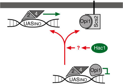
Our current understanding of INO1 activation is summarized in a model in Figure 7. The positive transcription activators Ino2 and Ino4 constitutively associate with the INO1 promoter, which is kept transcriptionally repressed by Opi1. We do not currently understand the mechanism by which Opi1 prevents activation. Activation of the UPR leads to the production of Hac1, which, by an unknown mechanism, promotes Opi1 dissociation from chromatin. We propose that Scs2 at the nuclear membrane binds to Opi1 released from the DNA and thus keeps Opi1 sequestered and prevented from rebinding. Indeed, overproduction of Scs2 bypasses the requirement for Hac1 in activation of INO1 transcription and allows _hac1_Δ cells to grow in the absence of inositol (Nikawa et al. 1995), supporting the role of Scs2 as a sink for Opi1 and suggesting that Opi1 may cycle between chromatin-bound and free states.
Figure 7. Model for INO1 Gene Recruitment and Transcriptional Activation.
Ino2 and Ino4 bind constitutively to the INO1 promoter. Under repressing conditions, Opi1 associates with chromatin to prevent activation, and the INO1 locus localizes to the nucleoplasm. Hac1 synthesis under UPR-inducing conditions promotes dissociation of Opi1 from chromatin. Scs2 binds to Opi1 at the nuclear membrane to stabilize the non-chromatin-bound state. Dissociation is coupled to recruitment of INO1 to the nuclear membrane, where transcriptional activation occurs.
Both Hac1 (Cox et al. 1997) and Scs2 (see Figure 1) are dispensable for INO1 activation in the absence of Opi1, suggesting that their role is to relieve Opi1 repression. However, our data suggest that Hac1 and Scs2 have distinct functions: While the absence of either protein prevents the dissociation of Opi1 from chromatin and the activation of INO1, we propose that the role of Hac1 is to promote dissociation and that of Scs2 is to prevent reassociation. This model explains why artificially tethering INO1 to the nuclear membrane suppresses the absence of Scs2 but not the absence of Hac1 (unpublished data): We propose that the environment of membrane-tethered INO1 promotes late steps in the transcription activation—such as chromatin remodeling, discussed above—permitting INO1 to be expressed upon transient Hac1-induced Opi1 dissociation. Therefore, we envision that dissociation of Opi1 from the INO1 promoter is coupled to the delivery of the gene to an environment near the nuclear membrane that is permissive for its activation.
The mechanistic role of Scs2 is currently not known. Its recently discovered function in promoting telomeric silencing (Craven and Petes 2001; Cuperus and Shore 2002) suggests that Scs2 may play a more global role in the regulation of transcription at the nuclear membrane. Scs2 contains a major sperm protein domain, named after a homologous protein in Ascaris suum sperm that forms a cytoskeletal structure and confers motility to sperm cells. It is thus tempting to speculate that Scs2 might similarly self-associate in the plane of the nuclear membrane, perhaps providing a two-dimensional matrix on which membrane-associated reactions could be organized. One suggestion from our data is that Scs2 may function as a local sink for Opi1. But it is also clear that other nuclear membrane components are likely to participate in the reaction. Opi1, for example, still localizes to nuclear membranes even in _scs2_Δ cells, indicating that another, yet-unidentified Opi1 binding partner must exist (Loewen et al. 2003; unpublished data). Similarly, artificial INO1 recruitment to the membrane via the FFAT motif suppresses the _scs2_Δ phenotype (see Figure 6)—i.e., it is sufficient to position INO1 in an environment permissive for its induction—yet the FFAT binding protein and the molecular nature of the permissive environment remain unknown.
Upon inducing the UPR, only 52% of the cells scored INO1 as membrane-associated (see Figure 5). Thus, under activating conditions, two types of cells are present in the population at any one time: those in which the INO1 gene is recruited to the membrane, and those in which the INO1 gene is dispersed throughout the nucleoplasm. This score correlated with the level of INO1 transcription; INO1 was membrane-associated in 68% of the cells in the opi1 mutant, which exhibits a correspondingly higher degree of activation than that observed in the wild-type strain. A quantitatively similar nuclear peripheral-nucleoplasmic distribution was observed upon activation of GAL genes (Casolari et al. 2004), suggesting that it may be a general feature of gene recruitment. There are at least two possible interpretations for the observed bimodal distributions. First, the distribution profiles might represent heterogeneity in the activation of INO1 among cells. In this case, activation of INO1 would be variable in individual cells exposed to identical conditions. Gene recruitment thus would stably trap INO1 in a permissive environment for activation, and the localization of INO1 would strictly correlate with its activation state. Alternatively, gene recruitment might alter the balance between two rapidly exchanging states; that is, stable membrane recruitment would not be required for activation. In this case, the observed distributions would represent snapshots of transient colocalization of INO1 with the nuclear membrane within a population of cells that are uniformly activating transcription. Dynamic measurements of gene recruitment and single cell activity assays will need to be developed to distinguish between these possibilities. But no matter which of these possibilities proves to be correct, gene recruitment emerges as a new mechanism regulating eukaryotic gene expression and may be crucial to the regulation of many genes.
Materials and Methods
Antibodies and reagents
Monoclonal anti- HA antibody HA11 was obtained from Babco (Berkeley, California, United States). Monoclonal anti-myc, anti-myc agarose and anti-HA agarose were from Santa Cruz Biotechnology (Santa Cruz, California, United States). Monoclonal anti-Pgk1, rabbit polyclonal anti-GFP, goat anti mouse IgG-Alexafluor 594, and goat anti-rabbit IgG Alexafluor 488 were from Molecular Probes (Eugene, Oregon, United States).
All restriction endonucleases and DNA modification enzymes were from New England Biolabs (Beverly, Massachusetts, United States). Unless indicated otherwise, all other chemicals and reagents were from Sigma (St. Louis, Missouri, United States).
Strains and plasmids
All yeast strains used in this study were derived from wild-type strain CRY1 (ade2–1 can1–100 his3–11,15 leu2–3,112 trp1–1 ura3–1 MAT_a). Tags and disruptions marked with either the kanr gene from E. coli or the His5 gene from S. pombe were introduced by recombination at the genomic loci as described (Longtine et al. 1998). Strains used in this study, with relevant differences indicated are JBY345 (OPI1–13myc::kanr), JBY350-r1 (scs2_Δ:: kanr), JBY359 (SCS2-HA:: kanr), JBY356–1A (opi1_Δ::LEU2),_ JBY356–1B (opi1_Δ::LEU2 scs2_Δ_:: kanr ),_ JBY356–1C (scs2_Δ:: kanr),_ JBY356–1D (wild-type control), JBY361 (scs2_Δ_TMD-HA:: kanr), JBY370_(INO2-HA3::His5+),_ JBY371 (INO4-HA3::His5+), JBY393 (INO4-myc::His5+ MAT_a),_ JBY397 (SEC63–13myc:: kanr INO1:LacO128:URA3 HIS3:LacI-GFP), JBY399 (SEC63–13myc::Kan^r INO1:LacO128:URA3 HIS3:LacI-FFAT-GFP), JBY401 (ino4_Δ::LEU2 SEC63–13myc::Kanr INO1:LacO128:URA3 HIS3:LacI-GFP MATα),_ JBY404 (opi1_Δ::LEU2 SEC63–13myc::Kanr INO1:LacO128:URA3 HIS3:LacI-GFP),_ JBY406 (opi1_Δ::LEU2 SEC63–13myc::Kanr INO1:LacO128:URA3 HIS3:LacI-FFAT-GFP),_ JBY409 (SEC63–13myc::Kanr URA3:LacO128:URA3 HIS3:LacI-GFP), JBY412 (INO2-myc::His5+), JBY 416 (hac1_Δ::URA3 SEC63–13myc::Kanr LacO128:INO1 HIS3:LacI-GFP)_.
Plasmid pRS315-Opi1-myc was created by first amplifying the OPI1-myc coding sequence and 686 bp upstream from the translational start site from strain JBY345 using the following primers: OPI1 promoter Up (5′-GGGAGATACAAACCATGAAG-3′) and OPI1 down (5′-ACTATACCTGAGAAAGCAACCTGACCTACAGG-3′). The resulting fragment was cloned into pCR2.1 using the Invitrogen (Carlsbad, California, United States) TOPO TA cloning kit. The OPI1-myc locus was then cloned into pRS315 as a HindIII-NotI fragment. Plasmid pASF144 expressing GFP-lacI has been described (Straight et al. 1996). Plasmid pGFP-FFAT-LacI was constructed by digesting pASF144 with EcoRI and ligating the fragment to the following hybridized oligonucleotides, encoding the FFAT motif from OPI1: LacI_FFAT1 (5′-AATTGGACGATGAGGAGTTTTTTGATGCCTCAGAGG-3′) and LacI_FFAT2 (5′-AATTCCTCTGAGGCATCAAAAAACTCCTCATCGTCC-3′). The orientation of the insert was confirmed by DNA sequencing. Both pAFS144 and pGFP-FFAT-LacI were digested with NheI, which cuts within the HIS3 gene, and transformed into yeast.
Plasmid p6INO1LacO128 was constructed as follows. The INO1 coding sequence, with 437 bp upstream and 758 bp downstream, was amplified from yeast genomic DNA using the following primers: INO1_promoter_Up (5′-GATGAGGCCGGTGCC-3′) and INO1_3′down (5′-AAGATTTCCTTCTTGGGCGC-3′), and cloned into pCR2.1 using the Invitrogen TOPO TA cloning kit, to produce pCR2.1-INO1. INO1 was moved from pCR2.1 into pRS306 as a KpnI fragment, to produce pRS306-INO1. The Lac operator array was then cloned from pAFS52 into pRS306-INO1 as a HindIII-XhoI fragment, to produce plasmid 10.2. Because the Lac operator fragment was smaller than had been reported (2.5 kb instead of 10 kb), presumably reflecting loss of Lac operator repeats by recombination, the Lac operator array was duplicated by digesting plasmid 10.2 with HindIII and SalI and introducing a second copy of the 2.5-kb HindIII-XhoI fragment, as described (Robinett et al. 1996). The resulting plasmid, p6INO1LacO128, has a 5-kb Lac operator array, corresponding to approximately 128 repeats of the lac operator. To integrate this plasmid at INO1, p6INO1LacO128 was digested with BglII, which cuts within the INO1 gene, and transformed into yeast.
The INO1 gene was removed from this plasmid to generate p6LacO128. This plasmid was used to integrate the Lac operator array at URA3 by digestion with StuI and transformation into yeast.
Immunoprecipitations
Cells were lysed using glass beads in IP buffer (50 mM Hepes-KOH pH 6.8, 150 mM potassium acetate, 2 mM magnesium acetate, and Complete Protease Inhibitors [Roche, Indianapolis, Indiana, United States]). The whole cell extract was used for coimmunoprecipitation of Ino2-myc and Ino4-HA. For immunoprecipitation of Opi1-myc, microsomes were pelleted by centrifugation for 10 min at 21,000 × g and resuspended in IP buffer. Triton X-100 was then added to either whole cell extract (Ino2-myc; final concentration of 1%) or the microsomal fraction (Opi1-myc; final concentration of 3%) and incubated for 30 min at 4 °C; detergent-insoluble material was then removed by centrifugation at 21,000 × g, 10 min. Anti-myc agarose was added to the supernatant and incubated 4 h at 4 °C, while rotating. For the experiment in Figure 1D, a fraction of the total was collected after antibody incubation. After agarose beads were pelleted, an equal fraction of the supernatant was collected. Beads were washed either five (see Figure 1B and 1D) or ten times (see Figure 1C) with IP buffer. A fraction of the final wash equal to the pellet fraction in Figure 1D was collected. After the final wash, proteins were eluted from the beads by heating in sample buffer and separating by SDS-PAGE (see Figure 1B and 1D). Trypsin digestion, gel extraction, and mass spectrometry of proteins that coimmunoprecipitated with Opi1 were performed by the HHMI Mass Spectrometry facility (University of California, Berkeley, United States).
Immunoblot and Northern blot analysis
For immunoblot analysis, 25 μg of crude protein, prepared using urea denaturing lysis buffer (Ruegsegger et al. 2001), was separated on Invitrogen NuPage polyacrylamide gels, transferred to nitrocellulose, and immunoblotted. RNA preparation, electrophoresis, and labeling of probes for Northern blot analysis has been described (Ruegsegger et al. 2001).
Immunofluorescence
Immunofluorescence was carried out as described (Redding et al. 1991), except that cells were harvested and fixed by incubation in 100% methanol at –20 °C for 20 min. Fixed, spheroplasted, detergent-extracted cells were probed with 1:200 monoclonal anti-myc (see Figures 1 and 5), 1:200 monoclonal anti-HA (see Figure 4), or 1:1000 rabbit polyclonal anti-GFP (see Figure 5). Secondary antibodies were diluted 1:200. Vectashield mounting medium (Vector Laboratories, Burlingame, California, United States) was applied to cells before sealing slides and visualizing using a Leica TCS NT confocal microscope (Leica, Wetzlar, Germany). For experiments localizing the GFP-Lac repressor, we first collected a single z slice through each cell that captured the brightest, most focused point of the GFP-visualized Lac operator array. This z slice was picked blind with respect to the nuclear membrane staining. Images in which this slice showed a clear nuclear membrane ring staining with a "hole" of nucleoplasm were then scored as follows: Cells in which the peak of the GFP-Lac repressor spot coincided with Sec63-myc nuclear membrane staining were scored as membrane-associated, and cells in which the peak of this spot was offset from nuclear membrane staining were scored as nucleoplasmic.
Chromatin immunoprecipitation
Chromatin immunoprecipitation was carried out on strains expressing endogenous levels of tagged Ino2, Ino4, and Opi1 as described (Strahl-Bolsinger et al. 1997), with the following modifications. The time of formaldehyde fixation was specific for each tagged protein. Strains expressing Ino2-HA were fixed for 15 min, strains expressing Ino4-HA were fixed for 60 min, and strains expressing Opi1-myc were fixed for 30 min. After lysis, cells were sonicated 15 times for 10 s at 30% power using a microtip on a Vibracell VCX 600 Watt sonicator (Sonics and Materials, Newtown, Connecticut, United States). After sonication, lysates were centrifuged 10 min at 21,000 × g to remove insoluble material and incubated for 4 h with anti-HA agarose or anti-myc agarose. After elution of immunoprecipitated DNA and reversal of crosslinks by heating to 65 °C for 8 h, DNA was recovered using Qiaquick columns from Qiagen (Alameda, California, United States). Eluted samples were analyzed by PCR using the following primers against the INO1 promoter or the URA3 gene: INO1_proUp2 (5′-GGAATCGAAAGTGTTGAATG-3′), INO1_proDown (5′-CCCGACAACAGAACAAGCC-3′), URAup (5′- GGGAGACGCATTGGGTCAAC-3′), and URADown (5′-GTTCTTTGGAGTTCAATGCGTCC-3′).
Real time quantitative PCR analysis
PCR reactions were carried out as described (Rogatsky et al. 2003) using a DNA Engine Opiticon 2 Real-Time PCR machine (MJ Research, Waltham, Massachusetts, United States), using 1/25 of the immunoprecipitation fraction and an equal volume of a 1:400 dilution of the input fraction as template. Primers used were: INO1up3 5′-ATTGCCTTTTTCTTCGTTCC-3′), INO1down2 (5′-CATTCAACACTTTCGATTCC-3′), URAup2 (5′-AGACGCATTGGGTCAAC-3′), and URAdown2 (5′-CTTCCCTTTGCAAATAGTCC-3′). Dilution of the input fraction from 1:25 to 1:12,800 in fourfold steps demonstrated that reactions were within the linear range of template. This dilution series was used as a standard curve of C(T) values versus relative template concentration for both primer sets. The concentration of the INO1 promoter and the URA3 gene were calculated using this standard curve. The ratio of INO1 promoter to URA3 was corrected for each sample to make the input ratio equal to 1.0.
Supporting Information
Accession Numbers
The GenBank accession numbers of the genes and proteins discussed in this paper are Ire1 (NP_116622), INO1 (NP_012382), Trl1 (NP_012448), Opi1 (NP_011843), Hac1 (NP_011946), Ino2 (NP_010408), Ino4 (NP_014533), Scs2 (NP_009461), Rap1 (NP_014183), URA3 (NP_010893), SSS1 (NP_010371), Pgk1 (NP_009938) lacI (NP_414879), and Ikaros (Q03267).
Acknowledgments
The authors are very grateful to all the members of the Walter lab, in particular Gustavo Pesce, Tobias Walther, Jess Leber, and Tomas Aragon, for helpful discussions and inspiration; Isabella Halama for help with immunofluorescence; Hans Lueke for help with ChIP experiments and real time quantitative PCR; Doris Fortin for help with confocal microscopy; Joel Credle for technical assistance; John Sedat for providing the Lac operator and the GFP-Lac repressor plasmids; and Keith Yamamoto for helpful comments on the manuscript. This work was supported by a Helen Hay Whitney Postdoctoral Fellowship (JHB), by the Sandler Family Opportunity Fund, and by grants from the National Institutes of Health to PW. PW is an Investigator of the Howard Hughes Medical Institute.
Abbreviations
ChIP
chromatin immunoprecipitation
ER
endoplasmic reticulum
FFAT
ER/nuclear membrane targeting motif containing two phenylalanines in an acidic tract
GFP
green fluorescent protein
HA
hemagglutinin
SEM
standard error of the mean
UASINO
upstream activating sequence responsive to inositol
UPR
unfolded protein response
Conflicts of interest. The authors have declared that no conflicts of interest exist.
Author contributions. JHB and PW conceived of the experiments. JHB performed the experiments. JHB and PW wrote the paper.
Academic Editor: Tom Misteli, National Cancer Institute
Citation: Brickner JH, Walter P (2004) Gene recruitment of the activated INO1 locus to the nuclear membrane. PLoS Biol 2(11): e342.
References
- Ambroziak J, Henry SA. INO2 and INO4 gene products, positive regulators of phospholipid biosynthesis in Saccharomyces cerevisiae, form a complex that binds to the INO1 promoter. J Biol Chem. 1994;269:15344–15349. [PubMed] [Google Scholar]
- Andrulis ED, Neiman AM, Zappulla DC, Sternglanz R. Perinuclear localization of chromatin facilitates transcriptional silencing. Nature. 1998;394:592–595. doi: 10.1038/29100. [DOI] [PubMed] [Google Scholar]
- Andrulis ED, Zappulla DC, Ansari A, Perrod S, Laiosa CV, et al. Esc1, a nuclear periphery protein required for Sir4-based plasmid anchoring and partitioning. Mol Cell Biol. 2002;22:8292–8301. doi: 10.1128/MCB.22.23.8292-8301.2002. [DOI] [PMC free article] [PubMed] [Google Scholar]
- Ashburner BP, Lopes JM. Regulation of yeast phospholipid biosynthetic gene expression in response to inositol involves two superimposed mechanisms. Proc Natl Acad Sci U S A. 1995a;92:9722–9726. doi: 10.1073/pnas.92.21.9722. [DOI] [PMC free article] [PubMed] [Google Scholar]
- Ashburner BP, Lopes JM. Autoregulated expression of the yeast INO2 and INO4 helix-loop-helix activator genes effects cooperative regulation on their target genes. Mol Cell Biol. 1995b;15:1709–1715. doi: 10.1128/mcb.15.3.1709. [DOI] [PMC free article] [PubMed] [Google Scholar]
- Bachhawat N, Ouyang Q, Henry SA. Functional characterization of an inositol-sensitive upstream activation sequence in yeast. A cis-regulatory element responsible for inositol-choline mediated regulation of phospholipid biosynthesis. J Biol Chem. 1995;270:25087–25095. doi: 10.1074/jbc.270.42.25087. [DOI] [PubMed] [Google Scholar]
- Blobel G. Gene gating: A hypothesis. Proc Natl Acad Sci U S A. 1985;82:8527–8529. doi: 10.1073/pnas.82.24.8527. [DOI] [PMC free article] [PubMed] [Google Scholar]
- Boveri T. Die blastermerenkerne von Ascaris megalocephala und die theorie der chromosomenindividualität. Arch Zellforschung. 1909;3:181. [Google Scholar]
- Bressan DA, Vazquez J, Haber JE. Mating type-dependent constraints on the mobility of the left arm of yeast chromosome III. J Cell Biol. 2004;164:361–371. doi: 10.1083/jcb.200311063. [DOI] [PMC free article] [PubMed] [Google Scholar]
- Brown KE, Baxter J, Graf D, Merkenschlager M, Fisher AG. Dynamic repositioning of genes in the nucleus of lymphocytes preparing for cell division. Mol Cell. 1999;3:207–217. doi: 10.1016/s1097-2765(00)80311-1. [DOI] [PubMed] [Google Scholar]
- Brown KE, Guest SS, Smale ST, Hahm K, Merkenschlager M, et al. Association of transcriptionally silent genes with Ikaros complexes at centromeric heterochromatin. Cell. 1997;91:845–854. doi: 10.1016/s0092-8674(00)80472-9. [DOI] [PubMed] [Google Scholar]
- Casolari JM, Brown CR, Komili S, West J, Hieronymous H, et al. Genome-wide localization of the nuclear transport machinery couples transcriptional status and nuclear organization. Cell. 2004;117:427–439. doi: 10.1016/s0092-8674(04)00448-9. [DOI] [PubMed] [Google Scholar]
- Cobb BS, Morales-Alcelay S, Kleiger G, Brown KE, Fisher AG, et al. Targeting of Ikaros to pericentromeric heterochromatin by direct DNA binding. Genes Dev. 2000;14:2146–2160. doi: 10.1101/gad.816400. [DOI] [PMC free article] [PubMed] [Google Scholar]
- Cockell M, Gasser SM. Nuclear compartments and gene regulation. Curr Opin Genet Dev. 1999;9:199–205. doi: 10.1016/S0959-437X(99)80030-6. [DOI] [PubMed] [Google Scholar]
- Cox JS, Walter P. A novel mechanism for regulating activity of a transcription factor that controls the unfolded protein response. Cell. 1996;87:391–404. doi: 10.1016/s0092-8674(00)81360-4. [DOI] [PubMed] [Google Scholar]
- Cox JS, Shamu CE, Walter P. Transcriptional induction of genes encoding endoplasmic reticulum resident proteins requires a transmembrane protein kinase. Cell. 1993;73:1197–1206. doi: 10.1016/0092-8674(93)90648-a. [DOI] [PubMed] [Google Scholar]
- Cox JS, Chapman RE, Walter P. The unfolded protein response coordinates the production of endoplasmic reticulum protein and endoplasmic reticulum membrane. Mol Biol Cell. 1997;8:1805–1814. doi: 10.1091/mbc.8.9.1805. [DOI] [PMC free article] [PubMed] [Google Scholar]
- Craven RJ, Petes TD. The Saccharomyces cerevisiae suppressor of choline sensitivity (SCS2) gene is a multicopy Suppressor of mec1 telomeric silencing defects. Genetics. 2001;158:145–154. doi: 10.1093/genetics/158.1.145. [DOI] [PMC free article] [PubMed] [Google Scholar]
- Csink AK, Henikoff S. Genetic modification of heterochromatic association and nuclear organization in Drosophila . Nature. 1996;381:529–531. doi: 10.1038/381529a0. [DOI] [PubMed] [Google Scholar]
- Cuperus G, Shore D. Restoration of silencing in Saccharomyces cerevisiae by tethering of a novel Sir2-interacting protein, Esc8. Genetics. 2002;162:633–645. doi: 10.1093/genetics/162.2.633. [DOI] [PMC free article] [PubMed] [Google Scholar]
- Dedon PC, Soults JA, Allis CD, Gorovsky MA. A simplified formaldehyde fixation and immunoprecipitation technique for studying protein-DNA interactions. Anal Biochem. 1991;197:83–90. doi: 10.1016/0003-2697(91)90359-2. [DOI] [PubMed] [Google Scholar]
- Dernburg AF, Broman KW, Fung JC, Marshall WF, Philips J, et al. Perturbation of nuclear architecture by long-distance chromosome interactions. Cell. 1996;85:745–759. doi: 10.1016/s0092-8674(00)81240-4. [DOI] [PubMed] [Google Scholar]
- Dietz M, Heyken WT, Hoppen J, Geburtig S, Schuller HJ. TFIIB and subunits of the SAGA complex are involved in transcriptional activation of phospholipid biosynthetic genes by the regulatory protein Ino2 in the yeast Saccharomyces cerevisiae . Mol Microbiol. 2003;48:1119–1130. doi: 10.1046/j.1365-2958.2003.03501.x. [DOI] [PubMed] [Google Scholar]
- Ebbert R, Birkmann A, Schuller HJ. The product of the SNF2/SWI2 paralogue INO80 of Saccharomyces cerevisiae required for efficient expression of various yeast structural genes is part of a high-molecular-weight protein complex. Mol Microbiol. 1999;32:741–751. doi: 10.1046/j.1365-2958.1999.01390.x. [DOI] [PubMed] [Google Scholar]
- Feuerbach F, Galy V, Trelles-Sticken E, Fromont-Racine M, Jacquier A, et al. Nuclear architecture and spatial positioning help establish transcriptional states of telomeres in yeast. Nat Cell Biol. 2002;4:214–221. doi: 10.1038/ncb756. [DOI] [PubMed] [Google Scholar]
- Galy V, Olivo-Marin JC, Scherthan H, Doye V, Rascalou N, et al. Nuclear pore complexes in the organization of silent telomeric chromatin. Nature. 2000;403:108–112. doi: 10.1038/47528. [DOI] [PubMed] [Google Scholar]
- Gasser SM. Visualizing chromatin dynamics in interphase nuclei. Science. 2002;296:1412–1416. doi: 10.1126/science.1067703. [DOI] [PubMed] [Google Scholar]
- Gavin AC, Bosche M, Krause R, Grandi P, Marzioch M, et al. Functional organization of the yeast proteome by systematic analysis of protein complexes. Nature. 2002;415:141–147. doi: 10.1038/415141a. [DOI] [PubMed] [Google Scholar]
- Gotta M, Laroche T, Formenton A, Maillet L, Scherthan H, et al. The clustering of telomeres and colocalization with Rap1, Sir3, and Sir4 proteins in wild-type Saccharomyces cerevisiae . J Cell Biol. 1996;134:1349–1363. doi: 10.1083/jcb.134.6.1349. [DOI] [PMC free article] [PubMed] [Google Scholar]
- Gottschling DE, Aparicio OM, Billington BL, Zakian VA. Position effect at S. cerevisiae telomeres: reversible repression of Pol II transcription. Cell. 1990;63:751–762. doi: 10.1016/0092-8674(90)90141-z. [DOI] [PubMed] [Google Scholar]
- Greenberg ML, Goldwasser P, Henry SA. Characterization of a yeast regulatory mutant constitutive for synthesis of inositol-1-phosphate synthase. Mol Gen Genet. 1982;186:157–163. doi: 10.1007/BF00331845. [DOI] [PubMed] [Google Scholar]
- Griac P. Regulation of yeast phospholipid biosynthetic genes in phosphatidylserine decarboxylase mutants. J Bacteriol. 1997;179:5843–5848. doi: 10.1128/jb.179.18.5843-5848.1997. [DOI] [PMC free article] [PubMed] [Google Scholar]
- Griac P, Swede MJ, Henry SA. The role of phosphatidylcholine biosynthesis in the regulation of the INO1 gene of yeast. J Biol Chem. 1996;271:25692–25698. doi: 10.1074/jbc.271.41.25692. [DOI] [PubMed] [Google Scholar]
- Hudak KA, Lopes JM, Henry SA. A pleiotropic phospholipid biosynthetic regulatory mutation in Saccharomyces cerevisiae is allelic to sin3 (sdi1, ume4, rpd1) . Genetics. 1994;136:475–483. doi: 10.1093/genetics/136.2.475. [DOI] [PMC free article] [PubMed] [Google Scholar]
- Isogai Y, Tjian R. Targeting genes and transcription factors to segregated nuclear compartments. Curr Opin Cell Biol. 2003;15:296–303. doi: 10.1016/s0955-0674(03)00052-8. [DOI] [PubMed] [Google Scholar]
- Kagiwada S, Zen R. Role of the yeast VAP homolog, Scs2p, in INO1 expression and phospholipid metabolism. J Biochem (Tokyo) 2003;133:515–522. doi: 10.1093/jb/mvg068. [DOI] [PubMed] [Google Scholar]
- Kagiwada S, Hosaka K, Murata M, Nikawa J, Takatsuki A. The Saccharomyces cerevisiae SCS2 gene product, a homolog of a synaptobrevin-associated protein, is an integral membrane protein of the endoplasmic reticulum and is required for inositol metabolism. J Bacteriol. 1998;180:1700–1708. doi: 10.1128/jb.180.7.1700-1708.1998. [DOI] [PMC free article] [PubMed] [Google Scholar]
- Kawahara T, Yanagi H, Yura T, Mori K. Endoplasmic reticulum stress-induced mRNA splicing permits synthesis of transcription factor Hac1p/Ern4p that activates the unfolded protein response. Mol Biol Cell. 1997;8:1845–1862. doi: 10.1091/mbc.8.10.1845. [DOI] [PMC free article] [PubMed] [Google Scholar]
- Kodaki T, Hosaka K, Nikawa J, Yamashita S. The SNF2/SWI2/GAM1/TYE3/RIC1 gene is involved in the coordinate regulation of phospholipid synthesis in Saccharomyces cerevisiae . J Biochem (Tokyo) 1995;117:362–368. doi: 10.1093/jb/117.2.362. [DOI] [PubMed] [Google Scholar]
- Laroche T, Martin SG, Gotta M, Gorham HC, Pryde FE, et al. Mutation of yeast Ku genes disrupts the subnuclear organization of telomeres. Curr Biol. 1998;8:653–656. doi: 10.1016/s0960-9822(98)70252-0. [DOI] [PubMed] [Google Scholar]
- Loewen CJ, Roy A, Levine TP. A conserved ER targeting motif in three families of lipid binding proteins and in Opi1p binds VAP. EMBO J. 2003;22:2025–2035. doi: 10.1093/emboj/cdg201. [DOI] [PMC free article] [PubMed] [Google Scholar]
- Loewy BS, Henry SA. The INO2 and INO4 loci of Saccharomyces cerevisiae are pleiotropic regulatory genes. Mol Cell Biol. 1984;4:2479–2485. doi: 10.1128/mcb.4.11.2479. [DOI] [PMC free article] [PubMed] [Google Scholar]
- Longtine MS, McKenzie A, 3rd, Demarini DJ, Shah NG, Wach A, et al. Additional modules for versatile and economical PCR-based gene deletion and modification in Saccharomyces cerevisiae . Yeast. 1998;14:953–961. doi: 10.1002/(SICI)1097-0061(199807)14:10<953::AID-YEA293>3.0.CO;2-U. [DOI] [PubMed] [Google Scholar]
- Lopes JM, Henry SA. Interaction of trans and cis regulatory elements in the INO1 promoter of Saccharomyces cerevisiae . Nucleic Acids Res. 1991;19:3987–3994. doi: 10.1093/nar/19.14.3987. [DOI] [PMC free article] [PubMed] [Google Scholar]
- Maillet L, Boscheron C, Gotta M, Marcand S, Gilson E, et al. Evidence for silencing compartments within the yeast nucleus: A role for telomere proximity and Sir protein concentration in silencer- mediated repression. Genes Dev. 1996;10:1796–1811. doi: 10.1101/gad.10.14.1796. [DOI] [PubMed] [Google Scholar]
- McGraw P, Henry SA. Mutations in the Saccharomyces cerevisiae opi3 gene: Effects on phospholipid methylation, growth and cross-pathway regulation of inositol synthesis. Genetics. 1989;122:317–330. doi: 10.1093/genetics/122.2.317. [DOI] [PMC free article] [PubMed] [Google Scholar]
- Mori K, Ma W, Gething MJ, Sambrook J. A transmembrane protein with a cdc2+/CDC28-related kinase activity is required for signaling from the ER to the nucleus. Cell. 1993;74:743–756. doi: 10.1016/0092-8674(93)90521-q. [DOI] [PubMed] [Google Scholar]
- Nikawa J, Murakami A, Esumi E, Hosaka K. Cloning and sequence of the SCS2 gene, which can suppress the defect of INO1 expression in an inositol auxotrophic mutant of Saccharomyces cerevisiae . J Biochem (Tokyo) 1995;118:39–45. doi: 10.1093/oxfordjournals.jbchem.a124889. [DOI] [PubMed] [Google Scholar]
- Patil C, Li H, Walter P. Gcn4p and novel upstream activating sequences regulate targets of the unfolded protein response. PLoS Biology. 2004;2:e246. doi: 10.1371/journal.pbio.0020246. [DOI] [PMC free article] [PubMed] [Google Scholar]
- Pollard KJ, Peterson CL. Role for ADA/GCN5 products in antagonizing chromatin-mediated transcriptional repression. Mol Cell Biol. 1997;17:6212–6222. doi: 10.1128/mcb.17.11.6212. [DOI] [PMC free article] [PubMed] [Google Scholar]
- Rabl C. Über Zeillteilung. Morphol Jahrbuch. 1885;10:214–330. [Google Scholar]
- Redding K, Holcomb C, Fuller RS. Immunolocalization of Kex2 protease identifies a putative late Golgi compartment in the yeast Saccharomyces cerevisiae . J Cell Biol. 1991;113:527–538. doi: 10.1083/jcb.113.3.527. [DOI] [PMC free article] [PubMed] [Google Scholar]
- Robinett CC, Straight A, Li G, Willhelm C, Sudlow G, et al. In vivo localization of DNA sequences and visualization of large-scale chromatin organization using lac operator/repressor recognition. J Cell Biol. 1996;135:1685–1700. doi: 10.1083/jcb.135.6.1685. [DOI] [PMC free article] [PubMed] [Google Scholar]
- Rodriguez-Navarro S, Fischer T, Luo MJ, Antunez O, Brettschneider S, et al. Sus1, a functional component of the SAGA histone acetylase complex and the nuclear pore-associated mRNA export machinery. Cell. 2004;116:75–86. doi: 10.1016/s0092-8674(03)01025-0. [DOI] [PubMed] [Google Scholar]
- Rogatsky I, Wang JC, Derynck MK, Nonaka DF, Khodabakhsh DB, et al. Target-specific utilization of transcriptional regulatory surfaces by the glucocorticoid receptor. Proc Natl Acad Sci U S A. 2003;100:13845–13850. doi: 10.1073/pnas.2336092100. [DOI] [PMC free article] [PubMed] [Google Scholar]
- Ruegsegger U, Leber JH, Walter P. Block of HAC1 mRNA translation by long-range base pairing is released by cytoplasmic splicing upon induction of the unfolded protein response. Cell. 2001;107:103–114. doi: 10.1016/s0092-8674(01)00505-0. [DOI] [PubMed] [Google Scholar]
- Rusche LN, Kirchmaier AL, Rine J. The establishment, inheritance, and function of silenced chromatin in Saccharomyces cerevisiae . Annu Rev Biochem. 2003;72:481–516. doi: 10.1146/annurev.biochem.72.121801.161547. [DOI] [PubMed] [Google Scholar]
- Schwank S, Hoffmann B, Schuller HJ. Influence of gene dosage and autoregulation of the regulatory genes INO2 and INO4 on inositol/choline-repressible gene transcription in the yeast Saccharomyces cerevisiae . Curr Genet. 1997;31:462–468. doi: 10.1007/s002940050231. [DOI] [PubMed] [Google Scholar]
- Schwank S, Ebbert R, Rautenstrauss K, Schweizer E, Schuller HJ. Yeast transcriptional activator INO2 interacts as an Ino2p/Ino4p basic helix-loop-helix heteromeric complex with the inositol/choline-responsive element necessary for expression of phospholipid biosynthetic genes in Saccharomyces cerevisiae . Nucleic Acids Res. 1995;23:230–237. doi: 10.1093/nar/23.2.230. [DOI] [PMC free article] [PubMed] [Google Scholar]
- Shamu CE, Walter P. Oligomerization and phosphorylation of the Ire1p kinase during intracellular signaling from the endoplasmic reticulum to the nucleus. EMBO J. 1996;15:3028–3039. [PMC free article] [PubMed] [Google Scholar]
- Shen X, Mizuguchi G, Hamiche A, Wu C. A chromatin remodelling complex involved in transcription and DNA processing. Nature. 2000;406:541–544. doi: 10.1038/35020123. [DOI] [PubMed] [Google Scholar]
- Shen X, Xiao H, Ranallo R, Wu WH, Wu C. Modulation of ATP-dependent chromatin-remodeling complexes by inositol polyphosphates. Science. 2003;299:112–114. doi: 10.1126/science.1078068. [DOI] [PubMed] [Google Scholar]
- Shirra MK, Patton-Vogt J, Ulrich A, Liuta-Tehlivets O, Kohlwein SD, et al. Inhibition of acetyl coenzyme A carboxylase activity restores expression of the INO1 gene in a snf1 mutant strain of Saccharomyces cerevisiae . Mol Cell Biol. 2001;21:5710–5722. doi: 10.1128/MCB.21.17.5710-5722.2001. [DOI] [PMC free article] [PubMed] [Google Scholar]
- Sidrauski C, Walter P. The transmembrane kinase Ire1p is a site-specific endonuclease that initiates mRNA splicing in the unfolded protein response. Cell. 1997;90:1031–1039. doi: 10.1016/s0092-8674(00)80369-4. [DOI] [PubMed] [Google Scholar]
- Sidrauski C, Cox JS, Walter P. tRNA ligase is required for regulated mRNA splicing in the unfolded protein response. Cell. 1996;87:405–413. doi: 10.1016/s0092-8674(00)81361-6. [DOI] [PubMed] [Google Scholar]
- Solomon MJ, Larsen PL, Varshavsky A. Mapping protein-DNA interactions in vivo with formaldehyde: Evidence that histone H4 is retained on a highly transcribed gene. Cell. 1988;53:937–947. doi: 10.1016/s0092-8674(88)90469-2. [DOI] [PubMed] [Google Scholar]
- Steger DJ, Haswell ES, Miller AL, Wente SR, O'Shea EK. Regulation of chromatin remodeling by inositol polyphosphates. Science. 2003;299:114–116. doi: 10.1126/science.1078062. [DOI] [PMC free article] [PubMed] [Google Scholar]
- Strahl-Bolsinger S, Hecht A, Luo K, Grunstein M. SIR2 and SIR4 interactions differ in core and extended telomeric heterochromatin in yeast. Genes Dev. 1997;11:83–93. doi: 10.1101/gad.11.1.83. [DOI] [PubMed] [Google Scholar]
- Straight AF, Belmont AS, Robinett CC, Murray AW. GFP tagging of budding yeast chromosomes reveals that protein-protein interactions can mediate sister chromatid cohesion. Curr Biol. 1996;6:1599–1608. doi: 10.1016/s0960-9822(02)70783-5. [DOI] [PubMed] [Google Scholar]
- Sugiyama M, Nikawa J. The Saccharomyces cerevisiae Isw2p-Itc1p complex represses INO1 expression and maintains cell morphology. J Bacteriol. 2001;183:4985–4993. doi: 10.1128/JB.183.17.4985-4993.2001. [DOI] [PMC free article] [PubMed] [Google Scholar]
- Sukegawa J, Blobel G. A nuclear pore complex protein that contains zinc finger motifs, binds DNA, and faces the nucleoplasm. Cell. 1993;72:29–38. doi: 10.1016/0092-8674(93)90047-t. [DOI] [PubMed] [Google Scholar]
- Travers KJ, Patil CK, Wodicka L, Lockhart DJ, Weissman JS, et al. Functional and genomic analyses reveal an essential coordination between the unfolded protein response and ER-associated degradation. Cell. 2000;101:249–258. doi: 10.1016/s0092-8674(00)80835-1. [DOI] [PubMed] [Google Scholar]
- Wagner C, Blank M, Strohmann B, Schuller HJ. Overproduction of the Opi1 repressor inhibits transcriptional activation of structural genes required for phospholipid biosynthesis in the yeast Saccharomyces cerevisiae . Yeast. 1999;15:843–854. doi: 10.1002/(SICI)1097-0061(199907)15:10A<843::AID-YEA424>3.0.CO;2-M. [DOI] [PubMed] [Google Scholar]
