Herpes Simplex Virus 1 Infection Activates the Endoplasmic Reticulum Resident Kinase PERK and Mediates eIF-2α Dephosphorylation by the γ134.5 Protein (original) (raw)
Abstract
The γ134.5 protein of herpes simplex virus (HSV) plays a crucial role in virus infection. Although the double-stranded RNA-dependent protein kinase (PKR) is activated during HSV infection, the γ134.5 protein inhibits the activity of PKR by mediating dephosphorylation of the translation initiation factor eIF-2α. Here we show that HSV infection also induces phosphorylation of an endoplasmic reticulum (ER) resident kinase PERK, a hallmark of ER stress response. The virus-induced phosphorylation of PERK is blocked by cycloheximide but not by phosphonoacetic acid, suggesting that the accumulation of viral proteins in the ER is essential. Notably, the maximal phosphorylation of PERK is delayed in PKR+/+ cells compared to that seen in PKR−/− cells. Further analysis indicates that hyperphosphorylation of eIF-2α caused by HSV is greater in PKR+/+ cells than in PKR−/− cells. However, expression of the γ134.5 protein suppresses the ER stress response caused by virus, dithiothreitol, and thapsigargin as measured by global protein synthesis. Interestingly, the expression of GADD34 stimulated by HSV infection parallels the status of eIF-2α phosphorylation. Together, these observations suggest that regulation of eIF-2α phosphorylation by the γ134.5 protein is an efficient way to antagonize the inhibitory activity of PKR as well as PERK during productive infection.
Virus infection of mammalian cells elicits cellular responses that limit or inhibit viral replication. A well-characterized host response involves the double-stranded RNA (dsRNA)-dependent protein kinase (PKR), which is a critical player of the interferon system (16). During virus infection, PKR is activated to phosphorylate the α subunit of translation initiation factor eIF-2 (eIF-2α). Phosphorylation of eIF-2α increases its affinity for guanine nucleotide exchange factor eIF-2B, thus sequestering eIF-2B in an inactive complex with phosphorylated eIF-2 and GDP (13, 14). Consequently, eIF-2B is not available to catalyze nucleotide exchange on nonphosphorylated eIF-2, which leads to the inhibition of protein synthesis. As a countermeasure, viruses have evolved a variety of mechanisms to inhibit the PKR response. For example, adenovirus VAI RNA, influenza virus-induced cellular p58, and hepatitis C virus NS5A bind to and block the activation of PKR (16). Poliovirus degrades PKR, whereas vaccinia virus E3L serves as a decoy of dsRNA and thus prevents the activation of PKR (16, 32).
Unlike other viruses, herpes simplex virus 1 (HSV-1) infection results in activation of PKR (7, 20). However, only in cells infected with γ134.5 null mutants is eIF-2α phosphorylated (7, 23). Therefore, replication of γ134.5 null mutants triggers translation inhibition (7, 9, 10). It has been documented that in wild-type virus-infected cells, the γ134.5 protein binds to protein phosphatase 1 (PP1), forming a high-molecular-weight complex that specifically dephosphorylates eIF-2α (5, 22, 23). This activity is linked to HSV resistance to interferon (4). Apparently, HSV has evolved a unique strategy to evade the antiviral action of PKR by targeting eIF-2α. As viral inhibition of PKR seems more economic before its activation, the question arises as to what biological advantage derives from the γ134.5 protein's acting on eIF-2α rather than on PKR directly.
The γ134.5 protein encoded by HSV-1(F) consists of 263 amino acids with a large amino-terminal domain, a linker or swivel region containing repeats of three amino acids (Ala-Thr-Pro), and a carboxyl-terminal domain (11, 12). The carboxyl domain is required to recruit PP1 and block translation shutoff during HSV-1 infection (3, 10, 21). This portion of the γ134.5 protein is homologous to the corresponding domain of cellular GADD34, which is expressed under conditions of DNA damage, growth arrest, differentiation, and apoptosis (24, 26, 39). GADD34 facilitates apoptosis induced by ionizing radiation or methyl methanesulfonate, and this activity is negatively regulated by Src kinase Lyn (17, 24). In addition, GADD34 negatively modulates transforming growth factor β signaling (33). Importantly, GADD34 controls stress-induced translation inhibition as well as gene expression under stress conditions in the endoplasmic reticulum (ER) (27, 29, 30). In light of these observations, it is notable that the carboxyl terminus of GADD34 functionally substitutes for the corresponding domain of the γ134.5 protein in the context of the HSV-1 genome (21, 23). Thus, the conserved carboxyl-terminal domains represent a functional module that may perform a common function in cellular processes, such as ER stress response.
The ER is a signal-transducing organelle continuously sensing intracellular changes triggered by diverse stimuli, which include heat shock, hypoxia, and virus infection. A key component is the ER resident kinase PERK (19, 34), which regulates the inhibition of translation initiation through phosphorylation of eIF-2α and the induction of genes encoding chaperones (18, 19, 27, 29, 30). These events are collectively defined as the integrated stress response (27). Moreover, PERK mediates ER stress-induced growth arrest (2). Obviously, viruses that use the ER as a part of their replication apparatus must cope with the ER stress response. Several RNA viruses have been reported to interfere with the ER stress response through different mechanisms, such as Japanese encephalitis virus, bovine viral diarrhea virus, and hepatitis C virus (25, 31, 36, 37). In this report, we show that HSV infection activates PERK that is dependent on viral protein production. Activation of PERK leads to eIF-2α phosphorylation in PKR+/+ and PKR−/− cells. However, in PKR+/+ cells the maximal PERK activation is delayed, and hyperphosphorylation of eIF-2α appears stronger with an early kinetics. Further, the γ134.5 protein blocks virus and dithiothreitol (DTT)- and thapsigargin (TG)-induced inhibition of translation involving PERK. Intriguingly, HSV-1-induced GADD34 expression parallels the status of eIF-2α phosphorylation. These data suggest that HSV infection triggers and then disarms cellular responses by the action of the γ134.5 protein in order to maximize viral replication.
MATERIALS AND METHODS
Cells and viruses.
HeLa, HEK 293, mouse 10T1/2, and NIH 3T3 cells were purchased from the American Type Culture Collection. PKR+/+ and PKR−/− mouse embryo fibroblasts were obtained from Bryan R. G. William. Cells were propagated in Dulbecco's modified Eagle's medium (DMEM) supplemented with 10% fetal bovine serum and 5% CO2. HSV-1(F) is a prototype HSV-1 strain used in this laboratory (15). Recombinant virus R3616 lacks a 1,000-bp fragment from the coding region of the γ134.5 gene (8). Recombinant virus H9813 has Val193Glu and Phe195Leu substitutions in the coding region of the γ134.5 gene (5).
Plasmids and transfection assays.
Plasmid pGF9912 expresses the wild-type γ134.5 protein, and pGF9913 expresses the γ134.5 mutant in which Val193Glu and Phe195Leu substitutions were made as described previously (6). For transient transfection, HEK 293 cells were grown to 80% confluency. An aliquot of plasmid DNA was mixed with Lipofectamine reagent (Invitrogen) and added to cells as suggested by the manufacturer. Twelve hours after incubation, the cells were placed in serum-containing medium and further incubated for 24 h at 37°C. The cells were then washed with serum-free DMEM and placed in fresh growth medium. After a 1-h incubation, DTT (Invitrogen) or TG (Sigma) was added to the medium at a final concentration of 2 mM or 0.5 μM, respectively. The incubation was continued with 5% CO2 at 37°C for an additional 1 h.
Immunoblot analysis.
Cells were rinsed, harvested, and solubilized in disruption buffer containing 50 mM Tris-HCl (pH 7.0), 5% 2-mercaptoethanol, 2% sodium dodecyl sulfate (SDS), and 2.75% sucrose. Samples were then sonicated, boiled, subjected to electrophoresis on denaturing 12% polyacrylamide gels, transferred to nitrocellulose membranes, blocked with 5% nonfat milk, and reacted with antibodies against γ134.5, phosphorylated PERK (Cell Signaling Technology, Inc.), PERK, phosphorylated eIF-2α (Biosource Inc.), or eIF-2α (Santa Cruz Biotechnology). The membranes were rinsed in phosphate-buffered saline (PBS) and reacted with either donkey anti-rabbit or anti-mouse immunoglobulin conjugated to horseradish peroxidase and developed with an enhanced chemiluminescence Western blot detection system kit (Amersham Pharmacia Biotechnology, Inc.).
[35S]methionine labeling.
Newly synthesized proteins were labeled in vivo by a 15-min pulse with [35S]methionine (50 μCi/ml; ICN) in DMEM lacking methionine but supplemented with 5% dialyzed fetal bovine serum. Labeled cells were rinsed with PBS and lysed in the cell culture plate. Cell lysates were then resolved by SDS-polyacrylamide gel electrophoresis (PAGE) and subjected to ponceau S staining and autoradiography (9).
eIF-2α phosphatase assays.
After transfection, cells were harvested, rinsed with PBS, resuspended in lysis buffer containing 10 mM HEPES (pH 7.6), 150 mM NaCl, 10 mM MgCl2, 0.2% Triton X-100, 10% glycerol, 0.5 mM phenylmethylsulfonyl fluoride, and 2 mM benzamidine, placed on ice for 30 min, and subjected to centrifugation to remove nuclei. The supernatant fluids were saved for analysis. eIF-2 was purified from rabbit reticulocytes and glutathione _S_-transferase-PKR fusion protein was expressed and purified from Escherichia coli BL21 cells as previously described (23). To prepare 32P-labeled eIF-2α, eIF-2 was incubated with glutathione _S_-transferase-PKR in buffer containing 20 mM Tris-HCl (pH 7.5), 40 mM KCl, 2.0 mM MgCl2, and 0.17 mM [γ-32P]ATP (10 Ci/mmol) for 30 min at 34°C. Aliquots of the above cell supernatants were then incubated with phosphorylated eIF-2α in buffer containing 20 mM Tris-HCl (pH 7.5), 40 mM KCl, 2.0 mM MgCl2, and 0.1 mM EDTA at 34°C for 2 min. The reaction was stopped by adding disruption buffer containing 50 mM Tris-HCl (pH 7.0), 5% 2-mercaptoethanol, 2% SDS, and 2.75% sucrose; the mixture was subjected to boiling for 3 min and SDS-12% PAGE, transferred onto a nitrocellulose membrane, and subjected to autoradiography (5, 23).
Plaque assay.
Monolayers of cells were infected with viruses at 0.05 PFU per cell and incubated at 37°C. At different time points postinfection, cells were harvested and freeze-thawed three times, and virus yield was determined on Vero cells (5).
Reverse transcription-PCR (RT-PCR).
Cells were either mock infected or infected with viruses at 10 PFU per cell. At 3, 7, and 24 h after infection, total RNA from infected cells was isolated by using the RNeasy kit (QIAGEN Inc.). Equal amounts of RNA from each sample were subjected to reverse transcription by using random hexanucleotide primers, as suggested by the manufacturer (Invitrogen Life Technology). cDNAs were then subjected to PCR amplification for GADD34 and 18S rRNA. The primers for 18s rRNA, used for internal control, were CGCAGCTAGGAATAATGGAA and TTATGACCCGCACTTACTGG. The primers for GADD34 were CCCAGACACATGGCCCCG and TTGTCTCAGGTCCTCCTTCC. PCR conditions consisted of 40 cycles, with each cycle run at 94°C for 15 sec, 55°C for 30 sec, and 72°C for 1 min. PCR products were separated on a 1.5% agarose gel, stained with ethidium bromide, and photographed by NucleoVision GelExpert system (Nucleotech Inc.).
RESULTS
The ER-resident kinase PERK is activated in cells infected with HSV-1.
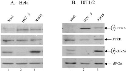
HSV infection activates PKR that mediates translation shutoff through eIF-2α phosphorylation. In the course of virus infection, the γ134.5 protein is expressed to recruit PP1 to and dephosphorylate eIF-2α (7, 22, 23). This process of cycling on and off raises the possibility that the γ134.5 protein has evolved to eliminate the inhibitory effects of multiple eIF-2α kinases activated by HSV. To test this hypothesis, we examined the phosphorylation state of PERK, an ER-resident kinase that is activated in response to ER stress. Specifically, monolayers of HeLa cells were either mock infected or infected with wild-type HSV-1(F) or the γ134.5 deletion mutant R3616 at 10 PFU per cell. At 24 h postinfection, cell lysates were processed for Western blot analysis with antibodies against PERK, phosphorylated PERK, eIF-2α, and phosphorylated eIF-2α. As illustrated in Fig. 1A, comparable levels of PERK were seen in both mock-infected and virus-infected cells (lanes 1 to 3). Under this experimental condition, phosphorylated PERK was not detected in mock-infected cells. However, phosphorylated PERK was found in cells infected with HSV-1(F) or R3616 (Fig. 1A, lanes 2 and 3). Since the viruses induced PERK activation, these results suggested that replication of HSV causes ER stress. In fact, phosphorylation of PERK was more pronounced in cells infected with HSV-1(F) than in cells infected with R3616. This phenotype is attributable to better replication of HSV-1(F) than of R3616. Similarly, PERK was activated by viruses in mouse 10T1/2 cells (Fig. 1B), NIH 3T3 cells, and mouse 3T6 cells (data not shown).
FIG. 1.
HSV-1 infection activates PERK kinase. (A) HeLa cells or (B) mouse 10T1/2 cells were mock infected or infected with wild-type virus HSV-1(F) or the γ134.5 deletion mutant R3616 at 10 PFU per cell. At 24 h postinfection, cells were harvested and lysates were prepared. Samples were then electrophoretically separated on denaturing 12% polyacrylamide gels and transferred to nitrocellulose membranes. The membranes were sequentially probed with antibodies against phosphorylated PERK (p-PERK), PERK, phosphorylated eIF2α (p-eIF-2α), and eIF-2α. The positions of the protein bands are indicated on the right.
As shown in Fig. 1A, although PERK was activated in virus-infected cells, increased eIF-2α phosphorylation was observed only in the absence of the γ134.5 protein. A weak band in mock-infected cells represents the basal level of phosphorylated eIF-2α. As eIF-2α was present at a similar level in both mock-infected and virus-infected cells, these results indicate that expression of the γ134.5 protein was responsible for eIF-2α dephosphorylation. Further analysis in mouse 10T1/2 cells yielded similar results (Fig. 1B). We conclude from these experiments that HSV infection activates PERK kinase. Moreover, expression of the γ134.5 protein promotes efficient dephosphorylation of eIF-2α even though PERK is activated during HSV infection.
The effects of phosphonoacetic acid (PAA) and cycloheximide on PERK activation in cells infected with viruses.
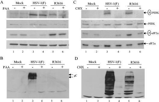
To explore the nature of PERK activation, we evaluated whether viral DNA replication affected phosphorylation of PERK. For this purpose, monolayers of NIH 3T3 cells were mock infected or infected with HSV-1(F) or the R3616 at 10 PFU per cell. At 1.5 h postinfection, cells were treated with PAA (400 μg/ml). At 24 h postinfection, cell lysates were processed for Western blot analysis with antibodies against phosphorylated PERK, PERK, eIF-2α and glycoprotein C (gC). Figure 2A shows that PERK was not phosphorylated in mock-infected cells with or without PAA treatment. Levels of PERK phosphorylation were higher in HSV-1(F)-infected cells than in R3616-infected cells. In virus-infected cells, PAA reduced PERK phosphorylation moderately (lanes 3 to 6). This seems evident in R3616-infected cells. As PAA effectively suppressed the expression of gC (a γ2 gene), the result suggests that viral DNA replication is inhibited (Fig. 2B). Thus, viral DNA replication-dependent and -independent processes are involved in activating PERK. In agreement with these results, an increase in eIF-2α phosphorylation was observed in cells infected with R3616 but not with HSV-1(F) (Fig. 2A, lanes 3 to 6). Notably, PAA also decreased eIF-2α phosphorylation slightly in R3616-infected cells (Fig. 2A, lanes 5 and 6). A basal level of eIF-2α phosphorylation was also detected in mock-infected cells. Western blot analysis indicated that levels of eIF-2α were comparable in mock- or virus-infected cells (Fig. 2A, lanes 1 to 6).
FIG. 2.
(A) The effect of PAA on HSV-induced PERK activation. Monolayers of mouse NIH 3T3 cells were infected with the indicated viruses at 10 PFU per cell. At 1.5 h postinfection, the cells were either left untreated or treated with PAA (400 μg/ml). At 24 h postinfection, cells were harvested and processed for Western blot analysis with antibodies against phosphorylated PERK (p-PERK), PERK, phosphorylated eIF2α (p-eIF-2α), and eIF-2α in the same membrane. (B) The effect of PAA on gC expression. Cell lysates in panel A were processed for Western blot analysis with antibodies against gC, whose expression is dependent on viral DNA replication. (C) HSV-induced PERK activation requires viral protein synthesis. Assay conditions are similar to those described in panel A except the protein synthesis inhibitor cycloheximide (CHX; 50 mg/ml) instead of PAA was added to cells 30 min prior to virus infection and was present throughout infection. (D) The effect of cycloheximide on viral protein synthesis. Cell lysates in panel C were processed for Western blot analysis with antibodies against HSV antibodies. The positions of protein bands are indicated at right.
We next examined whether the synthesis of viral proteins is required to activate PERK. To address this issue, cells were either left untreated or pretreated with cycloheximide (50 mg/ml) for 30 min. Cells were then mock infected or infected with viruses for 24 h. Western blot analysis was carried out as described above, and results are presented in Fig. 2C. In mock-infected cells, cycloheximide did not have any effect on PERK phosphorylation (lanes 1 and 2). HSV-1(F) or R3616 induced phosphorylation of PERK in the absence of cycloheximide (lanes 3 and 5). In correlation with these results, eIF-2α was strongly phosphorylated in cells infected with R3616 but not with HSV-1(F). In cells treated with cycloheximide, neither HSV-1(F) nor R3616 stimulated PERK phosphorylation (lanes 4 and 6). Accordingly, there was no increased eIF-2α phosphorylation in virus-infected cells compared with that seen in mock-infected cells. Further analysis indicated that PERK as well as eIF-2α is expressed at comparable levels (lanes 1 to 6). Western blot analysis with anti-HSV antibodies showed that cycloheximide treatment inhibited viral polypeptide production (Fig. 2D). Together, these data suggest that activation of PERK induced by HSV-1 infection requires viral protein synthesis.
HSV-1 infection induces phosphorylation of PERK and eIF-2α in PKR+/+ as well as in PKR−/− cells.
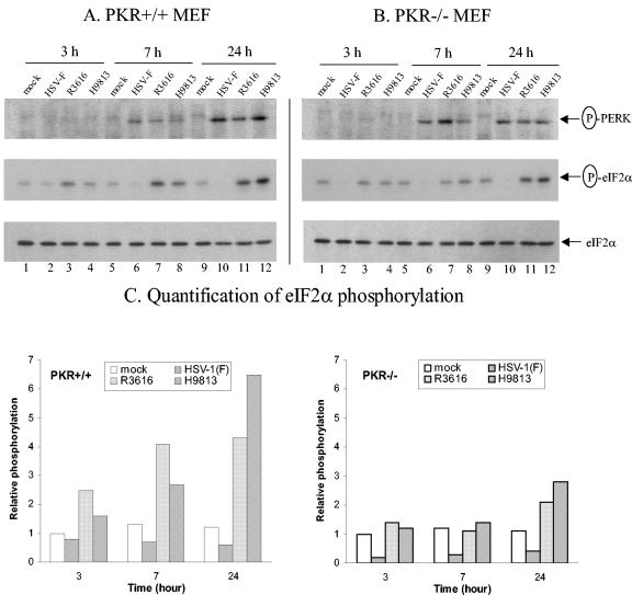
Previous studies demonstrated that HSV-1 infection activates PKR (7, 20). As the virus also activates PERK, we sought to determine the relationship between PERK, PKR, and eIF-2α. We first compared phosphorylation of PERK in PKR+/+ and PKR−/− mouse fibroblasts. Cells were mock-infected or infected with wild-type HSV-1(F), the γ134.5 deletion mutant R3616, or H9813 in which Vla193Glu and Phe195Leu substitutions were made in the PP1 binding motif of the γ134.5 protein. At different time points after infection, lysates of cells were processed for immunoblotting with antibodies against phosphorylated PERK, phosphorylated eIF-2α, and eIF-2α. As shown in Fig. 3A, in PKR+/+ cells, phosphorylated PERK was not detectable at 3 h postinfection (lanes 1 to 4). As virus infection proceeded, a low level of phosphorylated PERK was detected in virus-infected cells at 7 h after infection (lanes 6 to 8). Notably, PERK became strongly phosphorylated at a later stage of infection (24 h) (lanes 10 to 12). Figure 3B shows that in PKR−/− cells, a different pattern of PERK phosphorylation was observed (lanes 1 to 12). At 3 h after infection, phosphorylated PERK was not present. Nevertheless, PERK became strongly phosphorylated as early as 7 h after infection. At this time point, phosphorylation of PERK was more pronounced in PKR−/− cells than in PKR+/+ cells. This may relate to an enhanced viral replication in PKR−/− cells. Similarly, PERK remained strongly phosphorylated at 24 h after infection. These results indicate that the maximal phosphorylation of PERK was delayed in PKR+/+ cells compared to that in PKR−/− cells.
FIG. 3.
(A) Phosphorylation of PERK and eIF-2α in PKR+/+ mouse embryonic fibroblasts. Cells were infected with wild-type virus HSV-1(F), the γ134.5 deletion mutant R3616, or H9813 in which Val193 and Phe195 were replaced by Glu and Leu, respectively, at 10 PFU per cell at 37°C. At different time points after infection, cells were harvested, lysed, and subjected to Western blot analysis with antibodies against phosphorylated PERK, phosphorylated eIF-2α, and eIF-2α. (B) Phosphorylation of PERK and eIF-2α in PKR−/− mouse embryonic fibroblasts. Assays were carried out as described in panel A. The positions of protein bands are shown at right. (C) Quantitation of eIF-2α phosphorylation. Phosphorylated eIF-2α and total eIF-2α in panels A and B were quantitated by densitometry. Numbers were normalized to the total eIF-2α in each lane and expressed as eIF-2α phosphorylation relative to that in mock-infected cells at 3 h postinfection.
To determine the impact of virus infection, we also analyzed the phosphorylation state of eIF-2α. As shown in Fig. 3A and B, similar levels of eIF-2α were expressed in cells mock infected or infected with viruses. However, the phosphorylation of eIF-2α exhibited different patterns. In PKR+/+ cells mock infected or infected with HSV-1(F), little or no phosphorylated eIF-2α was seen throughout infection (Fig. 3A, lanes 1, 2, 5, 6, 9, and 10). In contrast, phosphorylated eIF-2α was detected in cells infected with R3616 or H9813, which increased at 7 and 24 h after infection, respectively. In PKR−/− cells, eIF-2α was not phosphorylated in HSV-1(F)-infected cells (Fig. 3B, lanes 2, 6, and 10), although a basal level of phosphorylated eIF-2α was seen in mock-infected cells. Phosphorylation of eIF-2α in cells infected with R3616 or H9813 was similar to that seen in mock-infected cells at 3 and 7 h postinfection. However, an increase in eIF-2α phosphorylation was observed at 24 h postinfection. To compare the extent of eIF-2α phosphorylation, phosphorylated eIF-2α was quantitated in PKR+/+ and PKR−/− cells with densitometry. Numbers were normalized to the total of eIF-2α in each lane and expressed as relative eIF-2α phosphorylation to that of mock-infected cells. Data summarized in Fig. 3C indicate that in PKR+/+ cells, as virus infection proceeded, R3616 or H9813 induced a progressive increase in eIF-2α phosphorylation. Phosphorylation of eIF-2α increased four- to sixfold at 24 h after infection. However, in PKR−/− cells, phosphorylation of eIF-2α essentially remained at the basal level at 3 or 7 h after infection. There was only a two- to threefold increase in eIF-2α phosphorylation at 24 h postinfection by R3616 or H9813. These results indicate that in response to virus infection, eIF-2α continued to be phosphorylated in the absence of PKR. However, the extent of eIF-2α phosphorylation was reduced and the kinetics of eIF-2α phosphorylation was delayed in PKR−/− cells compared to phosphorylation in PKR+/+ cells.
The growth of wild-type virus and γ134.5 mutants displayed differences in PKR+/+ and PKR−/− cells.
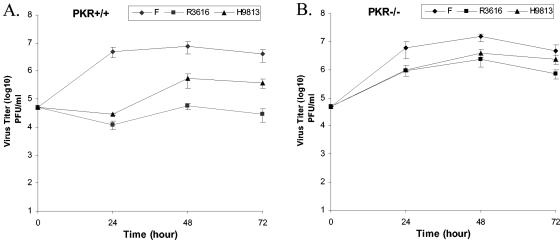
To evaluate viral replication, we carried out a multistep growth analysis in PKR+/+ as well as PKR−/− mouse embryo fibroblasts. Monolayers of cells were infected at 0.05 PFU per cell with wild-type HSV-1(F), the γ134.5 deletion mutant R3616, or H9813. Virus yields were measured over a course of 72 h of infection. As shown in Fig. 4A, in the PKR+/+ cell line, HSV-1(F) replicated to a titer of 5.0 × 10⁶ PFU/ml at 24 h postinfection and remained at a similar level throughout infection. In contrast, replication of R3616 was significantly reduced to a titer of 1.2 × 10⁴ PFU/ml at 24 h and remained at a similar level at 72 h. Similarly, replication of H9813 decreased to a titer of 2.4 × 10⁴ PFU/ml at 24 h. There was a recovery in virus growth for this mutant 48 h after infection, and the basis for this is unknown. In PKR−/− cell lines (Fig. 4B), HSV-1(F) replicated efficiently to a titer of 6.2 × 10⁶ PFU/ml at 24 h and reached a peak titer at 48 h. Consistent with a reduced level in eIF-2α phosphorylation, growth of R3616 and H9813 was improved drastically in PKR−/− cells to 9.5 × 10⁵ and 1.0 × 10⁶ PFU/ml, respectively, at 24 h. These results indicate that PKR plays a dominant role in limiting HSV-1 replication. However, it is notable that R3616 and H9813 replicated less well compared to HSV-1(F) in PKR−/− cells. There was a moderate decrease in virus growth for these mutants, suggesting that a PKR-independent cellular function contributes to the observed phenotype. Because phosphorylation of PERK coincided with eIF-2α phosphorylation at 24 h after infection in PKR−/− cells (Fig. 3B), these results suggest that PERK may contribute to a decrease in virus growth in PKR−/− cells.
FIG. 4.
Growth properties of HSV-1(F), R3616, and H9813 in PKR+/+ (A) and PKR−/− (B) mouse embryo fibroblasts. Monolayers of cells were infected with indicated viruses at 0.05 PFU per cell at 37°C. At various times postinfection, cells were harvested and freeze-thawed three times, and virus yield was determined on Vero cells. Duplicate samples were analyzed in parallel at each time point.
Expression of the γ134.5 protein alleviates the ER stress response induced by DTT and TG.
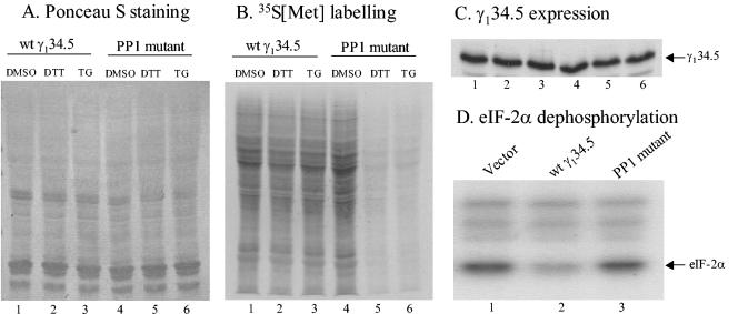
To further assess the role of the γ134.5 protein in the ER stress response, we asked whether the γ134.5 protein is able to alleviate the translation shutoff induced by DTT and TG. These compounds induce the ER stress that activates PERK and inhibits protein synthesis in a PERK-dependent manner (18, 29, 30). To address this issue, HEK 293 cells were transfected with the wild-type γ134.5 gene or a γ134.5 mutant in which Vla193Glu and Phe195Leu substitutions were made in the PP1 binding motif of the γ134.5 gene. This mutant is unable to mediate eIF-2α dephosphorylation (5). Thirty-six hours after transfection, cells were treated with DTT or TG. To determine ongoing protein synthesis, cells were labeled with [35S]methionine for 15 min, and lysates of cells were then subjected to SDS-PAGE and autoradiography. As shown in Fig. 5B, DTT or TG treatment induced a severe reduction of protein synthesis in cells transfected with the γ134.5 mutant (lanes 5 and 6). In contrast, the wild-type γ134.5-transfected cells exhibited normal protein synthesis in response to DTT and TG treatment (lanes 2 and 3). As expected, dimethyl sulfoxide had no detectable effect on protein synthesis (lanes 1 and 4). Ponceau S staining indicated equal loading of samples (Fig. 5A). Western blot analysis showed that both wild-type and the mutant γ134.5 proteins were expressed at comparable levels (Fig. 5C). Therefore, the wild-type but not the mutant γ134.5 protein is able to alleviate the PERK-mediated stress response in the cells. This activity correlated with the ability of the γ134.5 protein to mediate eIF2α dephosphorylation (Fig. 5D).
FIG. 5.
The γ134.5 protein of HSV-1 alleviates the ER stress response mediated by DTT or TG. 293 HEK cells were transfected with either pGF9912 expressing the wild-type γ134.5 protein or pGF9913 expressing the γ134.5 mutant in which Val193Glu and Phe195Leu substitutions were made by Lipofectamine reagent, as suggested by the manufacturer (Invitrogen). Thirty-six hours after transfection, cells were treated with dimethyl sulfoxide, DTT (2 mM; Invitrogen), or TG (0.5 μM; Sigma) for 1 h. Cells were then labeled with [35S]methionine (50 μCi/ml; ICN) in DMEM lacking methionine but supplemented with 5% fetal bovine serum for 15 min. Cell lysates were resolved on SDS-12% PAGE and subjected to ponceau S staining (A), autoradiography (B), and Western blot analysis with anti-γ134.5 antibody (C). (D) To measure eIF-2α phosphatase activity, cell lysates were prepared from the transfected cells and incubated with 32P-labeled eIF-2 as described in Materials and Methods. The reaction mixtures were then separated by SDS-12% PAGE electrophoresis and subjected to autoradiography.
HSV-1 infection transiently induces GADD34 expression.
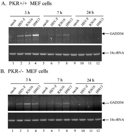
As presented in Fig. 3, although PERK was activated, phosphorylated eIF-2α remained at a lower level in cells infected with γ134.5 mutants at 7 h postinfection. Since PERK phosphorylates eIF-2α, which in turn induces the expression of GADD34 (29, 30), we examined whether HSV-1 infection induces the expression of GADD34. For this purpose, we measured the levels of GADD34 RNA transcripts in cells infected with viruses. Monolayers of cells were mock infected or infected with HSV-1(F), R3616, or H9813 at 10 PFU per cell. At different time points postinfection, total RNA was extracted and subjected to RT-PCR amplification specific for GADD34 mRNA. As indicated in Fig. 6A, in PKR+/+ cells, GADD34 mRNA was detected as early as 3 h postinfection in cells infected with either HSV-1(F), R3616, or H9813. The levels of GADD34 mRNA declined at 7 h and diminished at 24 h postinfection. Similar expression patterns were also observed in PKR−/− cells, as illustrated in Fig. 6B. These results thus indicate that HSV-1 infection stimulates GADD34 expression in the presence or absence of the γ134.5 protein. Moreover, deletion of PKR has no effect on the expression of GADD34 in HSV-1-infected cells. It is notable that virus induces the expression of GADD34 before PERK is phosphorylated, suggesting that an eIF-2α-independent mechanism is involved. Interestingly, the kinetics of GADD34 expression inversely correlates with an increase in eIF-2α phosphorylation. This is consistent with the ability of GADD34 to bind to PP1 and mediate eIF-2α dephosphorylation (23, 29).
FIG. 6.
HSV-1 infection induces GADD34 expression. PKR+/+ (A) and PKR−/− (B) cells were mock infected or infected with indicated viruses at 10 PFU per cell. At 3, 7, and 24 h postinfection, cells were harvested, and total RNA from infected cells was extracted by using the RNeasy kit (QIAGEN Inc.). Equal amounts of RNA from each sample were then subjected to RT-PCR amplification of GADD34. As a control, the cellular 18s rRNA was included in the assay. The PCR products were separated on a 1.5% agarose gel, stained with ethidium bromide, and photographed with NucleoVision GelExpert system (Nucleotech Inc.).
DISCUSSION
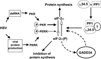
Regulation of the phosphorylation state of eIF-2α by HSV is a critical step during viral infection. In this process, multiple components or pathways are involved in determining the outcome of virus infection. Several lines of evidence indicated that PKR is activated by HSV to phosphorylate serine 51 on eIF-2α, which inhibits viral replication (4-7, 20, 38). While the role of PKR is well established, little is known about the function of PERK kinase during HSV infection. In this study, we find that PERK is activated in response to HSV infection. Our data are consistent with a model that HSV-1 infection triggers and then inhibits cellular responses. The dsRNA produced by HSV early in infection activates PKR. In addition, viral proteins accumulated during infection activate PERK in the ER. Activation of PKR and PERK leads to eIF-2α phosphorylation and, thereby, shutoff of protein synthesis. To maximize replication efficiency, HSV expresses the γ134.5 protein that redirects PP1 to dephosphorylate eIF-2α (Fig. 7).
FIG. 7.
A model depicting the role of the γ134.5 protein in inhibiting signaling through the integrated stress response pathway. HSV-1 infection triggers cellular responses involving PKR and PERK. The dsRNA produced by HSV early in infection activates PKR. In addition, viral proteins accumulated during infection activate PERK in the ER. Activation of PKR and PERK leads to eIF-2α phosphorylation and thereby shuts off protein synthesis. To block cellular responses, the γ134.5 protein recruits PP1, forming a complex that dephosphorylates eIF-2α. Phosphorylation of eIF-2α also activates the expression of GADD34, which serves as a feedback loop to mediate eIF-2α dephosphorylation by binding to PP1.
Recent work has demonstrated that PERK is a key mediator of ER stress signaling (18, 27, 29, 30, 35). Available evidence indicates that PERK normally exists as an inactive monomer held by ER chaperone proteins GRP78 and GRP94. Under stress conditions, PERK monomers oligomerize and autophosphorylate (1, 28). Accordingly, eIF-2α is phosphorylated by PERK to induce translation inhibition. The data presented in this report indicate that the infection of several cell lines with HSV-1 led to activation of PERK, as seen by an increase in autophosphorylation over the course of virus infection. Moreover, the phosphorylation of PERK was dependent on the synthesis of viral protein. We speculate that as PERK possesses an ER-luminal regulatory domain and a cytoplasmic kinase domain, processing or accumulation of viral proteins in the ER during infection may facilitate the oligomerization of PERK. One possibility is that HSV proteins associate with GRP78 or GRP94 and therefore release PERK from these chaperone proteins. Alternatively, HSV proteins may facilitate the rapid turnover of chaperone proteins. Previous studies indicated that the signal for the shutoff of protein synthesis in HSV-1-infected cells is linked to viral DNA synthesis (9). This is supported by the fact that PAA partially restores the shutoff of protein synthesis induced by HSV-1 infection (9). The present studies showed that PAA reduced but did not completely block the phosphorylation of PERK and eIF2α in HSV-infected cells. These results suggest that in addition to viral DNA synthesis, other viral processes may contribute to the phosphorylation of PERK and eIF2α. Further experiments are required to define the precise role of HSV proteins in PERK activation.
It is noteworthy that the kinetics of PERK phosphorylation exhibited differences in PKR+/+ and PKR−/− cells infected with viruses. Notably, the maximal phosphorylation of PERK by HSV-1 occurred later in PKR+/+ cells than in PKR−/− cells. Since PKR is activated early during HSV infection (7, 20), dsRNA produced by HSV is expected to activate PKR that phosphorylates eIF-2α and therefore limits viral replication. On the basis of this model, it is reasonable to observe that in PKR+/+ cells, PERK was only weakly phosphorylated at 7 h after infection but was maximally phosphorylated at 24 h after infection. In contrast, phosphorylation of PERK reached a maximum at 7 h after infection in PKR−/− cells. One explanation is that the rate of viral replication is slower in the presence of PKR. As synthesized viral gene products or newly produced virions accumulate, the level of ER stress increases to a threshold that triggers PERK activation. Therefore, it is likely that in normal cells PERK exerts its function later in virus infection.
Our experiments show that the γ134.5 protein functions to block the ER stress response. Although PERK was activated in cells infected with HSV-1, wild-type virus prevented eIF-2α phosphorylation throughout infection. This is further supported by the fact that when transfected into mammalian cells, the wild-type γ134.5 protein inhibited DTT- or TG-induced ER stress. It should be noted that at 7 h after infection, γ134.5 mutants stimulated an increase in eIF-2α phosphorylation in PKR+/+ cells but not in PKR−/− cells. Given that both PERK and PKR were activated at this time point, eIF-2α appeared to be preferentially phosphorylated by PKR. At 24 h after infection, γ134.5 mutants induced a significant increase in eIF-2α phosphorylation in PKR+/+ cells but only a moderate increase in eIF-2α phosphorylation in PKR−/− cells. Thus, phosphorylation of eIF-2α in PKR+/+ cells may reflect the compounding effect of PKR and PERK, whereas the increased phosphorylation of eIF-2α in PKR−/− cells may result from PERK. Therefore, PERK and PKR contribute to eIF-2α phosphorylation differentially. It is possible that PERK has a lower affinity for eIF-2α than PKR. Furthermore, the expression of GADD34 may mediate eIF-2α dephosphorylation. Further work is required to test these possibilities. Nevertheless, it should be stressed that the extent of eIF-2α phosphorylation correlated with decreased efficiency of viral replication (Fig. 6). While wild-type virus replicated efficiently, γ134.5 mutants replicated poorly in PKR+/+ cells. However, γ134.5 mutants replicated more efficiently in PKR−/− cells. These mutants replicated only 5- to 10-fold less compared to wild-type virus in PKR−/− cells. This moderate decrease in viral growth is likely to result from the activity of PERK and other eIF-2α kinases. These results are consistent with recent findings that the replication of HSV correlates with the regulation of eIF-2α phosphorylation, even though the activation of PERK was not analyzed (38).
Although the mechanisms of GADD34 expression remain unknown, phosphorylation of eIF-2α has been linked to the induction of GADD34 expression (29). In addition, an alternative pathway leads to GADD34 expression by the alkylating agent methyl methanesulfonate (29). Increased levels of GADD34 that occur in the stress response pathway are believed to serve as a feedback loop to retain protein translation and meanwhile to attenuate gene expression (27, 29, 30). In this context, it is interesting that GADD34 expression was rapidly induced shortly after HSV infection (3 h) in both PKR+/+ and PKR−/− cells. Because there is no increased eIF-2α phosphorylation at this time point, this suggests that HSV induces GADD34 expression that is independent of eIF-2α phosphorylation. As GADD34 also directs PP1 to dephosphorylate eIF-2α (23, 29), induction of GADD34 expression by HSV-1 may contribute to the delayed hyperphosphorylation of eIF-2α. The basis for the diminished GADD34 expression later in infection is unknown. As the 3′ untranslated region of GADD34 bears multiple AT3 motifs, these elements may destabilize mRNA (26). Alternatively, HSV gene products may regulate the degradation of GADD34 mRNA. Further experiments are needed to define the precise role of GADD34 in HSV-1 infection.
Several studies highlighted a role for the ER stress response in viral replication (25, 31, 36, 37). A cytopathic strain of bovine viral diarrhea virus causes ER stress that induces apoptosis through PERK activation (25). Similarly, Japanese encephalitis virus-induced ER stress participates in apoptosis via p38 kinase or CHOP (36). In addition, the E2 protein of hepatitis C virus binds to and inhibits PERK activation (31). The data presented in this work demonstrated that HSV replication induces ER stress involving PERK. As the HSV productive life cycle proceeds rapidly after infection, it triggers cellular responses that limit viral replication. During evolution HSV acquired the γ134.5 protein that functions to recruit PP1 to dephosphorylate eIF-2α and antagonizes the activities of both PKR and PERK.
Acknowledgments
We thank Bernard Roizman for HSV-1 strains and Gary Cohen and Roselyn Eisenberg for anti-gC antibody. We are grateful to Melissa Cerveny for critical reading of the manuscript.
This work was supported by grant AI 46665 (B.H.) from the National Institute of Allergy and Infectious Diseases.
REFERENCES
- 1.Bertolotti, A., Y. Zhang, L. M. Hendershot, H. P. Harding, and D. Ron. 2000. Dynamic interaction of BiP and ER stress transducers in the unfolded-protein response. Nat. Cell Biol. 2**:**326-332. [DOI] [PubMed] [Google Scholar]
- 2.Brewer, J. W., and J. A. Diehl. 2000. PERK mediates cell-cycle exit during the mammalian unfolded protein response. Proc. Natl. Acad. Sci. USA 97**:**12625-12630. [DOI] [PMC free article] [PubMed] [Google Scholar]
- 3.Cerveny, M., S. Hessefort, K. Yang, G. Cheng, M. Gross, and B. He. 2003. Amino acid substitutions in the effector domain of the γ134.5 protein of herpes simplex virus 1 have differential effects on viral response to interferon-α. Virology 307**:**290-300. [DOI] [PubMed] [Google Scholar]
- 4.Cheng, G., M. E. Brett, and B. He. 2001. Val193 and Phe195 of the γ134.5 protein of herpes simplex virus 1 are required for viral resistance to interferon α/β. Virology 290**:**115-120. [DOI] [PubMed] [Google Scholar]
- 5.Cheng, G., M. Gross, M. E. Brett, and B. He. 2001. AlaArg motif in the carboxyl terminus of the γ134.5 protein of herpes simplex virus type 1 is required for the formation of a high-molecular-weight complex that dephosphorylates eIF-2α. J. Virol. 75**:**3666-3674. [DOI] [PMC free article] [PubMed] [Google Scholar]
- 6.Cheng, G., K. Yang, and B. He. 2003. Dephosphorylation of eIF-2α mediated by the γ134.5 protein of herpes simplex virus type 1 is required for viral response to interferon but is not sufficient for efficient viral replication. J. Virol. 77**:**10154-10161. [DOI] [PMC free article] [PubMed] [Google Scholar]
- 7.Chou, J., J. J. Chen, M. Gross, and B. Roizman. 1995. Association of a M(r) 90,000 phosphoprotein with protein kinase PKR in cells exhibiting enhanced phosphorylation of translation initiation factor eIF-2α and premature shutoff of protein synthesis after infection with γ134.5-mutants of herpes simplex virus 1. Proc. Natl. Acad. Sci. USA 92**:**10516-10520. [DOI] [PMC free article] [PubMed] [Google Scholar]
- 8.Chou, J., E. R. Kern, R. J. Whitley, and B. Roizman. 1990. Mapping of herpes simplex virus-1 neurovirulence to γ134.5, a gene nonessential for growth in culture. Science 250**:**1262-1266. [DOI] [PubMed] [Google Scholar]
- 9.Chou, J., and B. Roizman. 1992. The γ134.5 gene of herpes simplex virus 1 precludes neuroblastoma cells from triggering total shutoff of protein synthesis characteristic of programmed cell death in neuronal cells. Proc. Natl. Acad. Sci. USA 89**:**3266-3270. [DOI] [PMC free article] [PubMed] [Google Scholar]
- 10.Chou, J., and B. Roizman. 1994. Herpes simplex virus 1 γ134.5 gene function, which blocks the host response to infection, maps in the homologous domain of the genes expressed during growth arrest and DNA damage. Proc. Natl. Acad. Sci. USA 91**:**5247-5251. [DOI] [PMC free article] [PubMed] [Google Scholar]
- 11.Chou, J., and B. Roizman. 1990. The herpes simplex virus 1 gene for ICP34.5, which maps in inverted repeats, is conserved in several limited-passage isolates but not in strain 17syn+. J. Virol. 64**:**1014-1020. [DOI] [PMC free article] [PubMed] [Google Scholar]
- 12.Chou, J., and B. Roizman. 1986. The terminal a sequence of the herpes simplex virus genome contains the promoter of a gene located in the repeat sequences of the L component. J. Virol. 57**:**629-637. [DOI] [PMC free article] [PubMed] [Google Scholar]
- 13.Clemens, M. J., K. G. Laing, I. W. Jeffrey, A. Schofield, T. V. Sharp, A. Elia, V. Matys, M. C. James, and V. J. Tilleray. 1994. Regulation of the interferon-inducible eIF-2 alpha protein kinase by small RNAs. Biochimie 76**:**770-778. [DOI] [PubMed] [Google Scholar]
- 14.de Haro, C., R. Mendez, and J. Santoyo. 1996. The eIF-2α kinases and the control of protein synthesis. FASEB J. 10**:**1378-1387. [DOI] [PubMed] [Google Scholar]
- 15.Ejercito, P. M., E. D. Kieff, and B. Roizman. 1968. Characterization of herpes simplex virus strains differing in their effects on social behaviour of infected cells. J. Gen. Virol. 2**:**357-364. [DOI] [PubMed] [Google Scholar]
- 16.Gale, M., Jr., and M. G. Katze. 1998. Molecular mechanisms of interferon resistance mediated by viral-directed inhibition of PKR, the interferon-induced protein kinase. Pharmacol. Ther. 78**:**29-46. [DOI] [PubMed] [Google Scholar]
- 17.Grishin, A. V., O. Azhipa, I. Semenov, and S. J. Corey. 2001. Interaction between growth arrest-DNA damage protein 34 and Src kinase Lyn negatively regulates genotoxic apoptosis. Proc. Natl. Acad. Sci. USA 98**:**10172-10177. [DOI] [PMC free article] [PubMed] [Google Scholar]
- 18.Harding, H. P., Y. Zhang, A. Bertolotti, H. Zeng, and D. Ron. 2000. Perk is essential for translational regulation and cell survival during the unfolded protein response. Mol. Cell 5**:**897-904. [DOI] [PubMed] [Google Scholar]
- 19.Harding, H. P., Y. Zhang, and D. Ron. 1999. Protein translation and folding are coupled by an endoplasmic-reticulum-resident kinase. Nature 397**:**271-274. [DOI] [PubMed] [Google Scholar]
- 20.He, B., J. Chou, R. Brandimarti, I. Mohr, Y. Gluzman, and B. Roizman. 1997. Suppression of the phenotype of γ134.5-herpes simplex virus 1: failure of activated RNA-dependent protein kinase to shut off protein synthesis is associated with a deletion in the domain of the α47 gene. J. Virol. 71**:**6049-6054. [DOI] [PMC free article] [PubMed] [Google Scholar]
- 21.He, B., J. Chou, D. A. Liebermann, B. Hoffman, and B. Roizman. 1996. The carboxyl terminus of the murine MyD116 gene substitutes for the corresponding domain of the γ134.5 gene of herpes simplex virus to preclude the premature shutoff of total protein synthesis in infected human cells. J. Virol. 70**:**84-90. [DOI] [PMC free article] [PubMed] [Google Scholar]
- 22.He, B., M. Gross, and B. Roizman. 1998. The γ134.5 protein of herpes simplex virus 1 has the structural and functional attributes of a protein phosphatase 1 regulatory subunit and is present in a high molecular weight complex with the enzyme in infected cells. J. Biol. Chem. 273**:**20737-20743. [DOI] [PubMed] [Google Scholar]
- 23.He, B., M. Gross, and B. Roizman. 1997. The γ134.5 protein of herpes simplex virus 1 complexes with protein phosphatase 1α to dephosphorylate the α subunit of the eukaryotic translation initiation factor 2 and preclude the shutoff of protein synthesis by double-stranded RNA-activated protein kinase. Proc. Natl. Acad. Sci. USA 94**:**843-848. [DOI] [PMC free article] [PubMed] [Google Scholar]
- 24.Hollander, M. C., Q. Zhan, I. Bae, and A. J. Fornace, Jr. 1997. Mammalian GADD34, an apoptosis- and DNA damage-inducible gene. J. Biol. Chem. 272**:**13731-13737. [DOI] [PubMed] [Google Scholar]
- 25.Jordan, R., L. Wang, T. M. Graczyk, T. M. Block, and P. R. Romano. 2002. Replication of a cytopathic strain of bovine viral diarrhea virus activates PERK and induces endoplasmic reticulum stress-mediated apoptosis of MDBK cells. J. Virol. 76**:**9588-9599. [DOI] [PMC free article] [PubMed] [Google Scholar]
- 26.Lord, K. A., B. Hoffman-Liebermann, and D. A. Liebermann. 1990. Sequence of MyD116 cDNA: a novel myeloid differentiation primary response gene induced by IL6. Nucleic Acids Res. 18**:**2823. [DOI] [PMC free article] [PubMed] [Google Scholar]
- 27.Lu, P. D., C. Jousse, S. J. Marciniak, Y. Zhang, I. Novoa, D. Scheuner, R. J. Kaufman, D. Ron, and H. P. Harding. 2004. Cytoprotection by pre-emptive conditional phosphorylation of translation initiation factor 2. EMBO J. 23**:**169-179. [DOI] [PMC free article] [PubMed] [Google Scholar]
- 28.Ma, K., K. M. Vattem, and R. C. Wek. 2002. Dimerization and release of molecular chaperone inhibition facilitate activation of eukaryotic initiation factor-2 kinase in response to endoplasmic reticulum stress. J. Biol. Chem. 277**:**18728-18735. [DOI] [PubMed] [Google Scholar]
- 29.Novoa, I., H. Zeng, H. P. Harding, and D. Ron. 2001. Feedback inhibition of the unfolded protein response by _GADD34_-mediated dephosphorylation of eIF2α. J. Cell Biol. 153**:**1011-1022. [DOI] [PMC free article] [PubMed] [Google Scholar]
- 30.Novoa, I., Y. Zhang, H. Zeng, R. Jungreis, H. P. Harding, and D. Ron. 2003. Stress-induced gene expression requires programmed recovery from translational repression. EMBO J. 22**:**1180-1187. [DOI] [PMC free article] [PubMed] [Google Scholar]
- 31.Pavio, N., P. R. Romano, T. M. Graczyk, S. M. Feinstone, and D. R. Taylor. 2003. Protein synthesis and endoplasmic reticulum stress can be modulated by the hepatitis C virus envelope protein E2 through the eukaryotic initiation factor 2α kinase PERK. J. Virol. 77**:**3578-3585. [DOI] [PMC free article] [PubMed] [Google Scholar]
- 32.Proud, C. G. 1995. PKR: a new name and new roles. Trends Biochem. Sci. 20**:**241-246. [DOI] [PubMed] [Google Scholar]
- 33.Shi, W. B., C. A. Sun, B. He, W. C. Xiong, X. M. Shi, D. C. Yao, and X. Cao. 2004. GADD34-PP1c recruited by smad7 dephosphorylates TGF-β type receptor. J. Cell Biol. 164**:**291-300. [DOI] [PMC free article] [PubMed] [Google Scholar]
- 34.Shi, Y., K. M. Vattem, R. Sood, J. An, J. Liang, L. Stramm, and R. C. Wek. 1998. Identification and characterization of pancreatic eukaryotic initiation factor 2α-subunit kinase, PEK, involved in translational control. Mol. Cell. Biol. 18**:**7499-7509. [DOI] [PMC free article] [PubMed] [Google Scholar]
- 35.Sood, R., A. C. Porter, K. Ma, L. A. Quilliam, and R. C. Wek. 2000. Pancreatic eukaryotic initiation factor-2α kinase (PEK) homologues in humans, Drosophila melanogaster and Caenorhabditis elegans that mediate translational control in response to endoplasmic reticulum stress. Biochem. J. 346**:**281-293. [PMC free article] [PubMed] [Google Scholar]
- 36.Su, H. L., C. L. Liao, and Y. L. Lin. 2002. Japanese encephalitis virus infection initiates endoplasmic reticulum stress and an unfolded protein response. J. Virol. 76**:**4162-4171. [DOI] [PMC free article] [PubMed] [Google Scholar]
- 37.Tardif, K. D., K. Mori, R. J. Kaufman, and A. Siddiqui. 2004. Hepatitis C virus suppresses the IRE1-XBP1 pathway of the unfolded protein response. J. Biol. Chem. 279**:**17158-17164. [DOI] [PubMed] [Google Scholar]
- 38.Ward, S. L., D. Scheuner, J. Poppers, R. J. Kaufman, I. Mohr, and D. A. Leib. 2003. In vivo replication of an ICP34.5 second-site suppressor mutant following corneal infection correlates with in vitro regulation of eIF2α phosphorylation. J. Virol. 77**:**4626-4634. [DOI] [PMC free article] [PubMed] [Google Scholar]
- 39.Zhan, Q., K. A. Lord, I. Alamo, Jr., M. C. Hollander, F. Carrier, D. Ron, K. W. Kohn, B. Hoffman, D. A. Liebermann, and A. J. Fornace, Jr. 1994. The gadd and MyD genes define a novel set of mammalian genes encoding acidic proteins that synergistically suppress cell growth. Mol. Cell. Biol. 14**:**2361-2371. [DOI] [PMC free article] [PubMed] [Google Scholar]
