The dual developmental origin of spinal cerebrospinal fluid-contacting neurons gives rise to distinct functional subtypes (original) (raw)
- Article
- Open access
- Published: 07 April 2017
- Laura Desban1,
- Johanna Gomez ORCID: orcid.org/0000-0002-9557-13531,
- Jenna R. Sternberg1,
- Andrew Prendergast1,
- Dominique Langui1,
- Feng B. Quan1,2,
- Hugo Marnas1,
- Thomas O. Auer3 nAff4,
- Jean-Paul Rio1,
- Filippo Del Bene ORCID: orcid.org/0000-0001-8551-28463,
- Pierre-Luc Bardet1 &
- …
- Claire Wyart ORCID: orcid.org/0000-0002-1668-49751
Scientific Reports volume 7, Article number: 719 (2017)Cite this article
- 3381 Accesses
- 39 Citations
- 2 Altmetric
- Metrics details
Subjects
Abstract
Chemical and mechanical cues from the cerebrospinal fluid (CSF) can affect the development and function of the central nervous system (CNS). How such cues are detected and relayed to the CNS remains elusive. Cerebrospinal fluid-contacting neurons (CSF-cNs) situated at the interface between the CSF and the CNS are ideally located to convey such information to local networks. In the spinal cord, these GABAergic neurons expressing the PKD2L1 channel extend an apical extension into the CSF and an ascending axon in the spinal cord. In zebrafish and mouse spinal CSF-cNs originate from two distinct progenitor domains characterized by distinct cascades of transcription factors. Here we ask whether these neurons with different developmental origins differentiate into cells types with different functional properties. We show in zebrafish larva that the expression of specific markers, the morphology of the apical extension and axonal projections, as well as the neuronal targets contacted by CSF-cN axons, distinguish the two CSF-cN subtypes. Altogether our study demonstrates that the developmental origins of spinal CSF-cNs give rise to two distinct functional populations of sensory neurons. This work opens novel avenues to understand how these subtypes may carry distinct functions related to development of the spinal cord, locomotion and posture.
Introduction
The cerebrospinal fluid (CSF) is a complex solution circulating around the central nervous system (CNS). It has classically been assumed that the CSF ensures the homeostasis of the CNS[1](/articles/s41598-017-00350-1#ref-CR1 "Iliff, J. J. et al. A paravascular pathway facilitates CSF flow through the brain parenchyma and the clearance of interstitial solutes, including amyloid beta. Sci Transl Med 4, 147ra111, doi: 10.1126/scitranslmed.30037484/147/147ra111
               (2012)."). Multiple studies indicate that the CSF also conveys signals affecting the development and output functions of the CNS, such as feeding, sleep, and locomotion[2](#ref-CR2 "Pappenheimer, J. R., Miller, T. B. & Goodrich, C. A. Sleep-promoting effects of cerebrospinal fluid from sleep-deprived goats. Proc Natl Acad Sci USA
                       58, 513–517 (1967)."),[3](#ref-CR3 "Martin, F. H., Seoane, J. R. & Baile, C. A. Feeding in satiated sheep elicited by intraventricular injections of CSF from fasted sheep. Life Sci
                       13, 177–184 (1973)."),[4](#ref-CR4 "Lerner, R. A. et al. Cerebrodiene: a brain lipid isolated from sleep-deprived cats. Proc Natl Acad Sci USA
                       91, 9505–9508 (1994)."),[5](#ref-CR5 "Nishino, S. et al. Low cerebrospinal fluid hypocretin (Orexin) and altered energy homeostasis in human narcolepsy. Ann Neurol
                       50, 381–388 (2001)."),[6](/articles/s41598-017-00350-1#ref-CR6 "Xie, L. et al. Sleep drives metabolite clearance from the adult brain. Science
                       342, 373–377, doi:
                10.1126/science.1241224342/6156/373

               (2013)."). These observations suggest that chemical or mechanical cues from the CSF may act on neurons in the brain and spinal cord. Cerebrospinal fluid-contacting neurons (CSF-cNs) are precisely located at the interface between the CSF and the neuronal circuits[7](/articles/s41598-017-00350-1#ref-CR7 "Vigh, B., Vigh-Teichmann, I. & Aros, B. Special dendritic and axonal endings formed by the cerebrospinal fluid contacting neurons of the spinal cord. Cell Tissue Res
                       183, 541–552 (1977)."), [8](/articles/s41598-017-00350-1#ref-CR8 "Vigh, B. & Vigh-Teichmann, I. Actual problems of the cerebrospinal fluid-contacting neurons. Microsc Res Tech
                       41, 57–83, doi:10.1002/(SICI)1097-0029(19980401)41:1<57::AID-JEMT6>3.0.CO;2-R (1998)."). In the vertebrate spinal cord, CSF-cNs reside around the central canal[9](#ref-CR9 "Kolmer, W. Das “Sagittalorgan” der Wirbeltiere. Zeitschrift für Anatomie und Entwicklungsgeschichte
                       60, 652–717 (1921)."),[10](#ref-CR10 "Agduhr, E. Über ein zentrales Sinnesorgan (?) bei den Vertebraten. Zeitschrift für Anatomie und Entwicklungsgeschichte
                       66, 223–360 (1922)."),[11](#ref-CR11 "Huang, A. L. et al. The cells and logic for mammalian sour taste detection. Nature
                       442, 934–938, doi:
                10.1038/nature05084

               (2006)."),[12](#ref-CR12 "Wyart, C. et al. Optogenetic dissection of a behavioural module in the vertebrate spinal cord. Nature
                       461, 407–410, doi:
                10.1038/nature08323nature08323

               (2009)."),[13](#ref-CR13 "Djenoune, L. et al. Investigation of spinal cerebrospinal fluid-contacting neurons expressing PKD2L1: evidence for a conserved system from fish to primates. Front Neuroanat
                       8, 26, doi:
                10.3389/fnana.2014.00026

               (2014)."),[14](#ref-CR14 "Jalalvand, E., Robertson, B., Wallen, P., Hill, R. H. & Grillner, S. Laterally projecting cerebrospinal fluid-contacting cells in the lamprey spinal cord are of two distinct types. J Comp Neurol
                       522, Spc1, doi:
                10.1002/cne.23584

               (2014)."),[15](/articles/s41598-017-00350-1#ref-CR15 "Fidelin, K. et al. State-Dependent Modulation of Locomotion by GABAergic Spinal Sensory Neurons. Curr Biol
                       25, 3035–3047, doi:
                10.1016/j.cub.2015.09.070

               (2015).") and project an apical extension in contact with the CSF[8](/articles/s41598-017-00350-1#ref-CR8 "Vigh, B. & Vigh-Teichmann, I. Actual problems of the cerebrospinal fluid-contacting neurons. Microsc Res Tech
                       41, 57–83, doi:10.1002/(SICI)1097-0029(19980401)41:1<57::AID-JEMT6>3.0.CO;2-R (1998)."), [16](#ref-CR16 "LaMotte, C. C. Vasoactive intestinal polypeptide cerebrospinal fluid-contacting neurons of the monkey and cat spinal central canal. J Comp Neurol
                       258, 527–541, doi:
                10.1002/cne.902580405

               (1987)."),[17](#ref-CR17 "Stoeckel, M. E. et al. Cerebrospinal fluid-contacting neurons in the rat spinal cord, a gamma-aminobutyric acidergic system expressing the P2X2 subunit of purinergic receptors, PSA-NCAM, and GAP-43 immunoreactivities: light and electron microscopic study. J Comp Neurol
                       457, 159–174, doi:
                10.1002/cne.10565

               (2003)."),[18](/articles/s41598-017-00350-1#ref-CR18 "Marichal, N., Garcia, G., Radmilovich, M., Trujillo-Cenoz, O. & Russo, R. E. Enigmatic central canal contacting cells: immature neurons in “standby mode”? J Neurosci
                       29, 10010–10024, doi:
                10.1523/JNEUROSCI.6183-08.200929/32/10010

               (2009).") and have a locally-projecting axon[14](/articles/s41598-017-00350-1#ref-CR14 "Jalalvand, E., Robertson, B., Wallen, P., Hill, R. H. & Grillner, S. Laterally projecting cerebrospinal fluid-contacting cells in the lamprey spinal cord are of two distinct types. J Comp Neurol
                       522, Spc1, doi:
                10.1002/cne.23584

               (2014)."), [19](/articles/s41598-017-00350-1#ref-CR19 "Christenson, J., Bongianni, F., Grillner, S. & Hokfelt, T. Putative GABAergic input to axons of spinal interneurons and primary sensory neurons in the lamprey spinal cord as shown by intracellular Lucifer yellow and GABA immunohistochemistry. Brain Res
                       538, 313–318 (1991)."), [20](/articles/s41598-017-00350-1#ref-CR20 "Megias, M., Alvarez-Otero, R. & Pombal, M. A. Calbindin and calretinin immunoreactivities identify different types of neurons in the adult lamprey spinal cord. J Comp Neurol
                       455, 72–85, doi:
                10.1002/cne.10473

               (2003).") that contacts other spinal neurons[12](/articles/s41598-017-00350-1#ref-CR12 "Wyart, C. et al. Optogenetic dissection of a behavioural module in the vertebrate spinal cord. Nature
                       461, 407–410, doi:
                10.1038/nature08323nature08323

               (2009)."), [15](/articles/s41598-017-00350-1#ref-CR15 "Fidelin, K. et al. State-Dependent Modulation of Locomotion by GABAergic Spinal Sensory Neurons. Curr Biol
                       25, 3035–3047, doi:
                10.1016/j.cub.2015.09.070

               (2015)."), [21](/articles/s41598-017-00350-1#ref-CR21 "Christenson, J., Alford, S., Grillner, S. & Hokfelt, T. Co-localized GABA and somatostatin use different ionic mechanisms to hyperpolarize target neurons in the lamprey spinal cord. Neurosci Lett
                       134, 93-97, doi:0304-3940(91)90516-V (1991).").
One essential step to understand spinal CSF-cN functions lies in identifying specific markers for these cells. In this regard, the polycystic kidney disease 2 like 1 (PKD2L1) channel also called TRPP3[22](/articles/s41598-017-00350-1#ref-CR22 "Delmas, P. Polycystins: from mechanosensation to gene regulation. Cell 118, 145–148, doi: 10.1016/j.cell.2004.07.007S0092867404006671
               (2004)."), which belongs to the Transient Potential Receptor (TRP) family, appears as a robust candidate to label CSF-cNs[11](/articles/s41598-017-00350-1#ref-CR11 "Huang, A. L. et al. The cells and logic for mammalian sour taste detection. Nature
                       442, 934–938, doi:
                10.1038/nature05084

               (2006)."), [13](/articles/s41598-017-00350-1#ref-CR13 "Djenoune, L. et al. Investigation of spinal cerebrospinal fluid-contacting neurons expressing PKD2L1: evidence for a conserved system from fish to primates. Front Neuroanat
                       8, 26, doi:
                10.3389/fnana.2014.00026

               (2014)."), [23](/articles/s41598-017-00350-1#ref-CR23 "Orts-Del’immagine, A. et al. Properties of subependymal cerebrospinal fluid contacting neurones in the dorsal vagal complex of the mouse brainstem. J Physiol
                       590, 3719–3741, doi:
                10.1113/jphysiol.2012.227959jphysiol.2012.227959

               (2012)."), [24](/articles/s41598-017-00350-1#ref-CR24 "Orts Del’Immagine, A. et al. A single polycystic kidney disease 2-like 1 channel opening acts as a spike generator in cerebrospinal fluid-contacting neurons of adult mouse brainstem. Neuropharmacology, doi:
                10.1016/j.neuropharm.2015.07.030

               (2015)."). PKD2L1, originally identified as the sour taste receptor in the taste buds, has been found in the spinal CSF-cNs of mouse[11](/articles/s41598-017-00350-1#ref-CR11 "Huang, A. L. et al. The cells and logic for mammalian sour taste detection. Nature
                       442, 934–938, doi:
                10.1038/nature05084

               (2006)."), [13](/articles/s41598-017-00350-1#ref-CR13 "Djenoune, L. et al. Investigation of spinal cerebrospinal fluid-contacting neurons expressing PKD2L1: evidence for a conserved system from fish to primates. Front Neuroanat
                       8, 26, doi:
                10.3389/fnana.2014.00026

               (2014)."), [25](/articles/s41598-017-00350-1#ref-CR25 "Orts-Del’immagine, A. et al. Morphology, Distribution and Phenotype of Polycystin Kidney Disease 2-like 1-Positive Cerebrospinal Fluid Contacting Neurons in The Brainstem of Adult Mice. PLoS One
                       9, e87748, doi:
                10.1371/journal.pone.0087748PONE-D-13-31038

               (2014)."), [26](/articles/s41598-017-00350-1#ref-CR26 "Petracca, Y. L. et al. The late and dual origin of cerebrospinal fluid-contacting neurons in the mouse spinal cord. Development
                       143, 880–891, doi:
                10.1242/dev.129254

               (2016)."), zebrafish and macaques[13](/articles/s41598-017-00350-1#ref-CR13 "Djenoune, L. et al. Investigation of spinal cerebrospinal fluid-contacting neurons expressing PKD2L1: evidence for a conserved system from fish to primates. Front Neuroanat
                       8, 26, doi:
                10.3389/fnana.2014.00026

               (2014)."). The opening of the PKD2L1 channel is modulated by variations in pH[27](/articles/s41598-017-00350-1#ref-CR27 "Ishimaru, Y. et al. Transient receptor potential family members PKD1L3 and PKD2L1 form a candidate sour taste receptor. Proc Natl Acad Sci USA
                       103, 12569–12574, doi:
                10.1073/pnas.0602702103

               (2006).") and osmolarity[28](/articles/s41598-017-00350-1#ref-CR28 "Shimizu, T., Janssens, A., Voets, T. & Nilius, B. Regulation of the murine TRPP3 channel by voltage, pH, and changes in cell volume. Pflugers Arch
                       457, 795–807, doi:
                10.1007/s00424-008-0558-6

               (2009)."). Although the physiological variations of pH and osmolarity in the CSF are not well known, these observations suggest that CSF-cNs could be interoceptors modulated by chemical and/or mechanical cues from the CSF[23](/articles/s41598-017-00350-1#ref-CR23 "Orts-Del’immagine, A. et al. Properties of subependymal cerebrospinal fluid contacting neurones in the dorsal vagal complex of the mouse brainstem. J Physiol
                       590, 3719–3741, doi:
                10.1113/jphysiol.2012.227959jphysiol.2012.227959

               (2012).").
There is evidence that spinal CSF-cNs do not constitute a homogeneous population of neurons. In particular, these cells originate from distinct progenitor domains and are specified differentially by several cascades of transcription factors[26](/articles/s41598-017-00350-1#ref-CR26 "Petracca, Y. L. et al. The late and dual origin of cerebrospinal fluid-contacting neurons in the mouse spinal cord. Development 143, 880–891, doi: 10.1242/dev.129254
               (2016)."), [29](#ref-CR29 "Park, H. C., Shin, J. & Appel, B. Spatial and temporal regulation of ventral spinal cord precursor specification by Hedgehog signaling. Development
                       131, 5959–5969, doi:
                10.1242/dev.01456

               (2004)."),[30](#ref-CR30 "Yang, L., Rastegar, S. & Strahle, U. Regulatory interactions specifying Kolmer-Agduhr interneurons. Development
                       137, 2713–2722, doi:
                10.1242/dev.048470dev.048470

               (2010)."),[31](#ref-CR31 "England, S., Batista, M. F., Mich, J. K., Chen, J. K. & Lewis, K. E. Roles of Hedgehog pathway components and retinoic acid signalling in specifying zebrafish ventral spinal cord neurons. Development
                       138, 5121–5134, doi:
                10.1242/dev.066159138/23/5121

               (2011)."),[32](/articles/s41598-017-00350-1#ref-CR32 "Huang, P., Xiong, F., Megason, S. G. & Schier, A. F. Attenuation of Notch and Hedgehog signaling is required for fate specification in the spinal cord. PLoS Genet
                       8, e1002762, doi:
                10.1371/journal.pgen.1002762PGENETICS-D-11-02695

               (2012)."). In zebrafish, CSF-cNs are subdivided into the ventral population, referred to as KA”, originating from the progenitor domain p3 and a dorsal population, referred to as KA’, originating from pMN[29](/articles/s41598-017-00350-1#ref-CR29 "Park, H. C., Shin, J. & Appel, B. Spatial and temporal regulation of ventral spinal cord precursor specification by Hedgehog signaling. Development
                       131, 5959–5969, doi:
                10.1242/dev.01456

               (2004)."), [30](/articles/s41598-017-00350-1#ref-CR30 "Yang, L., Rastegar, S. & Strahle, U. Regulatory interactions specifying Kolmer-Agduhr interneurons. Development
                       137, 2713–2722, doi:
                10.1242/dev.048470dev.048470

               (2010)."). In mouse, spinal CSF-cNs were recently shown to originate from the p3 and p2 progenitor domains[26](/articles/s41598-017-00350-1#ref-CR26 "Petracca, Y. L. et al. The late and dual origin of cerebrospinal fluid-contacting neurons in the mouse spinal cord. Development
                       143, 880–891, doi:
                10.1242/dev.129254

               (2016)."). In addition, some secreted compounds have been previously reported in a restricted number of spinal CSF-cNs, such as the somatostatin[33](/articles/s41598-017-00350-1#ref-CR33 "Buchanan, J. T., Brodin, L., Hokfelt, T., Van Dongen, P. A. & Grillner, S. Survey of neuropeptide-like immunoreactivity in the lamprey spinal cord. Brain Res
                       408, 299–302, doi:0006-8993(87)90392-1 (1987)."), [34](/articles/s41598-017-00350-1#ref-CR34 "Lopez, J. M. et al. Distribution of somatostatin-like immunoreactivity in the brain of the caecilian Dermophis mexicanus (Amphibia: Gymnophiona): comparative aspects in amphibians. J Comp Neurol
                       501, 413–430, doi:
                10.1002/cne.21244

               (2007).") or serotonin[35](/articles/s41598-017-00350-1#ref-CR35 "Sims, T. J. The development of monamine-containing neurons in the brain and spinal cord of the salamander, Ambystoma mexicanum. J Comp Neurol
                       173, 319–336, doi:
                10.1002/cne.901730208

               (1977)."), [36](/articles/s41598-017-00350-1#ref-CR36 "Chiba, A. & Oka, S. Serotonin-immunoreactive structures in the central nervous system of the garfish Lepisosteus productus (Semionotiformes, Osteichthyes). Neurosci Lett
                       261, 73–76, doi:S0304-3940(98)01011-8 (1999).").
The zebrafish has emerged as an ideal model organism to study the development[13](/articles/s41598-017-00350-1#ref-CR13 "Djenoune, L. et al. Investigation of spinal cerebrospinal fluid-contacting neurons expressing PKD2L1: evidence for a conserved system from fish to primates. Front Neuroanat 8, 26, doi: 10.3389/fnana.2014.00026
               (2014)."), [29](/articles/s41598-017-00350-1#ref-CR29 "Park, H. C., Shin, J. & Appel, B. Spatial and temporal regulation of ventral spinal cord precursor specification by Hedgehog signaling. Development
                       131, 5959–5969, doi:
                10.1242/dev.01456

               (2004)."), [30](/articles/s41598-017-00350-1#ref-CR30 "Yang, L., Rastegar, S. & Strahle, U. Regulatory interactions specifying Kolmer-Agduhr interneurons. Development
                       137, 2713–2722, doi:
                10.1242/dev.048470dev.048470

               (2010)."), the morphology and physiological roles of CSF-cNs _in vivo_ due to its transparency at early stages[12](/articles/s41598-017-00350-1#ref-CR12 "Wyart, C. et al. Optogenetic dissection of a behavioural module in the vertebrate spinal cord. Nature
                       461, 407–410, doi:
                10.1038/nature08323nature08323

               (2009)."), [15](/articles/s41598-017-00350-1#ref-CR15 "Fidelin, K. et al. State-Dependent Modulation of Locomotion by GABAergic Spinal Sensory Neurons. Curr Biol
                       25, 3035–3047, doi:
                10.1016/j.cub.2015.09.070

               (2015)."), [37](/articles/s41598-017-00350-1#ref-CR37 "Böhm, U. L. et al. CSF-contacting neurons regulate locomotion by relaying mechanical stimuli to spinal circuits. Nat Commun
                       7, 10866, doi:
                10.1038/ncomms10866

               (2016)."). Yet, few functional markers of CSF-cNs related to their sensory or secretory functions have been identified in this species. Here, we investigate whether the two types of spinal CSF-cNs defined by distinct developmental origins can be discriminated by morphology, projection on neuronal targets and expression of secreted compounds. Using a quantitative measure of cell shape, we show that ventral and dorsal CSF-cNs have differently shaped apical extensions as well as different axonal projections and consequently project onto distinct neuronal targets within the spinal cord. By electron microscopy (EM), we found that both populations of CSF-cNs exhibit large granular vesicles in accordance with secretory properties. We demonstrate that these two cell types express distinct modulators and peptides: while ventral CSF-cNs transiently express serotonin, dorsal CSF-cNs express the _sst1_._1_ somatostatin paralog. We observed that the Pkd2l1 channel is not required for the differentiation of CSF-cN axonal projections, for the expression of serotonin or somatostatin. Altogether, our results show that spinal CSF-cNs constitute two distinct functional cell types that differ in apical and axonal morphology, neuronal targets within the spinal cord as well as in the transient expression of secreted compounds.
Results
CSF-cNs are neurosecretory cells with an apical extension into the central canal
We characterized CSF-cN ultrastructure in transverse sections of the 2.5 days post fertilization (dpf) zebrafish spinal cord by transmission electron microscopy (TEM). Without specific markers, we observed cells around the central canal that bore an apical extension with microvilli (Supplemental Fig. 1). To demonstrate these cells were CSF-cNs, we transiently expressed the engineered genetically-encoded peroxidase APEX2 under the pkd2l1 promoter (see Materials and Methods, Supplemental Fig. 2). In APEX2-TagRFP+ larvae, we performed diaminobenzidine (DAB) staining (see Material and Methods, and Supplemental Fig. 3). DAB was detected only within CSF-cNs in the spinal cord, making the cells appear darker (Supplemental Fig. 3b). The specific DAB staining (Figs 1 and 2) revealed that CSF-cNs were either round (Figs 1a and 2a-b) or pear-shaped (Fig. 1b). Ventral and dorsal CSF-cNs bear an apical extension with several microvilli directed toward the lumen of the central canal (Figs 1c and 2c). Among actin-based microvilli, we observed, as previously described in one cell[37](/articles/s41598-017-00350-1#ref-CR37 "Böhm, U. L. et al. CSF-contacting neurons regulate locomotion by relaying mechanical stimuli to spinal circuits. Nat Commun 7, 10866, doi: 10.1038/ncomms10866
               (2016)."), a single cilium directed toward the CSF (Fig. [1d](/articles/s41598-017-00350-1#Fig1), n = 12 ventral CSF-cNs, Fig. [2d](/articles/s41598-017-00350-1#Fig2), n = 3 dorsal CSF-cNs). This cilium bears two central microtubule singlets along the axoneme (Figs [1d](/articles/s41598-017-00350-1#Fig1) and [2d-e](/articles/s41598-017-00350-1#Fig2)), often seen in a kinocilium[38](/articles/s41598-017-00350-1#ref-CR38 "Flock, A. & Duvall, A. J. 3rd The Ultrastructure of the Kinocilium of the Sensory Cells in the Inner Ear and Lateral Line Organs. J Cell Biol
                       25, 1–8 (1965)."), [39](/articles/s41598-017-00350-1#ref-CR39 "Kindt, K. S., Finch, G. & Nicolson, T. Kinocilia mediate mechanosensitivity in developing zebrafish hair cells. Dev Cell
                       23, 329–341, doi:
                10.1016/j.devcel.2012.05.022S1534-5807(12)00249-3

               (2012)."). In APEX2+ CSF-cNs, we observed large granular vesicles (Figs [1e](/articles/s41598-017-00350-1#Fig1) and [2f](/articles/s41598-017-00350-1#Fig2)), suggesting that these cells either release or uptake peptides or neuromodulators into or from the CSF. Interestingly, we observed one axo-somatic symmetric synaptic contact at the basal pole of a CSF-cN (Fig. [1f](/articles/s41598-017-00350-1#Fig1)), suggesting that CSF-cNs receive inhibitory synaptic inputs. Indeed, asymmetric synapses usually contain glutamate, are non-GABA immunoreactive and are therefore considered excitatory, while symmetric synapses contain GABA and are considered inhibitory[40](#ref-CR40 "DeFelipe, J. Types of neurons, synaptic connections and chemical characteristics of cells immunoreactive for calbindin-D28K, parvalbumin and calretinin in the neocortex. J Chem Neuroanat
                       14, 1–19, doi:S0891061897100138 (1997)."),[41](#ref-CR41 "Fiala, J. C., Feinberg, M., Popov, V. & Harris, K. M. Synaptogenesis via dendritic filopodia in developing hippocampal area CA1. J Neurosci
                       18, 8900–8911 (1998)."),[42](/articles/s41598-017-00350-1#ref-CR42 "Megias, M., Emri, Z., Freund, T. F. & Gulyas, A. I. Total number and distribution of inhibitory and excitatory synapses on hippocampal CA1 pyramidal cells. Neuroscience
                       102, 527–540, doi:S0306-4522(00)00496-6 (2001)."). Altogether, our TEM data show that both populations of CSF-cNs exhibit properties of sensory and secretory cells.
Figure 1
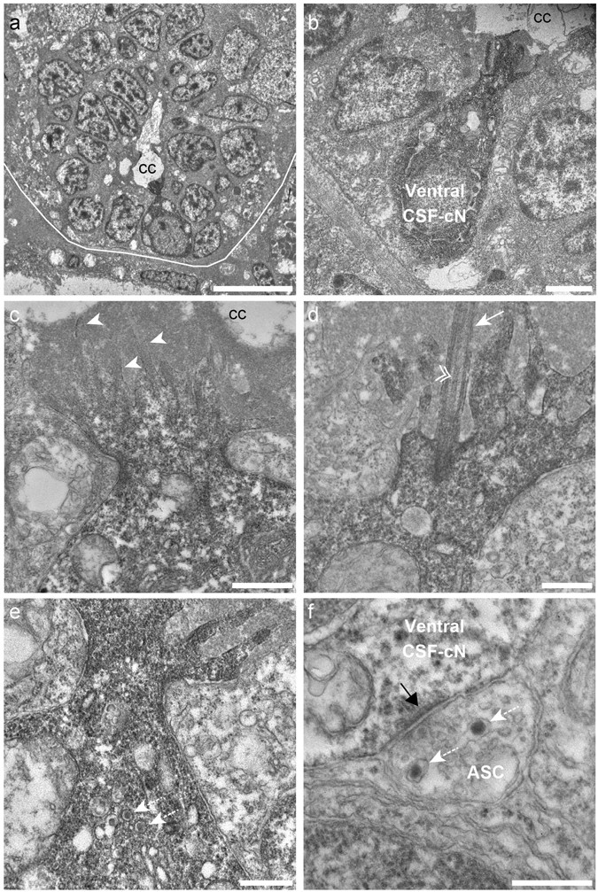
Ventral CSF-cNs exhibit an apical extension composed of microvilli and a kinocilium in the spinal cord. (a) Transverse section of the spinal cord showing restricted deposition of DAB in a ventral CSF-cN. (b) Overall view of a DAB+ ventral CSF-cN contacting the central canal (cc) and surrounded by ependymal cells. (c) Ventral CSF-cNs project at the apical pole an extension toward the central canal bearing several microvilli (arrowheads). (d) Within this extension is located a cilium (arrows) with two central microtubule along the axoneme (double arrowhead), suggesting a motile cilium. Large granular vesicles (LGV) are observed in the cytoplasm (e, dotted arrows) and axo-somatic synaptic contacts in the basal pole (f, ASC). Note the symmetry of the synaptic contact (black arrow) is reminiscent of an inhibitory synapse. Note the presence of LGV in the axon (f, dotted arrows). Scale bar = 10 μm (a), 2 µm (b), 1 μm (c), 500 nm (d,e) and 400 nm (f).
Figure 2
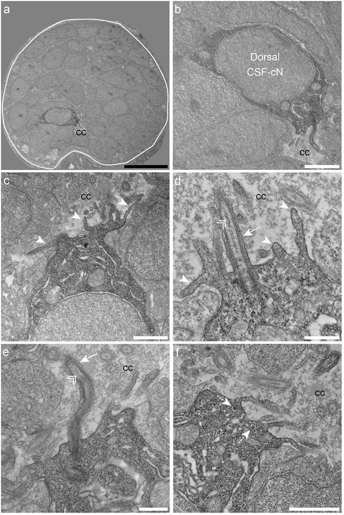
Dorsal spinal CSF-cNs also exhibit ultrastructural properties of sensory neurons. (a) Transverse section of the spinal cord showing restricted deposition of DAB in a dorsal CSF-cN. (b) Overall view of a DAB+ dorsal CSF-cN contacting the central canal (cc). (c,d) Dorsal CSF-cNs bear at the apical pole multiple microvilli (arrowheads). (d,e) In the apical pole is located a cilium (arrow) with two central microtubule singlets along the axoneme (double arrowhead), reminiscent of a motile cilium. (f) Dorsal CSF-cN also exhibit LGV distributed in the cytoplasm (dotted arrows). Scale bar = 10 μm (a), 2 μm (b), 1 µm (c,f) and 500 nm (d,e).
CSF-cNs display different shapes of apical extensions and axonal projections
As a quantitative analysis of CSF-cN morphology is difficult using serial EM, we turned to fluorescence for a quantitative comparison of the apical extension and the axonal projections of dorsal and ventral CSF-cNs. First, we tested whether these cells differ in the morphology of their apical extension. We investigated the shape of the CSF-cN apical extension by labeling the membrane of CSF-cNs in Tg(pkd2l1:Gal4;UAS:TagRFPCAAX;cmlc2:eGFP)icm22 larvae or F-actin itself in Tg(pkd2l1:Gal4;UAS:LifeAct-GFP;cryaa:V)icm28 larvae or larvae transiently expressing (pkd2l1:Gal4) and (UAS:LifeAct-TagRFP;cryaa:C). Using these different approaches, we found that the shape of the apical extension differs between ventral and dorsal CSF-cNs at 3 and 6 dpf (Fig. 3a,b and Material and Methods). The apical extension of dorsal CSF-cNs (Fig. 3a,b) was more extended along the border of the central canal than for ventral cells (Fig. 3a,b). The apical extension in ventral CSF-cNs was overall more compact with a small proportion displaying a more extended apical extension similar to dorsal CSF-cNs (13.3%, n = 6 out of 45 ventral CSF-cNs, Fig. 3a, empty arrow). Next, to assess whether ventral and dorsal CSF-cNs differ in their axonal projections, we injected the (pkd2l1-TagRFP) DNA construct to label single cells (Supplemental Fig. 4a) and reconstructed their axon (Fig. 3c, Supplemental Fig. 4b,c). All CSF-cN axonal projections were ventral, ipsilateral and ascending, yet they were heterogeneous along the rostrocaudal axis of the spinal cord (n = 54, Fig. 3c). We measured significant differences between the axonal projections of the two populations of CSF-cNs in terms of the area of the axonal arborization, the length of the axon, the dorso-ventral coverage within the spinal cord and the number of axonal branches (Fig. 3d). The larger axonal length, arborization and number of branches in ventral CSF-cNs compared to dorsal cells suggest these ventral cells possibly differentiate before dorsal ones. Nonetheless, the fact that the dorsal and ventral populations of CSF-cNs project on different domains along the dorso-ventral axis of the spinal cord suggests that these two populations may target different neuronal types within the ventral spinal cord.
Figure 3
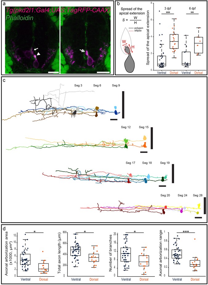
Morphological analysis of CSF-cNs reveals heterogeneous shapes of apical extension and axonal projections. (a) Transverse sections showing ventral and dorsal TagRFP-CAAX+ CSF-cNs (magenta) at 3 dpf reflecting the diversity of morphologies of the apical extension. The apical extension of all dorsal CSF-cNs spreads along the central canal border (arrow) while most ventral CSF-cNs (86.7%) form compact extensions (arrowhead). The small remaining subpopulation of ventral CSF-cNs exhibits the typical spread of dorsal apical extensions (arrow with empty head; Phalloidin staining, green). (b) Schematics of the analysis of the apical extension performed on each cell and statistical analysis comparing the size of the apical extension between ventral and dorsal CSF-cNs at 3 dpf (n = 45 versus 21) and 6 dpf (n = 14 versus 10). The apical extension of dorsal CSF-cNs extends more than for ventral CSF-cNs (two-sample t-tests, p < 5 · 10−7) and this difference persists at later stages (6 dpf, p < 0.002). (c) The reconstruction from dorsal (light shade) and ventral (dark shade) CSF-cNs from different segments (Seg) illustrates the diversity of axonal morphologies CSF-cNs between the two types along the spinal cord (n = 11 for each type). Vertical black bars represent the dorso-ventral limits of the spinal cord. Cells are positioned according to their dorso-ventral (D-V) position with dorsal edge set to 1 and ventral to 0. (d) Comparison of ventral and dorsal CSF-cNs for axonal arborization area, total axon length, number of branches and axonal arborization dorso-ventral range (n = 39 and 15 cells respectively). Ventral CSF-cNs have a wider axonal arborization (p < 0.003), a longer axon (p = 0.0014), reach more ventral domains of the spinal cord (p < 9 · 10−4), and cover a larger dorso-ventral (D-V) range (p < 2 · 10−4) with more axonal branches (p < 0.02). Two-sample t-tests were performed to compare the two populations. Scale bar = 10 μm (a) and 20 µm (c).
Distinct neuronal targets for ventral and dorsal CSF-cNs within the spinal cord
Differences in the axonal projection of ventral and dorsal CSF-cNs suggest these cells may project onto distinct targets within the spinal cord. Our lab recently demonstrated that CSF-cNs project onto caudal primary (CaP) motor neurons and commissural primary ascending (CoPA) sensory interneurons within the escape circuit[43](/articles/s41598-017-00350-1#ref-CR43 "Hubbard, J. M. et al. Intraspinal Sensory Neurons Provide Powerful Inhibition to Motor Circuits Ensuring Postural Control during Locomotion. Curr Biol 26, 2841–2853, doi: 10.1016/j.cub.2016.08.026
               (2016).") as well as onto V0-v interneurons within the slow swimming circuit[15](/articles/s41598-017-00350-1#ref-CR15 "Fidelin, K. et al. State-Dependent Modulation of Locomotion by GABAergic Spinal Sensory Neurons. Curr Biol
                       25, 3035–3047, doi:
                10.1016/j.cub.2015.09.070

               (2015)."). We investigated which CSF-cN type projects onto these targets using mosaic labeling in transgenic lines labeling CaP (_Tg_(_parg_ _mnet2_ _\-GFP_)[43](/articles/s41598-017-00350-1#ref-CR43 "Hubbard, J. M. et al. Intraspinal Sensory Neurons Provide Powerful Inhibition to Motor Circuits Ensuring Postural Control during Locomotion. Curr Biol
                       26, 2841–2853, doi:
                10.1016/j.cub.2016.08.026

               (2016)."), [44](/articles/s41598-017-00350-1#ref-CR44 "Balciunas, D. et al. Enhancer trapping in zebrafish using the Sleeping Beauty transposon. BMC Genomics
                       5, 62, doi:
                10.1186/1471-2164-5-62

               (2004).")), V0-v (_Tg_(_vglut2a_:_lox_:_DsRed_:_lox_:_GFP_)[15](/articles/s41598-017-00350-1#ref-CR15 "Fidelin, K. et al. State-Dependent Modulation of Locomotion by GABAergic Spinal Sensory Neurons. Curr Biol
                       25, 3035–3047, doi:
                10.1016/j.cub.2015.09.070

               (2015)."), [45](/articles/s41598-017-00350-1#ref-CR45 "Koyama, M., Kinkhabwala, A., Satou, C., Higashijima, S. & Fetcho, J. Mapping a sensory-motor network onto a structural and functional ground plan in the hindbrain. Proc Natl Acad Sci USA
                       108, 1170–1175, doi:
                10.1073/pnas.1012189108

               (2011).")) or CoPA (_Tg_(_tbx16-GFP_)[43](/articles/s41598-017-00350-1#ref-CR43 "Hubbard, J. M. et al. Intraspinal Sensory Neurons Provide Powerful Inhibition to Motor Circuits Ensuring Postural Control during Locomotion. Curr Biol
                       26, 2841–2853, doi:
                10.1016/j.cub.2016.08.026

               (2016)."), [46](/articles/s41598-017-00350-1#ref-CR46 "Wells, S., Nornes, S. & Lardelli, M. Transgenic zebrafish recapitulating tbx16 gene early developmental expression. PLoS One
                       6, e21559, doi:
                10.1371/journal.pone.0021559

               (2011).")). We observed that only ventral CSF-cNs form the stereotypical basket structure around the soma of CaP motor neurons (Fig. [4a](/articles/s41598-017-00350-1#Fig4), n = 7 out of 8 ventral CSF-cNs found projecting onto CaP and 0 out of 5 dorsal CSF-cNs). In contrast, we found only dorsal CSF-cNs contacting ventrolateral glutamatergic cells, putatively V0-v based on their location and our previous findings[15](/articles/s41598-017-00350-1#ref-CR15 "Fidelin, K. et al. State-Dependent Modulation of Locomotion by GABAergic Spinal Sensory Neurons. Curr Biol
                       25, 3035–3047, doi:
                10.1016/j.cub.2015.09.070

               (2015)."), [47](#ref-CR47 "Hale, M. E., Ritter, D. A. & Fetcho, J. R. A confocal study of spinal interneurons in living larval zebrafish. J Comp Neurol
                       437, 1–16 (2001)."),[48](#ref-CR48 "McLean, D. L., Fan, J., Higashijima, S., Hale, M. E. & Fetcho, J. R. A topographic map of recruitment in spinal cord. Nature
                       446, 71–75, doi:
                10.1038/nature05588

               (2007)."),[49](/articles/s41598-017-00350-1#ref-CR49 "Satou, C., Kimura, Y. & Higashijima, S. Generation of multiple classes of V0 neurons in zebrafish spinal cord: progenitor heterogeneity and temporal control of neuronal diversity. J Neurosci
                       32, 1771–1783, doi:
                10.1523/JNEUROSCI.5500-11.2012

               (2012).") (Fig. [4b](/articles/s41598-017-00350-1#Fig4), n = 3 out of 3 dorsal CSF-cNs and 0 out of 5 ventral CSF-cNs). Interestingly, we observed that both ventral and dorsal CSF-cNs project onto CoPA sensory interneurons (Fig. [4c,d](/articles/s41598-017-00350-1#Fig4), n = 4 dorsal and 5 ventral CSF-cNs). Altogether, our observations demonstrate a complex connectivity pattern from CSF-cNs onto their neuronal targets in the spinal cord. While some targets receive projections from both CSF-cN types (such as CoPA sensory interneurons), others seem to only receive inputs from either ventral (CaP motor neurons) or dorsal (ventrolateral glutamatergic neurons, most likely V0-v interneurons) CSF-cNs.
Figure 4
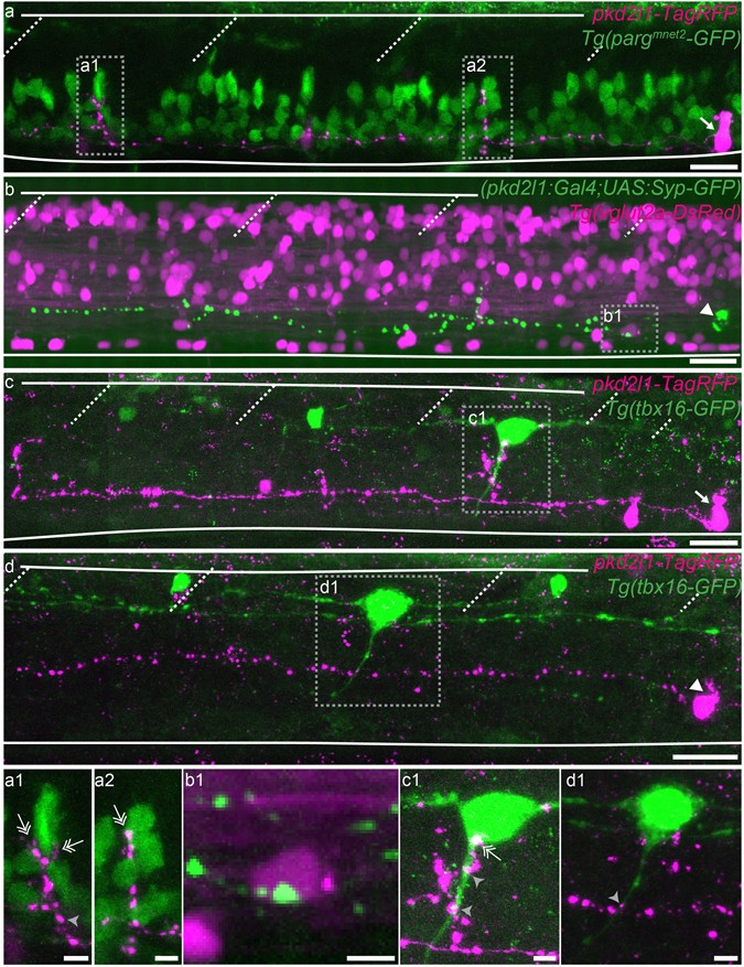
Ventral and dorsal CSF-cNs project onto distinct neuronal populations. (a–d) Z projection stacks showing contact from ventral and dorsal CSF-cNs onto different spinal targets. (a) Lateral view of a ventral CSF-cN (magenta, arrow) contacting 2 CaP motor neurons (identified based on their location within the segment) labelled in green in the Tg(parg mnet2 -GFP) transgenic line (a1,a2, double headed arrows). (b) Dorsal CSF-cN (green, arrowhead) contacting a putative V0-v interneuron (magenta, based on its dorso-ventral and lateral location) in the Tg(vglut2a:DsRed) transgenic line. (c,d) Ventral (c, arrow) and dorsal (d, arrowhead) CSF-cNs (magenta) contact CoPA sensory interneurons (green) labelled in the Tg(tbx16-GFP) transgenic line. Boxes with close-ups highlight contacts between the CSF-cN and its target. Scale bar = 20 µm (a–d) and 5 µm (a1–d1).
Investigation of secreted compounds expressed in dorsal and ventral CSF-cNs
We tested whether ventral and dorsal CSF-cNs differentially express secreted compounds previously reported in a restricted number of CSF-cNs, namely somatostatin[14](/articles/s41598-017-00350-1#ref-CR14 "Jalalvand, E., Robertson, B., Wallen, P., Hill, R. H. & Grillner, S. Laterally projecting cerebrospinal fluid-contacting cells in the lamprey spinal cord are of two distinct types. J Comp Neurol 522, Spc1, doi: 10.1002/cne.23584
               (2014)."), [21](/articles/s41598-017-00350-1#ref-CR21 "Christenson, J., Alford, S., Grillner, S. & Hokfelt, T. Co-localized GABA and somatostatin use different ionic mechanisms to hyperpolarize target neurons in the lamprey spinal cord. Neurosci Lett
                       134, 93-97, doi:0304-3940(91)90516-V (1991)."), [33](/articles/s41598-017-00350-1#ref-CR33 "Buchanan, J. T., Brodin, L., Hokfelt, T., Van Dongen, P. A. & Grillner, S. Survey of neuropeptide-like immunoreactivity in the lamprey spinal cord. Brain Res
                       408, 299–302, doi:0006-8993(87)90392-1 (1987)."), [34](/articles/s41598-017-00350-1#ref-CR34 "Lopez, J. M. et al. Distribution of somatostatin-like immunoreactivity in the brain of the caecilian Dermophis mexicanus (Amphibia: Gymnophiona): comparative aspects in amphibians. J Comp Neurol
                       501, 413–430, doi:
                10.1002/cne.21244

               (2007).") and serotonin[35](/articles/s41598-017-00350-1#ref-CR35 "Sims, T. J. The development of monamine-containing neurons in the brain and spinal cord of the salamander, Ambystoma mexicanum. J Comp Neurol
                       173, 319–336, doi:
                10.1002/cne.901730208

               (1977)."), [36](/articles/s41598-017-00350-1#ref-CR36 "Chiba, A. & Oka, S. Serotonin-immunoreactive structures in the central nervous system of the garfish Lepisosteus productus (Semionotiformes, Osteichthyes). Neurosci Lett
                       261, 73–76, doi:S0304-3940(98)01011-8 (1999)."). By taking advantage of _pkd2l1_ expression in CSF-cNs[13](/articles/s41598-017-00350-1#ref-CR13 "Djenoune, L. et al. Investigation of spinal cerebrospinal fluid-contacting neurons expressing PKD2L1: evidence for a conserved system from fish to primates. Front Neuroanat
                       8, 26, doi:
                10.3389/fnana.2014.00026

               (2014)."), we used the _Tg_(_pkd2l1_:_GCaMP5G_)_icm07_ transgenic line to selectively target CSF-cNs[37](/articles/s41598-017-00350-1#ref-CR37 "Böhm, U. L. et al. CSF-contacting neurons regulate locomotion by relaying mechanical stimuli to spinal circuits. Nat Commun
                       7, 10866, doi:
                10.1038/ncomms10866

               (2016)."). We demonstrated the specificity of this line using fluorescent _in situ_ hybridization (FISH) for _pkd2l1_ mRNA coupled to GFP immunohistochemistry (IHC) to amplify the endogenous GCaMP5G signal from 24 hours post fertilization (hpf) to 5 dpf (Supplemental Fig. [5](/articles/s41598-017-00350-1#MOESM1)).
In zebrafish, somatostatin immunoreactivity has previously been reported[12](/articles/s41598-017-00350-1#ref-CR12 "Wyart, C. et al. Optogenetic dissection of a behavioural module in the vertebrate spinal cord. Nature 461, 407–410, doi: 10.1038/nature08323nature08323
               (2009)."), but 6 different paralogs exist (_sst1_._1_, _sst1_._2_, _sst2_, _sst3_, _sst5_ and _sst6_)[50](/articles/s41598-017-00350-1#ref-CR50 "Tostivint, H., Lihrmann, I. & Vaudry, H. New insight into the molecular evolution of the somatostatin family. Mol Cell Endocrinol
                       286, 5–17, doi:
                10.1016/j.mce.2008.02.029S0303-7207(08)00115-9

               (2008)."), [51](/articles/s41598-017-00350-1#ref-CR51 "Tostivint, H., Quan, F. B., Bougerol, M., Kenigfest, N. B. & Lihrmann, I. Impact of gene/genome duplications on the evolution of the urotensin II and somatostatin families. Gen Comp Endocrinol
                       188, 110–117, doi:
                10.1016/j.ygcen.2012.12.015S0016-6480(13)00005-1

               (2013)."). Testing the expression of all of them, we only detected expression of _sst1_._1_ in CSF-cNs. Expression of _sst1_._1_ was observed only in dorsal CSF-cNs (Fig. [5a,b](/articles/s41598-017-00350-1#Fig5), arrows) in the rostral part of the spinal cord, from segment 1 to 13 at 24, 48, 72 hpf and in the adult spinal cord (data _not shown_). Since _sst1_._1_ had been reported as expressed transiently in motor neurons from 19 hpf until 55 hpf[52](/articles/s41598-017-00350-1#ref-CR52 "Devos, N. et al. Differential expression of two somatostatin genes during zebrafish embryonic development. Mech Dev
                       115, 133–137, doi:S0925477302000825 (2002)."), we showed using FISH for _sst1_._1_ with GFP IHC in 24 hpf _Tg_(_mnx1_:_GFP_) embryos[53](/articles/s41598-017-00350-1#ref-CR53 "Flanagan-Steet, H., Fox, M. A., Meyer, D. & Sanes, J. R. Neuromuscular synapses can form in vivo by incorporation of initially aneural postsynaptic specializations. Development
                       132, 4471–4481, doi:
                10.1242/dev.02044

               (2005).") that _sst1_._1_ expression was excluded from motor neurons (Fig. [5c](/articles/s41598-017-00350-1#Fig5)). Altogether, our results show that _sst1_._1_ is expressed in dorsal CSF-cNs and absent in ventral ones at early stages and restricted as well to a subpopulation of CSF-cNs in the adult. Next, we tested whether zebrafish CSF-cNs were serotoninergic by performing double IHC for 5-HT and GFP on _Tg_(_pkd2l1_:_GCaMP5G_)_icm07_ embryos and larvae from 24 hpf to 72 hpf. At 24 hpf, no 5-HT immunostaining was detected in the zebrafish spinal cord (_data not shown_). At 48 hpf, we detected 5-HT in ventral CSF-cNs (Fig. [5d](/articles/s41598-017-00350-1#Fig5), arrowheads) in the rostral part of the spinal cord (from segment 1 to 24). This expression was restricted to a subset of ventral CSF-cNs (Fig. [5d](/articles/s41598-017-00350-1#Fig5)). The proportion of 5-HT+ CSF-cNs at 48 hpf decreased along the rostrocaudal axis of the spinal cord (segments 3 to 6: 88.6 ± 16.0%, 96 cells; segments 10 to 13: 73.2 ± 27.3%, 100 cells; segments 23 to 26: 31.2 ± 33.3%, 94 cells). At 3 dpf, although 5-HT was still expressed along the entire spinal cord in other cells, the staining was absent in CSF-cNs in the rostral spinal cord (Fig. [5e](/articles/s41598-017-00350-1#Fig5)). In conclusion, dorsal CSF-cNs express _sst1_._1_ while a subpopulation of ventral CSF-cNs population is transiently serotoninergic. Hence, these two populations originating from different progenitor pools differentiate into distinct cell types expressing specific secreted compounds.
Figure 5
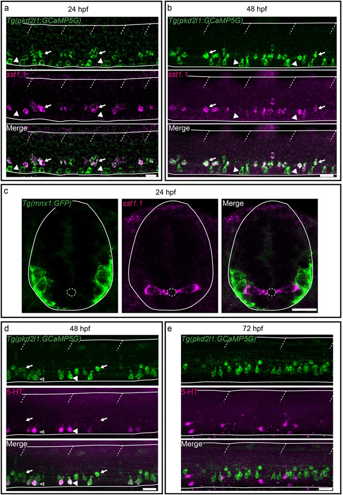
Secreted factors distinguish dorsal and ventral CSF-cNs: while dorsal express the somatostatin paralog sst1.1, ventral CSF-cNs express 5-HT. (a–c) Lateral views of the spinal cord show that sst1.1 expression is restricted to dorsal CSF-cNs (arrows, FISH for sst1.1 (magenta) coupled to GFP IHC (green) on Tg(pkd2l1:GCaMP5G)) (a,b) and Tg(mnx1:GFP) (c) embryos and larvae at 24 hpf (a,c) and 48 hpf (b). (c) Transverse sections show that sst1.1 (magenta) is not expressed in motor neurons (green) as previously suggested (Devos et al.52). (d,e) IHC for 5-HT (magenta) and GFP (green) on Tg(pkd2l1:GCaMP5G) transgenic larvae at 48 hpf (d) and 72 hpf (e). (d) At 48 hpf, most ventral CSF-cNs express 5-HT (arrowhead, compared to negative cells shown with empty arrowhead). Note that dorsal CSF-cNs (arrows) are not labelled by 5-HT. (e) At 72 hpf, ventral CSF-cNs are not serotoninergic anymore in the rostral part of the spinal cord. Horizontal lines represent the limits of the spinal cord and slash dashed lines represent somite boundaries. Small dotted ellipses represent the limit of the central canal. Scale bars = 20 μm.
pkd2l1 null mutation does not impact on the differentiation of spinal CSF-cNs
We previously showed that the PKD2L1 channel is expressed in spinal CSF-cNs across multiple vertebrate species[13](/articles/s41598-017-00350-1#ref-CR13 "Djenoune, L. et al. Investigation of spinal cerebrospinal fluid-contacting neurons expressing PKD2L1: evidence for a conserved system from fish to primates. Front Neuroanat 8, 26, doi: 10.3389/fnana.2014.00026
               (2014).") and that the channel appears necessary to mediate detection of spinal bending by CSF-cNs in the larva[37](/articles/s41598-017-00350-1#ref-CR37 "Böhm, U. L. et al. CSF-contacting neurons regulate locomotion by relaying mechanical stimuli to spinal circuits. Nat Commun
                       7, 10866, doi:
                10.1038/ncomms10866

               (2016)."). In _pkd2l1_ mutants, we observed defects in locomotion that we interpreted as being caused by the lack of sensory responses in CSF-cNs[37](/articles/s41598-017-00350-1#ref-CR37 "Böhm, U. L. et al. CSF-contacting neurons regulate locomotion by relaying mechanical stimuli to spinal circuits. Nat Commun
                       7, 10866, doi:
                10.1038/ncomms10866

               (2016)."). To strengthen the conclusions of this previous study, we sought to precisely assess whether CSF-cNs develop properly in the _pkd2l1_ mutants. We found that in mutants, CSF-cNs were still GABAergic, ventral ones were still serotoninergic, that the number of CSF-cNs was not impacted, and that the morphology of their axonal projections was not different from WT (Supplemental Fig. [6](/articles/s41598-017-00350-1#MOESM1)). We also wanted to determine whether the functional connection between ventral CSF-cNs and CaPs[43](/articles/s41598-017-00350-1#ref-CR43 "Hubbard, J. M. et al. Intraspinal Sensory Neurons Provide Powerful Inhibition to Motor Circuits Ensuring Postural Control during Locomotion. Curr Biol
                       26, 2841–2853, doi:
                10.1016/j.cub.2016.08.026

               (2016)."), [54](/articles/s41598-017-00350-1#ref-CR54 "Sternberg, J. R. et al. Optimization of a Neurotoxin to Investigate the Contribution of Excitatory Interneurons to Speed Modulation In Vivo. Curr Biol
                       26, 2319–2328, doi:
                10.1016/j.cub.2016.06.037

               (2016).") was impacted and found that light-mediated activation of mutants CSF-cNs was still able to induce monosynaptic inhibitory postsynaptic currents in CaPs (Supplemental Fig. [7](/articles/s41598-017-00350-1#MOESM1)). Altogether, our results show that _pkd2l1_ is not required for CSF-cN differentiation.
Discussion
Spinal CSF-cNs were previously shown to originate from distinct progenitor domains characterized by distinct pools of transcription factors in the embryo[26](/articles/s41598-017-00350-1#ref-CR26 "Petracca, Y. L. et al. The late and dual origin of cerebrospinal fluid-contacting neurons in the mouse spinal cord. Development 143, 880–891, doi: 10.1242/dev.129254
               (2016)."), [29](/articles/s41598-017-00350-1#ref-CR29 "Park, H. C., Shin, J. & Appel, B. Spatial and temporal regulation of ventral spinal cord precursor specification by Hedgehog signaling. Development
                       131, 5959–5969, doi:
                10.1242/dev.01456

               (2004)."), [30](/articles/s41598-017-00350-1#ref-CR30 "Yang, L., Rastegar, S. & Strahle, U. Regulatory interactions specifying Kolmer-Agduhr interneurons. Development
                       137, 2713–2722, doi:
                10.1242/dev.048470dev.048470

               (2010)."). Here we demonstrate that these two domains give rise to two cell types of CSF-cNs with probably distinct functional properties at the larval stages (Supplemental Fig. [8](/articles/s41598-017-00350-1#MOESM1)). First, the morphology of these two cell types differs both at the apical extension level as well as at the axonal level. Furthermore, we show that these differences in axonal projections lead to differences in neuronal targets reached in the spinal cord. Second, our ultrastructure data indicate that both ventral and dorsal CSF-cNs bear dense granules under the apical extension, suggesting possible roles in release or uptake of compounds from the CSF. We show that ventral and dorsal populations express distinct secreted factors, in particular 5-HT and _sst1_._1_, during development. Finally we show that Pkd2l1 is not necessary for the differentiation of CSF-cNs. Altogether, our results demonstrate that the two cell types of CSF-cNs previously identified with different developmental origins segregate into distinct populations bearing specific functional properties.
Due to their peculiar apical extension contacting the CSF, it had been hypothesized that CSF-cNs may release compounds into the CSF7, 55. The presence of large granular vesicles (LGV) within CSF-cNs in our EM data, combined with expression of sst1.1 and 5-HT for dorsal and ventral CSF-cNs respectively, reinforces this hypothesis.
Previous works have shown that CSF-cNs are chemosensory[11](/articles/s41598-017-00350-1#ref-CR11 "Huang, A. L. et al. The cells and logic for mammalian sour taste detection. Nature 442, 934–938, doi: 10.1038/nature05084
               (2006)."), [24](/articles/s41598-017-00350-1#ref-CR24 "Orts Del’Immagine, A. et al. A single polycystic kidney disease 2-like 1 channel opening acts as a spike generator in cerebrospinal fluid-contacting neurons of adult mouse brainstem. Neuropharmacology, doi:
                10.1016/j.neuropharm.2015.07.030

               (2015)."), [56](/articles/s41598-017-00350-1#ref-CR56 "Bushman, J. D., Ye, W. & Liman, E. R. A proton current associated with sour taste: distribution and functional properties. FASEB J
                       29, 3014–3026, doi:
                10.1096/fj.14-265694fj.14-265694

               (2015)."), [57](/articles/s41598-017-00350-1#ref-CR57 "Jalalvand, E., Robertson, B., Tostivint, H., Wallen, P. & Grillner, S. The Spinal Cord Has an Intrinsic System for the Control of pH. Curr Biol
                       26, 1346–1351, doi:
                10.1016/j.cub.2016.03.048

               (2016).") as well as mechanosensory cells[37](/articles/s41598-017-00350-1#ref-CR37 "Böhm, U. L. et al. CSF-contacting neurons regulate locomotion by relaying mechanical stimuli to spinal circuits. Nat Commun
                       7, 10866, doi:
                10.1038/ncomms10866

               (2016)."), [43](/articles/s41598-017-00350-1#ref-CR43 "Hubbard, J. M. et al. Intraspinal Sensory Neurons Provide Powerful Inhibition to Motor Circuits Ensuring Postural Control during Locomotion. Curr Biol
                       26, 2841–2853, doi:
                10.1016/j.cub.2016.08.026

               (2016)."), [58](/articles/s41598-017-00350-1#ref-CR58 "Jalalvand, E., Robertson, B., Wallen, P. & Grillner, S. Ciliated neurons lining the central canal sense both fluid movement and pH through ASIC3. Nat Commun
                       7, 10002, doi:
                10.1038/ncomms10002

               (2016)."). In zebrafish larvae, we previously showed that dorsal CSF-cNs respond to lateral bending of the spinal cord[37](/articles/s41598-017-00350-1#ref-CR37 "Böhm, U. L. et al. CSF-contacting neurons regulate locomotion by relaying mechanical stimuli to spinal circuits. Nat Commun
                       7, 10866, doi:
                10.1038/ncomms10866

               (2016).") while ventral CSF-cNs respond to longitudinal contractions[43](/articles/s41598-017-00350-1#ref-CR43 "Hubbard, J. M. et al. Intraspinal Sensory Neurons Provide Powerful Inhibition to Motor Circuits Ensuring Postural Control during Locomotion. Curr Biol
                       26, 2841–2853, doi:
                10.1016/j.cub.2016.08.026

               (2016)."). The difference in shape of the apical extension between dorsal and ventral CSF-cNs may reflect a structural difference relevant for either their mechanosensory functions (direction of maximum sensitivity to bending of the tissue or CSF flow) or their secretory functions (with possibly larger surface area for dorsal CSF-cNs).
There are indications that spinal CSF-cNs harbor different morphologies in other species as well. In rat, spinal GABAergic CSF-cNs were first classified into three subtypes according to the shape of their soma59. Four morphological types of CSF-cNs were then described based on the shape of their soma, their axonal projection, and on the expression of the peptide Met-Enk-Arg-Gly-Leu60. These indications together with our data reveal a high level of heterogeneity among spinal CSF-cNs. Future studies will investigate the role of CSF-cN structural differences in sensory and secretory functions.
Using a single cell labelling approach, we show that ventral CSF-cNs have on average a longer and broader axonal arborization and cover a higher dorso-ventral spinal cord range with more axonal branches than dorsal CSF-cNs. These differences in axonal projections suggest the two CSF-cN populations might have distinct targets within the spinal cord. We had shown that CSF-cNs form active GABAergic synapses onto glutamatergic descending V0-v interneurons[15](/articles/s41598-017-00350-1#ref-CR15 "Fidelin, K. et al. State-Dependent Modulation of Locomotion by GABAergic Spinal Sensory Neurons. Curr Biol 25, 3035–3047, doi: 10.1016/j.cub.2015.09.070
               (2015).") as well as CaP primary motor neurons and CoPA sensory interneurons[43](/articles/s41598-017-00350-1#ref-CR43 "Hubbard, J. M. et al. Intraspinal Sensory Neurons Provide Powerful Inhibition to Motor Circuits Ensuring Postural Control during Locomotion. Curr Biol
                       26, 2841–2853, doi:
                10.1016/j.cub.2016.08.026

               (2016)."). Here we show that only ventral CSF-cNs form the characteristic basket-like contact onto CaP soma while we only found dorsal CSF-cNs contacting ventrolateral glutamatergic neurons, most likely V0-v interneurons[47](#ref-CR47 "Hale, M. E., Ritter, D. A. & Fetcho, J. R. A confocal study of spinal interneurons in living larval zebrafish. J Comp Neurol
                       437, 1–16 (2001)."),[48](#ref-CR48 "McLean, D. L., Fan, J., Higashijima, S., Hale, M. E. & Fetcho, J. R. A topographic map of recruitment in spinal cord. Nature
                       446, 71–75, doi:
                10.1038/nature05588

               (2007)."),[49](/articles/s41598-017-00350-1#ref-CR49 "Satou, C., Kimura, Y. & Higashijima, S. Generation of multiple classes of V0 neurons in zebrafish spinal cord: progenitor heterogeneity and temporal control of neuronal diversity. J Neurosci
                       32, 1771–1783, doi:
                10.1523/JNEUROSCI.5500-11.2012

               (2012).") previously shown to receive inputs from CSF-cNs[15](/articles/s41598-017-00350-1#ref-CR15 "Fidelin, K. et al. State-Dependent Modulation of Locomotion by GABAergic Spinal Sensory Neurons. Curr Biol
                       25, 3035–3047, doi:
                10.1016/j.cub.2015.09.070

               (2015)."). Interestingly CoPA sensory interneurons received innervation from both dorsal and ventral CSF-cNs. Our results suggest that targets of the slow swimming circuits receive innervation from dorsal CSF-cNs while targets of the fast swimming circuit receive innervation from ventral CSF-cNs, and sensory interneurons involved in processing feedback receive innervation from both ventral and dorsal CSF-sNs. Further investigations will be necessary to establish whether other targets of the slow and fast swimming circuits follow the same dichotomy, receiving inputs from dorsal and ventral CSF-cNs respectively.
Multiple markers have been found in subsets of CSF-cNs across vertebrate species: somatostatin[12](/articles/s41598-017-00350-1#ref-CR12 "Wyart, C. et al. Optogenetic dissection of a behavioural module in the vertebrate spinal cord. Nature 461, 407–410, doi: 10.1038/nature08323nature08323
               (2009)."), [14](/articles/s41598-017-00350-1#ref-CR14 "Jalalvand, E., Robertson, B., Wallen, P., Hill, R. H. & Grillner, S. Laterally projecting cerebrospinal fluid-contacting cells in the lamprey spinal cord are of two distinct types. J Comp Neurol
                       522, Spc1, doi:
                10.1002/cne.23584

               (2014)."), [21](/articles/s41598-017-00350-1#ref-CR21 "Christenson, J., Alford, S., Grillner, S. & Hokfelt, T. Co-localized GABA and somatostatin use different ionic mechanisms to hyperpolarize target neurons in the lamprey spinal cord. Neurosci Lett
                       134, 93-97, doi:0304-3940(91)90516-V (1991)."), [33](/articles/s41598-017-00350-1#ref-CR33 "Buchanan, J. T., Brodin, L., Hokfelt, T., Van Dongen, P. A. & Grillner, S. Survey of neuropeptide-like immunoreactivity in the lamprey spinal cord. Brain Res
                       408, 299–302, doi:0006-8993(87)90392-1 (1987)."), [34](/articles/s41598-017-00350-1#ref-CR34 "Lopez, J. M. et al. Distribution of somatostatin-like immunoreactivity in the brain of the caecilian Dermophis mexicanus (Amphibia: Gymnophiona): comparative aspects in amphibians. J Comp Neurol
                       501, 413–430, doi:
                10.1002/cne.21244

               (2007)."), dopamine[61](#ref-CR61 "Barreiro-Iglesias, A., Villar-Cervino, V., Anadon, R. & Rodicio, M. C. Descending brain-spinal cord projections in a primitive vertebrate, the lamprey: cerebrospinal fluid-contacting and dopaminergic neurons. J Comp Neurol
                       511, 711–723, doi:
                10.1002/cne.21863

               (2008)."),[62](#ref-CR62 "Rodicio, M. C., Villar-Cervino, V., Barreiro-Iglesias, A. & Anadon, R. Colocalization of dopamine and GABA in spinal cord neurones in the sea lamprey. Brain Res Bull
                       76, 45–49, doi:
                10.1016/j.brainresbull.2007.10.062S0361-9230(07)00399-1

               (2008)."),[63](#ref-CR63 "Roberts, B. L., Maslam, S., Scholten, G. & Smit, W. Dopaminergic and GABAergic cerebrospinal fluid-contacting neurons along the central canal of the spinal cord of the eel and trout. J Comp Neurol
                       354, 423–437, doi:
                10.1002/cne.903540310

               (1995)."),[64](/articles/s41598-017-00350-1#ref-CR64 "Acerbo, M. J., Hellmann, B. & Gunturkun, O. Catecholaminergic and dopamine-containing neurons in the spinal cord of pigeons: an immunohistochemical study. J Chem Neuroanat
                       25, 19–27, doi:S0891061802000728 (2003).") and serotonin[35](/articles/s41598-017-00350-1#ref-CR35 "Sims, T. J. The development of monamine-containing neurons in the brain and spinal cord of the salamander, Ambystoma mexicanum. J Comp Neurol
                       173, 319–336, doi:
                10.1002/cne.901730208

               (1977)."), [36](/articles/s41598-017-00350-1#ref-CR36 "Chiba, A. & Oka, S. Serotonin-immunoreactive structures in the central nervous system of the garfish Lepisosteus productus (Semionotiformes, Osteichthyes). Neurosci Lett
                       261, 73–76, doi:S0304-3940(98)01011-8 (1999)."), [65](/articles/s41598-017-00350-1#ref-CR65 "Parent, A. & Northcutt, R. G. The monoamine-containing neurons in the brain of the garfish, Lepisosteus osseus. Brain Res Bull
                       9, 189–204 (1982)."). The expression of most markers was reported in a restricted fraction of CSF-cNs. Here we tested whether some of these markers showed restricted expression patterns among CSF-cNs. We found that ventral CSF-cNs were at 48 hpf transiently serotoninergic as previously described in other species[35](/articles/s41598-017-00350-1#ref-CR35 "Sims, T. J. The development of monamine-containing neurons in the brain and spinal cord of the salamander, Ambystoma mexicanum. J Comp Neurol
                       173, 319–336, doi:
                10.1002/cne.901730208

               (1977)."), [36](/articles/s41598-017-00350-1#ref-CR36 "Chiba, A. & Oka, S. Serotonin-immunoreactive structures in the central nervous system of the garfish Lepisosteus productus (Semionotiformes, Osteichthyes). Neurosci Lett
                       261, 73–76, doi:S0304-3940(98)01011-8 (1999)."), [65](/articles/s41598-017-00350-1#ref-CR65 "Parent, A. & Northcutt, R. G. The monoamine-containing neurons in the brain of the garfish, Lepisosteus osseus. Brain Res Bull
                       9, 189–204 (1982).") in accordance with findings from Montgomery _et al_.[66](/articles/s41598-017-00350-1#ref-CR66 "Montgomery, J. E., Wiggin, T. D., Rivera-Perez, L. M., Lillesaar, C. & Masino, M. A. Intraspinal serotonergic neurons consist of two, temporally distinct populations in developing zebrafish. Dev Neurobiol
                       76, 673–687, doi:
                10.1002/dneu.22352

               (2016)."), in zebrafish. In addition, we demonstrated that, even at 48 hpf, not all ventral CSF-cNs expressed 5-HT. This suggests that there may be different subtypes of ventral CSF-cNs based on 5-HT expression. Moreover, Montgomery _et al_.[66](/articles/s41598-017-00350-1#ref-CR66 "Montgomery, J. E., Wiggin, T. D., Rivera-Perez, L. M., Lillesaar, C. & Masino, M. A. Intraspinal serotonergic neurons consist of two, temporally distinct populations in developing zebrafish. Dev Neurobiol
                       76, 673–687, doi:
                10.1002/dneu.22352

               (2016)."), also showed that the rate-limiting enzyme involved in 5-HT synthesis _tryptophan hydroxylase 1_ a (_tph1a_)[67](/articles/s41598-017-00350-1#ref-CR67 "Bellipanni, G., Rink, E. & Bally-Cuif, L. Cloning of two tryptophan hydroxylase genes expressed in the diencephalon of the developing zebrafish brain. Mech Dev
                       119 Suppl 1, S215–220, doi:S0925477303001199 (2002)."), [68](/articles/s41598-017-00350-1#ref-CR68 "Teraoka, H. et al. Hedgehog and Fgf signaling pathways regulate the development of tphR-expressing serotonergic raphe neurons in zebrafish embryos. J Neurobiol
                       60, 275–288, doi:
                10.1002/neu.20023

               (2004).") is expressed in the ventral spinal cord at 24 and 48 hpf suggesting that ventral CSF-cNs could constitute a transient source of serotonin at early stages. One possible explanation for the disappearance of 5-HT in ventral CSF-cNs at later stages could be the emergence of descending fibers from 5-HT+ neurons originating from the brainstem raphe nuclei from 48 hpf onwards[67](/articles/s41598-017-00350-1#ref-CR67 "Bellipanni, G., Rink, E. & Bally-Cuif, L. Cloning of two tryptophan hydroxylase genes expressed in the diencephalon of the developing zebrafish brain. Mech Dev
                       119 Suppl 1, S215–220, doi:S0925477303001199 (2002)."). 5-HT from descending fibers has been previously shown in mammals to suppress the monoaminergic expression of CSF-cNs[69](#ref-CR69 "Branchereau, P., Chapron, J. & Meyrand, P. Descending 5-hydroxytryptamine raphe inputs repress the expression of serotonergic neurons and slow the maturation of inhibitory systems in mouse embryonic spinal cord. J Neurosci
                       22, 2598–2606, doi:20026199 (2002)."),[70](#ref-CR70 "Allain, A. E., Segu, L., Meyrand, P. & Branchereau, P. Serotonin controls the maturation of the GABA phenotype in the ventral spinal cord via 5-HT1b receptors. Ann N Y Acad Sci
                       1198, 208–219, doi:
                10.1111/j.1749-6632.2010.05433.x

               (2010)."),[71](/articles/s41598-017-00350-1#ref-CR71 "Wienecke, J. et al. Spinal cord injury enables aromatic L-amino acid decarboxylase cells to synthesize monoamines. J Neurosci
                       34, 11984–12000, doi:
                10.1523/JNEUROSCI.3838-13.2014

               (2014)."). The physiological relevance of the transient 5-HT expression among this ventral population of CSF-cNs remains to be elucidated.
Regarding the origin of somatostatin in zebrafish CSF-cNs, we found that among the six SST paralogs, sst1.1 was the only one expressed. We demonstrated that the expression of this gene is specific and restricted to the dorsal population of CSF-cNs, identifying for the first time a specific marker of dorsal CSF-cNs. We also demonstrated that this peptide is excluded from motor neurons contrary to what previous studies suggested52. The physiological relevance of sst1.1 expression by dorsal CSF-cNs is not known. In mammals and lamprey, it has been shown that somatostatin can reduce locomotor frequency[58](/articles/s41598-017-00350-1#ref-CR58 "Jalalvand, E., Robertson, B., Wallen, P. & Grillner, S. Ciliated neurons lining the central canal sense both fluid movement and pH through ASIC3. Nat Commun 7, 10002, doi: 10.1038/ncomms10002
               (2016)."), [72](/articles/s41598-017-00350-1#ref-CR72 "Barriere, G., Bertrand, S. & Cazalets, J. R. Peptidergic neuromodulation of the lumbar locomotor network in the neonatal rat spinal cord. Peptides
                       26, 277–286, doi:
                10.1016/j.peptides.2004.09.002

               (2005)."), [73](/articles/s41598-017-00350-1#ref-CR73 "Miles, G. B. & Sillar, K. T. Neuromodulation of vertebrate locomotor control networks. Physiology (Bethesda)
                       26, 393–411, doi:
                10.1152/physiol.00013.201126/6/393

               (2011)."). The release of SST by dorsal CSF-cNs could impact the frequency of locomotor events along with the release of neuropeptides of the UII family, _urp1_ and _urp2_ by ventral CSF-cNs only[74](/articles/s41598-017-00350-1#ref-CR74 "Quan, F. B. et al. Comparative distribution and in vitro activities of the urotensin II-related peptides URP1 and URP2 in zebrafish: evidence for their colocalization in spinal cerebrospinal fluid-contacting neurons. PLoS One
                       10, e0119290, doi:
                10.1371/journal.pone.0119290PONE-D-14-36037

               (2015).") as previously suggested by the presence in coho salmon of a UII-like immunoreactive ventral population of CSF-cNs and a distinct somatostatinergic one[75](/articles/s41598-017-00350-1#ref-CR75 "Yulis, C. R. & Lederis, K. Relationship between urotensin II- and somatostatin-immunoreactive spinal cord neurons of Catostomus commersoni and Oncorhynchus kisutch (Teleostei). Cell Tissue Res
                       254, 539–542 (1988)."). Taken together, our results confirm that ventral and dorsal CSF-cNs express distinct peptides and neuromodulators. The transient nature of the expression of 5-HT suggest that it could have developmental roles to be further investigated.
Material and Methods
Animal care
Zebrafish (Danio rerio) adults and larvae were maintained and raised on a 14/10 hour light cycle. Fish lines used in this study are referenced in Table 1. Water was regulated at 28.5 °C, conductivity at 500 μS and pH at 7.4. All embryos and larvae under 6 dpf were anesthetized in 0.02% tricaine methane sulfonate (MS 222) (Sandoz, Levallois-Perret, France) and euthanized in 0.2% MS 222 prior to fixation. Experimental and animal protocols were approved by the Institut du Cerveau et de la Moelle Épinière in agreement with the French National Ethics Committee (Comité National de Réflexion Éthique sur l’Éxpérimentation Animale; Ce5/2011/056) and European regulations.Genotype protocol for the pkd2l1 icm02 mutants was described in ref. [37](/articles/s41598-017-00350-1#ref-CR37 "Böhm, U. L. et al. CSF-contacting neurons regulate locomotion by relaying mechanical stimuli to spinal circuits. Nat Commun 7, 10866, doi: 10.1038/ncomms10866
(2016).") (Böhm _et al_. 2016). Table 1 Transgenic lines used in our study.
Generation of transgenic lines
In order to generate the Tg(UAS:TagRFP-CAAX;cmlc2:eGFP)icm22 line, the TagRFP-CAAX sequence was amplified using a TagRFP-forward (5′-CCCGGGATCCACATGGTGTCTAAGGGCGAAG-3′) and reverse primer (5′-GATCGCGGCCGCTCAGGAGAGCACACACTTGCAGCTCATGCAGCCGGGGCCACTCTCATCAGGAGGGTTCAGCTTATTAAGTTTGTGCCC-3′) and inserted into the pME-MCS vector[76](/articles/s41598-017-00350-1#ref-CR76 "Kwan, K. M. et al. The Tol2kit: a multisite gateway-based construction kit for Tol2 transposon transgenesis constructs. Dev Dyn 236, 3088–3099, doi: 10.1002/dvdy.21343
               (2007).") via BamHI/NotI restriction digestion. The resulting _pME-TagRFP-CAAX_ vector was recombined via a Gateway reaction (MultiSite Gateway Three-Fragment Vector Construction Kit) with _p5E-4nrUAS_ [77](/articles/s41598-017-00350-1#ref-CR77 "Auer, T. O. et al. Deletion of a kinesin I motor unmasks a mechanism of homeostatic branching control by neurotrophin-3. Elife
                       4,
                10.7554/eLife.05061

               (2015)."), _p3E-pA_ and _pDest-Tol2_; _cmlc2_:_eGFP_ [76](/articles/s41598-017-00350-1#ref-CR76 "Kwan, K. M. et al. The Tol2kit: a multisite gateway-based construction kit for Tol2 transposon transgenesis constructs. Dev Dyn
                       236, 3088–3099, doi:
                10.1002/dvdy.21343

               (2007).") resulting in (_p4nrUAS_:_TagRFP-CAAX-pA-Tol2_;_cmcl2_:_eGFP_). Using a similar approach, the _pME\_LifeAct-GFP_ plasmid was generated from the plasmid _pCS2\_LifeAct-GFP_ (kind gift from Nicolas David) using the SP6 promoter sequencing primer (5′-ATTTAGGTGACACTATAG-3′) and inserted into the pME\_MCS vector via a BamHI/NotI restriction digestion. The final three-way gateway reaction used the _pME\_LifeAct-GFP_, _pDest\_cryaa_:_V_ and _p5_′_E\_polyA_ plasmids to generate the (_UAS_:_LifeAct-GFP_;_cryaa_:_V_) construct. Microinjection of these plasmids was performed with Tol2 mRNA (25 ng/µl) following standard protocols. Transgenic founder fish _Tg_(_UAS_:_TagRFP-CAAX_;_cmlc2_:_eGFP_)_icm22_ where screened based on GFP expression in the heart. These two lines were also screened based on transactivation of their respective transgene when crossed with various Gal4 lines.
Plasmid design
In order to investigate the CSF-cNs ultrastructure, we took advantage of the engineered peroxidase APEX2[78](/articles/s41598-017-00350-1#ref-CR78 "Lam, S. S. et al. Directed evolution of APEX2 for electron microscopy and proximity labeling. Nat Methods 12, 51–54, doi: 10.1038/nmeth.3179nmeth.3179
(2015)."). We generated a three-fragment Gateway recombining reaction (Invitrogen, Carlsbad, CA, USA) to design the (_UAS_:_APEX2-TagRFP_) construct. This plasmid was injected into single-cell stage _Tg_(_pkd2l1_:_Gal4_)_icm10_ embryos at 60 ng/µl or co-injected with the (_pkd2l1_:_Gal4_)_icm10_ construct into _Tg_(_pkd2l1_:_GaMP5G_)_icm07_ singe-cell stage embryo. To generate the (_UAS_:_LifeAct-TagRFP_;_cryaa_:_C_) DNA construct, we extracted the coding sequence of the tagged protein LifeAct-TagRFP from the plasmid (_mTagRFP-T- LifeAct-7_) (Addgene #54586, kind gift from Michael Davidson) by PCR using a forward (5'-GGGGACAAGTTTGTACAAAAAAGCAGGCTAGATCTCTGCCACCATGGGCGTGGCCGACTTGATC-3') and a reverse (5'-GGGGACCACTTTGTACAAGAAAGCTGGGTACTAGTTTACTTGTACAGCTCGTCCATGCC-3') primer. LifeAct-TagRFP was then inserted into the plasmid _pDONR221_ by a BP reaction to produce the _pME\_LifeAct-TagRFP_ plasmid and a three-way gateway reaction was performed using _pME\_LifeAct-TagRFP_, _pDest\_cryaa_:_C_, _p5_′_E\_10XUAS_ and _p3_′_E\_polyA_ plasmids to produce the (_UAS_:_LifeAct-TagRFP_;_cryaa_:_C_) construct.Electron microscopy
To label CSF-cNs for electron microscopy, we followed the procedure described in refs [37](/articles/s41598-017-00350-1#ref-CR37 "Böhm, U. L. et al. CSF-contacting neurons regulate locomotion by relaying mechanical stimuli to spinal circuits. Nat Commun 7, 10866, doi: 10.1038/ncomms10866
               (2016)."), [78](/articles/s41598-017-00350-1#ref-CR78 "Lam, S. S. et al. Directed evolution of APEX2 for electron microscopy and proximity labeling. Nat Methods
                       12, 51–54, doi:
                10.1038/nmeth.3179nmeth.3179

               (2015).") and Supplemental Figs [2](/articles/s41598-017-00350-1#MOESM1) and [3](/articles/s41598-017-00350-1#MOESM1). 2.5 dpf larvae selected for TagRFP expression in CSF-cNs were primarily anaesthetized in 0.02% tricaine methane sulfonate (MS 222) then euthanized in 0.2% tricaine prior to fixation in 2% glutaraldehyde in 100 mM sodium cacodylate buffer pH 7.4 to which 2 mM of CaCl2 was added for 45 min. Whole embryos were then rinsed in the same buffer 5 × 2 min each. Functional aldehyde excess was blocked by treating embryos for 5 min with 20 mM glycine in the sodium cacodylate buffer. Other rinses in buffer (5 × 2 min) were then carried out. The APEX2 peroxidase was further revealed in diaminobenzidine (DAB, 0.5 mg/ml) to which 10 mM hydrogen peroxide (30%) was added (see Lam _et al_.), in 100 mM cacodylate buffer. After 5 min, the reaction was stopped by rinsing embryos twice in cold buffer before post fixation in 2% osmium tetroxide (OsO4) in the same buffer for 45 min. Embryos were then rinsed in cold distilled water, stained in 2% uranyl acetate in distilled water overnight in a cold chamber. Animals were returned to room temperature, rinsed in distilled water, then dehydrated in a graded series of ethanol, cleared in acetone, and embedded in epoxy resin (EMBed812, Electron Microscopy Science, France). Embryos were oriented coronally in resin molds cured at 60 °C for 48 hr. Whole embryos were imaged using a Leica DMRB microscope to assess the presence of DAB positive CSF-cNs and determine their location within the animals. Embryos were first cut in 1 µm semi-thin sections with an ultramicrotome (Ultracut E, Leica). Sections were picked up every µm, stained with toluidine blue. Semi-thin sectioning was performed until the first level of DAB+ CSF-cN cell appears. Then, serial ultra-thin sections (\~70 nm thick) were collected onto copper grids (about 8 sections per grid). 12 ventral CSF-cNs, and 3 dorsal CSF-cNs were analyzed. They were contrasted in urany-less solution (Delta Microscopies, France) for 1–2 min, rinsed in distilled water, dried for at least 1 hr. Observations were made with an Hitachi HT 7700 electron microscope operating at 70 kV. Electron micrographs from DAB+ CSF-cNs were taken at low (×6200), medium (×22,000) and high (×53,000) magnifications, using an integrated AMT XR41-B camera (2048 × 2048 pixels). In all the images displayed, dorsal is up and rostral is left.
Analysis of the apical dendritic extension of CSF-cNs
To image the apical dendritic extension at a high resolution, we used different labeling strategies relying on transverse sections of the spinal cord. At 3 dpf, we used the stable Tg(pkd2l1:Gal4;UAS:TagRFP-CAAX;cmlc2:eGFP) transgenic line with membrane TagRFP and the transient expression of (pkd2l1:Gal4)[15](/articles/s41598-017-00350-1#ref-CR15 "Fidelin, K. et al. State-Dependent Modulation of Locomotion by GABAergic Spinal Sensory Neurons. Curr Biol 25, 3035–3047, doi: 10.1016/j.cub.2015.09.070
               (2015).") and (_UAS_:_LifeAct-TagRFP_;_cryaa_:_C_) DNA constructs injected into _Tg_(_pkd2l1_:_GCaMP5_)_icm07_ eggs at the one cell stage to label F-actin with LifeAct[79](/articles/s41598-017-00350-1#ref-CR79 "Riedl, J. et al. Lifeact: a versatile marker to visualize F-actin. Nat Methods
                       5, 605–607, doi:
                10.1038/nmeth.1220

               (2008)."). At 6 dpf, we took advantage of the stable _Tg_(_pkd2l1_:_Gal4_;_UAS_:_LifeAct-GFP_;_cryaa_:_V_)_icm28_ line. 3 and 6 dpf larvae were screened for expression in CSF-cNs and fixed using 4% paraformaldehyde (PFA) during 4 hours at 4 °C and immunostained as described below. We sliced 50 µm-thick transverse sections on larvae mounted in 3% low melting point agarose using a vibratome (HM 650 V Microtome, Thermo Scientific). For each cell, Z stacks (step size 0.25 µm) were acquired to image the entire apical extension and maximum projection was performed in Fiji[80](/articles/s41598-017-00350-1#ref-CR80 "Schindelin, J. et al. Fiji: an open-source platform for biological-image analysis. Nat Methods
                       9, 676–682, doi:
                10.1038/nmeth.2019nmeth.2019

               (2012)."). The shape of the apical extension (S) was calculated as the ratio of the width (W, basal extension along the central canal) over the height (H, vertical extension within the central canal). W and H were extracted by drawing a polygon around the apical extension using the polygon tool in Fiji and estimated by the parallel and perpendicular axis, respectively, of the best fitting ellipse (Fig. [4b](/articles/s41598-017-00350-1#Fig4)).
Single cell labeling
To assess whether pkd2l1 mutation led to a disruption of CSF-cN axonal refinement, we injected at 25 ng/μl the (pkd2l1-TagRFP) construct generated with a three-fragment Gateway recombineering reaction into single-cell stage embryos from pkd2l1 icm02/+ incrosses (see ref. [15](/articles/s41598-017-00350-1#ref-CR15 "Fidelin, K. et al. State-Dependent Modulation of Locomotion by GABAergic Spinal Sensory Neurons. Curr Biol 25, 3035–3047, doi: 10.1016/j.cub.2015.09.070
               (2015).")). 3 dpf larvae selected for single CSF-cN expression were fixed with 4% PFA for 3 hours and immunostained following standard procedures. Genotyping of the larvae was performed after immunostaining. To assess the connectivity of ventral and dorsal CSF-cNs, the same procedure was followed when the (_pkd2l1-TagRFP_) construct has been injected in the _Tg_(_parg_ _mnet2_ _\-GFP_)[44](/articles/s41598-017-00350-1#ref-CR44 "Balciunas, D. et al. Enhancer trapping in zebrafish using the Sleeping Beauty transposon. BMC Genomics
                       5, 62, doi:
                10.1186/1471-2164-5-62

               (2004).") and _Tg_(_tbx16-GFP_)[46](/articles/s41598-017-00350-1#ref-CR46 "Wells, S., Nornes, S. & Lardelli, M. Transgenic zebrafish recapitulating tbx16 gene early developmental expression. PLoS One
                       6, e21559, doi:
                10.1371/journal.pone.0021559

               (2011).") _lines_ and the (_pkd2l1_:_Gal4_) _with_ (_UAS_:_synaptophysin-GFP_)[81](/articles/s41598-017-00350-1#ref-CR81 "Meyer, M. P. & Smith, S. J. Evidence from in vivo imaging that synaptogenesis guides the growth and branching of axonal arbors by two distinct mechanisms. J Neurosci
                       26, 3604–3614, doi:
                10.1523/JNEUROSCI.0223-06.2006

               (2006).") constructs in the _Tg_(_vglut2a_._lox-DsRed-lox-GFP_)_line_ [45](/articles/s41598-017-00350-1#ref-CR45 "Koyama, M., Kinkhabwala, A., Satou, C., Higashijima, S. & Fetcho, J. Mapping a sensory-motor network onto a structural and functional ground plan in the hindbrain. Proc Natl Acad Sci USA
                       108, 1170–1175, doi:
                10.1073/pnas.1012189108

               (2011)."). Some of these animals have been imaged live.
Analysis of the axonal arborization of isolated CSF-cNs
To label and trace individual CSF-cNs, we followed the same procedure as described in ref. [15](/articles/s41598-017-00350-1#ref-CR15 "Fidelin, K. et al. State-Dependent Modulation of Locomotion by GABAergic Spinal Sensory Neurons. Curr Biol 25, 3035–3047, doi: 10.1016/j.cub.2015.09.070
(2015)."), in Supplemental Information and in Supplemental Fig. [4](/articles/s41598-017-00350-1#MOESM1).Fluorescent in situ (FISH)
The pkd2l1 ISH probe was generated as previously described[13](/articles/s41598-017-00350-1#ref-CR13 "Djenoune, L. et al. Investigation of spinal cerebrospinal fluid-contacting neurons expressing PKD2L1: evidence for a conserved system from fish to primates. Front Neuroanat 8, 26, doi: 10.3389/fnana.2014.00026
               (2014)."). The _sst1_._1_ plasmid originates from the Argenton lab, Padova, Italy[52](/articles/s41598-017-00350-1#ref-CR52 "Devos, N. et al. Differential expression of two somatostatin genes during zebrafish embryonic development. Mech Dev
                       115, 133–137, doi:S0925477302000825 (2002)."), [82](/articles/s41598-017-00350-1#ref-CR82 "Argenton, F., Zecchin, E. & Bortolussi, M. Early appearance of pancreatic hormone-expressing cells in the zebrafish embryo. Mech Dev
                       87, 217–221, doi:S0925-4773(99)00151-3 (1999)."). _pkd2l1_ and _sst1_._1_ plasmids were respectively linearized with NotI and SalI. Digoxigenin (DIG)- and fluorescein (Fluo)-labeled probes were synthesized using SP6 RNA polymerase with the RNA Labeling Kit (Roche Applied Science, Basel, Switzerland) to generate both _pkd2l1_ and _sst1_._1_ antisense probes. All probes were purified using the mini Quick Spin RNA Column (Roche, Basel, Switzerland). Whole-mount ISH were performed as previously described[13](/articles/s41598-017-00350-1#ref-CR13 "Djenoune, L. et al. Investigation of spinal cerebrospinal fluid-contacting neurons expressing PKD2L1: evidence for a conserved system from fish to primates. Front Neuroanat
                       8, 26, doi:
                10.3389/fnana.2014.00026

               (2014)."), [83](/articles/s41598-017-00350-1#ref-CR83 "Parmentier, C. et al. Occurrence of two distinct urotensin II-related peptides in zebrafish provides new insight into the evolutionary history of the urotensin II gene family. Endocrinology
                       152, 2330–2341, doi:
                10.1210/en.2010-1500en.2010-1500

               (2011).") on embryos or larvae fixed in 4% PFA in phosphate buffered saline (PBS) overnight at 4 °C.
Immunohistochemistry (IHC)
Procedures for IHC were described in Supplemental Information.
FISH coupled to IHC
Procedures were described in ref. [13](/articles/s41598-017-00350-1#ref-CR13 "Djenoune, L. et al. Investigation of spinal cerebrospinal fluid-contacting neurons expressing PKD2L1: evidence for a conserved system from fish to primates. Front Neuroanat 8, 26, doi: 10.3389/fnana.2014.00026
(2014)."). Briefly, _pkd2l1_ and _sst1_._1_ FISH were performed prior to IHC of green fluorescent protein (GFP): embryos and larvae were washed and immunostained with the chicken anti-GFP antibody (1:500 dilution, Abcam ab13970, Cambridge, UK) overnight at 4 °C, and then incubated with the corresponding Alexa-conjugated secondary antibodies IgG (1:500, Invitrogen A11039, Carlsbad, CA, USA) combined with DAPI (2.5 μg/mL, Invitrogen D3571, Carlsbad, CA, USA).Cell counting
To compare the density of markers investigated, we systematically imaged three regions along the rostrocaudal axis of the fish: segments 3–6 (referred as rostral), 10–13 (referred as middle and displayed in all Figures) and 23–26 (referred as caudal).
Imaging
Images were acquired using an Olympus FV1000 confocal microscope equipped with a 20 and 40x water and 60X oil immersion objectives using the 405, 473 and 543 nm laser lines or using an upright microscope (Examiner Z1, Zeiss) equipped with a spinning disk head (CSU-X1, Yokogawa) and a modular laser light source (LasterStack, 3i Intelligent Imaging Innovations). To determine the overlap of GFP in the Tg(pkd2l1:GCaMP5G)icm07 transgenic embryos and larvae with pkd2l1, sst1.1 FISH or GABA and 5-HT IHC, fish were mounted laterally in 1, 5% agarose covered of Vectashield Mounting Medium (Vectorlabs, CA, USA). To analyze apical extensions, slices were transferred into Vectashield mounting medium as well (Vectorlab, CA, USA).
Electrophysiology and optogenetic stimulation
Procedures for optogenetic stimulation of CSF-cNs and electrophysiological recordings of CaP motor neurons are described in Supplemental Information and Supplemental Fig. 7.
Statistics
We used Student’s t-tests for the morphological comparison of ventral versus dorsal CSF-cNs in WT and pkd2l1 icm02/icm02 mutants and two-way ANOVAs to test the interaction between the genotypes and the cells investigated. The level of significance was p < 0.05 for all datasets. p values are represented as the following: (*)p < 0.05; (***)p < 0.001; (****)p < 0.0001.
References
- Iliff, J. J. et al. A paravascular pathway facilitates CSF flow through the brain parenchyma and the clearance of interstitial solutes, including amyloid beta. Sci Transl Med 4, 147ra111, doi:10.1126/scitranslmed.30037484/147/147ra111 (2012).
Article PubMed PubMed Central Google Scholar - Pappenheimer, J. R., Miller, T. B. & Goodrich, C. A. Sleep-promoting effects of cerebrospinal fluid from sleep-deprived goats. Proc Natl Acad Sci USA 58, 513–517 (1967).
Article ADS CAS PubMed PubMed Central Google Scholar - Martin, F. H., Seoane, J. R. & Baile, C. A. Feeding in satiated sheep elicited by intraventricular injections of CSF from fasted sheep. Life Sci 13, 177–184 (1973).
Article CAS PubMed Google Scholar - Lerner, R. A. et al. Cerebrodiene: a brain lipid isolated from sleep-deprived cats. Proc Natl Acad Sci USA 91, 9505–9508 (1994).
Article ADS CAS PubMed PubMed Central Google Scholar - Nishino, S. et al. Low cerebrospinal fluid hypocretin (Orexin) and altered energy homeostasis in human narcolepsy. Ann Neurol 50, 381–388 (2001).
Article CAS PubMed Google Scholar - Xie, L. et al. Sleep drives metabolite clearance from the adult brain. Science 342, 373–377, doi:10.1126/science.1241224342/6156/373 (2013).
Article ADS CAS PubMed Google Scholar - Vigh, B., Vigh-Teichmann, I. & Aros, B. Special dendritic and axonal endings formed by the cerebrospinal fluid contacting neurons of the spinal cord. Cell Tissue Res 183, 541–552 (1977).
Article CAS PubMed Google Scholar - Vigh, B. & Vigh-Teichmann, I. Actual problems of the cerebrospinal fluid-contacting neurons. Microsc Res Tech 41, 57–83, doi:10.1002/(SICI)1097-0029(19980401)41:1<57::AID-JEMT6>3.0.CO;2-R (1998).
Article CAS PubMed Google Scholar - Kolmer, W. Das “Sagittalorgan” der Wirbeltiere. Zeitschrift für Anatomie und Entwicklungsgeschichte 60, 652–717 (1921).
Article Google Scholar - Agduhr, E. Über ein zentrales Sinnesorgan (?) bei den Vertebraten. Zeitschrift für Anatomie und Entwicklungsgeschichte 66, 223–360 (1922).
Article Google Scholar - Huang, A. L. et al. The cells and logic for mammalian sour taste detection. Nature 442, 934–938, doi:10.1038/nature05084 (2006).
Article ADS CAS PubMed PubMed Central Google Scholar - Wyart, C. et al. Optogenetic dissection of a behavioural module in the vertebrate spinal cord. Nature 461, 407–410, doi:10.1038/nature08323nature08323 (2009).
Article ADS CAS PubMed PubMed Central Google Scholar - Djenoune, L. et al. Investigation of spinal cerebrospinal fluid-contacting neurons expressing PKD2L1: evidence for a conserved system from fish to primates. Front Neuroanat 8, 26, doi:10.3389/fnana.2014.00026 (2014).
Article PubMed PubMed Central Google Scholar - Jalalvand, E., Robertson, B., Wallen, P., Hill, R. H. & Grillner, S. Laterally projecting cerebrospinal fluid-contacting cells in the lamprey spinal cord are of two distinct types. J Comp Neurol 522, Spc1, doi:10.1002/cne.23584 (2014).
Article PubMed Google Scholar - Fidelin, K. et al. State-Dependent Modulation of Locomotion by GABAergic Spinal Sensory Neurons. Curr Biol 25, 3035–3047, doi:10.1016/j.cub.2015.09.070 (2015).
Article CAS PubMed Google Scholar - LaMotte, C. C. Vasoactive intestinal polypeptide cerebrospinal fluid-contacting neurons of the monkey and cat spinal central canal. J Comp Neurol 258, 527–541, doi:10.1002/cne.902580405 (1987).
Article CAS PubMed Google Scholar - Stoeckel, M. E. et al. Cerebrospinal fluid-contacting neurons in the rat spinal cord, a gamma-aminobutyric acidergic system expressing the P2X2 subunit of purinergic receptors, PSA-NCAM, and GAP-43 immunoreactivities: light and electron microscopic study. J Comp Neurol 457, 159–174, doi:10.1002/cne.10565 (2003).
Article PubMed Google Scholar - Marichal, N., Garcia, G., Radmilovich, M., Trujillo-Cenoz, O. & Russo, R. E. Enigmatic central canal contacting cells: immature neurons in “standby mode”? J Neurosci 29, 10010–10024, doi:10.1523/JNEUROSCI.6183-08.200929/32/10010 (2009).
Article CAS PubMed PubMed Central Google Scholar - Christenson, J., Bongianni, F., Grillner, S. & Hokfelt, T. Putative GABAergic input to axons of spinal interneurons and primary sensory neurons in the lamprey spinal cord as shown by intracellular Lucifer yellow and GABA immunohistochemistry. Brain Res 538, 313–318 (1991).
Article CAS PubMed Google Scholar - Megias, M., Alvarez-Otero, R. & Pombal, M. A. Calbindin and calretinin immunoreactivities identify different types of neurons in the adult lamprey spinal cord. J Comp Neurol 455, 72–85, doi:10.1002/cne.10473 (2003).
Article CAS PubMed Google Scholar - Christenson, J., Alford, S., Grillner, S. & Hokfelt, T. Co-localized GABA and somatostatin use different ionic mechanisms to hyperpolarize target neurons in the lamprey spinal cord. Neurosci Lett 134, 93-97, doi:0304-3940(91)90516-V (1991).
- Delmas, P. Polycystins: from mechanosensation to gene regulation. Cell 118, 145–148, doi:10.1016/j.cell.2004.07.007S0092867404006671 (2004).
Article CAS PubMed Google Scholar - Orts-Del’immagine, A. et al. Properties of subependymal cerebrospinal fluid contacting neurones in the dorsal vagal complex of the mouse brainstem. J Physiol 590, 3719–3741, doi:10.1113/jphysiol.2012.227959jphysiol.2012.227959 (2012).
Article PubMed PubMed Central Google Scholar - Orts Del’Immagine, A. et al. A single polycystic kidney disease 2-like 1 channel opening acts as a spike generator in cerebrospinal fluid-contacting neurons of adult mouse brainstem. Neuropharmacology, doi:10.1016/j.neuropharm.2015.07.030 (2015).
- Orts-Del’immagine, A. et al. Morphology, Distribution and Phenotype of Polycystin Kidney Disease 2-like 1-Positive Cerebrospinal Fluid Contacting Neurons in The Brainstem of Adult Mice. PLoS One 9, e87748, doi:10.1371/journal.pone.0087748PONE-D-13-31038 (2014).
Article ADS PubMed PubMed Central Google Scholar - Petracca, Y. L. et al. The late and dual origin of cerebrospinal fluid-contacting neurons in the mouse spinal cord. Development 143, 880–891, doi:10.1242/dev.129254 (2016).
Article CAS PubMed PubMed Central Google Scholar - Ishimaru, Y. et al. Transient receptor potential family members PKD1L3 and PKD2L1 form a candidate sour taste receptor. Proc Natl Acad Sci USA 103, 12569–12574, doi:10.1073/pnas.0602702103 (2006).
Article ADS CAS PubMed PubMed Central Google Scholar - Shimizu, T., Janssens, A., Voets, T. & Nilius, B. Regulation of the murine TRPP3 channel by voltage, pH, and changes in cell volume. Pflugers Arch 457, 795–807, doi:10.1007/s00424-008-0558-6 (2009).
Article CAS PubMed Google Scholar - Park, H. C., Shin, J. & Appel, B. Spatial and temporal regulation of ventral spinal cord precursor specification by Hedgehog signaling. Development 131, 5959–5969, doi:10.1242/dev.01456 (2004).
Article CAS PubMed Google Scholar - Yang, L., Rastegar, S. & Strahle, U. Regulatory interactions specifying Kolmer-Agduhr interneurons. Development 137, 2713–2722, doi:10.1242/dev.048470dev.048470 (2010).
Article CAS PubMed Google Scholar - England, S., Batista, M. F., Mich, J. K., Chen, J. K. & Lewis, K. E. Roles of Hedgehog pathway components and retinoic acid signalling in specifying zebrafish ventral spinal cord neurons. Development 138, 5121–5134, doi:10.1242/dev.066159138/23/5121 (2011).
Article CAS PubMed PubMed Central Google Scholar - Huang, P., Xiong, F., Megason, S. G. & Schier, A. F. Attenuation of Notch and Hedgehog signaling is required for fate specification in the spinal cord. PLoS Genet 8, e1002762, doi:10.1371/journal.pgen.1002762PGENETICS-D-11-02695 (2012).
Article CAS PubMed PubMed Central Google Scholar - Buchanan, J. T., Brodin, L., Hokfelt, T., Van Dongen, P. A. & Grillner, S. Survey of neuropeptide-like immunoreactivity in the lamprey spinal cord. Brain Res 408, 299–302, doi:0006-8993(87)90392-1 (1987).
- Lopez, J. M. et al. Distribution of somatostatin-like immunoreactivity in the brain of the caecilian Dermophis mexicanus (Amphibia: Gymnophiona): comparative aspects in amphibians. J Comp Neurol 501, 413–430, doi:10.1002/cne.21244 (2007).
Article CAS PubMed Google Scholar - Sims, T. J. The development of monamine-containing neurons in the brain and spinal cord of the salamander, Ambystoma mexicanum. J Comp Neurol 173, 319–336, doi:10.1002/cne.901730208 (1977).
Article CAS PubMed Google Scholar - Chiba, A. & Oka, S. Serotonin-immunoreactive structures in the central nervous system of the garfish Lepisosteus productus (Semionotiformes, Osteichthyes). Neurosci Lett 261, 73–76, doi:S0304-3940(98)01011-8 (1999).
- Böhm, U. L. et al. CSF-contacting neurons regulate locomotion by relaying mechanical stimuli to spinal circuits. Nat Commun 7, 10866, doi:10.1038/ncomms10866 (2016).
Article PubMed PubMed Central Google Scholar - Flock, A. & Duvall, A. J. 3rd The Ultrastructure of the Kinocilium of the Sensory Cells in the Inner Ear and Lateral Line Organs. J Cell Biol 25, 1–8 (1965).
Article CAS PubMed PubMed Central Google Scholar - Kindt, K. S., Finch, G. & Nicolson, T. Kinocilia mediate mechanosensitivity in developing zebrafish hair cells. Dev Cell 23, 329–341, doi:10.1016/j.devcel.2012.05.022S1534-5807(12)00249-3 (2012).
Article CAS PubMed PubMed Central Google Scholar - DeFelipe, J. Types of neurons, synaptic connections and chemical characteristics of cells immunoreactive for calbindin-D28K, parvalbumin and calretinin in the neocortex. J Chem Neuroanat 14, 1–19, doi:S0891061897100138 (1997).
- Fiala, J. C., Feinberg, M., Popov, V. & Harris, K. M. Synaptogenesis via dendritic filopodia in developing hippocampal area CA1. J Neurosci 18, 8900–8911 (1998).
CAS PubMed Google Scholar - Megias, M., Emri, Z., Freund, T. F. & Gulyas, A. I. Total number and distribution of inhibitory and excitatory synapses on hippocampal CA1 pyramidal cells. Neuroscience 102, 527–540, doi:S0306-4522(00)00496-6 (2001).
- Hubbard, J. M. et al. Intraspinal Sensory Neurons Provide Powerful Inhibition to Motor Circuits Ensuring Postural Control during Locomotion. Curr Biol 26, 2841–2853, doi:10.1016/j.cub.2016.08.026 (2016).
Article CAS PubMed Google Scholar - Balciunas, D. et al. Enhancer trapping in zebrafish using the Sleeping Beauty transposon. BMC Genomics 5, 62, doi:10.1186/1471-2164-5-62 (2004).
Article PubMed PubMed Central Google Scholar - Koyama, M., Kinkhabwala, A., Satou, C., Higashijima, S. & Fetcho, J. Mapping a sensory-motor network onto a structural and functional ground plan in the hindbrain. Proc Natl Acad Sci USA 108, 1170–1175, doi:10.1073/pnas.1012189108 (2011).
Article ADS CAS PubMed PubMed Central Google Scholar - Wells, S., Nornes, S. & Lardelli, M. Transgenic zebrafish recapitulating tbx16 gene early developmental expression. PLoS One 6, e21559, doi:10.1371/journal.pone.0021559 (2011).
Article ADS CAS PubMed PubMed Central Google Scholar - Hale, M. E., Ritter, D. A. & Fetcho, J. R. A confocal study of spinal interneurons in living larval zebrafish. J Comp Neurol 437, 1–16 (2001).
Article CAS PubMed Google Scholar - McLean, D. L., Fan, J., Higashijima, S., Hale, M. E. & Fetcho, J. R. A topographic map of recruitment in spinal cord. Nature 446, 71–75, doi:10.1038/nature05588 (2007).
Article ADS CAS PubMed Google Scholar - Satou, C., Kimura, Y. & Higashijima, S. Generation of multiple classes of V0 neurons in zebrafish spinal cord: progenitor heterogeneity and temporal control of neuronal diversity. J Neurosci 32, 1771–1783, doi:10.1523/JNEUROSCI.5500-11.2012 (2012).
Article CAS PubMed Google Scholar - Tostivint, H., Lihrmann, I. & Vaudry, H. New insight into the molecular evolution of the somatostatin family. Mol Cell Endocrinol 286, 5–17, doi:10.1016/j.mce.2008.02.029S0303-7207(08)00115-9 (2008).
Article CAS PubMed Google Scholar - Tostivint, H., Quan, F. B., Bougerol, M., Kenigfest, N. B. & Lihrmann, I. Impact of gene/genome duplications on the evolution of the urotensin II and somatostatin families. Gen Comp Endocrinol 188, 110–117, doi:10.1016/j.ygcen.2012.12.015S0016-6480(13)00005-1 (2013).
Article CAS PubMed Google Scholar - Devos, N. et al. Differential expression of two somatostatin genes during zebrafish embryonic development. Mech Dev 115, 133–137, doi:S0925477302000825 (2002).
- Flanagan-Steet, H., Fox, M. A., Meyer, D. & Sanes, J. R. Neuromuscular synapses can form in vivo by incorporation of initially aneural postsynaptic specializations. Development 132, 4471–4481, doi:10.1242/dev.02044 (2005).
Article CAS PubMed Google Scholar - Sternberg, J. R. et al. Optimization of a Neurotoxin to Investigate the Contribution of Excitatory Interneurons to Speed Modulation In Vivo. Curr Biol 26, 2319–2328, doi:10.1016/j.cub.2016.06.037 (2016).
Article CAS PubMed Google Scholar - Vigh, B. & Vigh-Teichmann, I. Structure of the medullo-spinal liquor-contacting neuronal system. Acta Biol Acad Sci Hung 22, 227–243 (1971).
Article CAS PubMed Google Scholar - Bushman, J. D., Ye, W. & Liman, E. R. A proton current associated with sour taste: distribution and functional properties. FASEB J 29, 3014–3026, doi:10.1096/fj.14-265694fj.14-265694 (2015).
Article CAS PubMed PubMed Central Google Scholar - Jalalvand, E., Robertson, B., Tostivint, H., Wallen, P. & Grillner, S. The Spinal Cord Has an Intrinsic System for the Control of pH. Curr Biol 26, 1346–1351, doi:10.1016/j.cub.2016.03.048 (2016).
Article CAS PubMed Google Scholar - Jalalvand, E., Robertson, B., Wallen, P. & Grillner, S. Ciliated neurons lining the central canal sense both fluid movement and pH through ASIC3. Nat Commun 7, 10002, doi:10.1038/ncomms10002 (2016).
Article ADS CAS PubMed PubMed Central Google Scholar - Barber, R. P., Vaughn, J. E. & Roberts, E. The cytoarchitecture of GABAergic neurons in rat spinal cord. Brain Res 238, 305–328, doi:0006-8993(82)90107-X (1982).
- Shimosegawa, T. et al. An immunohistochemical study of methionine-enkephalin-Arg6-Gly7-Leu8-like immunoreactivity-containing liquor-contacting neurons (LCNs) in the rat spinal cord. Brain Res 379, 1–9, doi:0006-8993(86)90249-0 (1986).
- Barreiro-Iglesias, A., Villar-Cervino, V., Anadon, R. & Rodicio, M. C. Descending brain-spinal cord projections in a primitive vertebrate, the lamprey: cerebrospinal fluid-contacting and dopaminergic neurons. J Comp Neurol 511, 711–723, doi:10.1002/cne.21863 (2008).
Article PubMed Google Scholar - Rodicio, M. C., Villar-Cervino, V., Barreiro-Iglesias, A. & Anadon, R. Colocalization of dopamine and GABA in spinal cord neurones in the sea lamprey. Brain Res Bull 76, 45–49, doi:10.1016/j.brainresbull.2007.10.062S0361-9230(07)00399-1 (2008).
Article CAS PubMed Google Scholar - Roberts, B. L., Maslam, S., Scholten, G. & Smit, W. Dopaminergic and GABAergic cerebrospinal fluid-contacting neurons along the central canal of the spinal cord of the eel and trout. J Comp Neurol 354, 423–437, doi:10.1002/cne.903540310 (1995).
Article CAS PubMed Google Scholar - Acerbo, M. J., Hellmann, B. & Gunturkun, O. Catecholaminergic and dopamine-containing neurons in the spinal cord of pigeons: an immunohistochemical study. J Chem Neuroanat 25, 19–27, doi:S0891061802000728 (2003).
- Parent, A. & Northcutt, R. G. The monoamine-containing neurons in the brain of the garfish, Lepisosteus osseus. Brain Res Bull 9, 189–204 (1982).
Article CAS PubMed Google Scholar - Montgomery, J. E., Wiggin, T. D., Rivera-Perez, L. M., Lillesaar, C. & Masino, M. A. Intraspinal serotonergic neurons consist of two, temporally distinct populations in developing zebrafish. Dev Neurobiol 76, 673–687, doi:10.1002/dneu.22352 (2016).
Article CAS PubMed Google Scholar - Bellipanni, G., Rink, E. & Bally-Cuif, L. Cloning of two tryptophan hydroxylase genes expressed in the diencephalon of the developing zebrafish brain. Mech Dev 119 Suppl 1, S215–220, doi:S0925477303001199 (2002).
- Teraoka, H. et al. Hedgehog and Fgf signaling pathways regulate the development of tphR-expressing serotonergic raphe neurons in zebrafish embryos. J Neurobiol 60, 275–288, doi:10.1002/neu.20023 (2004).
Article CAS PubMed PubMed Central Google Scholar - Branchereau, P., Chapron, J. & Meyrand, P. Descending 5-hydroxytryptamine raphe inputs repress the expression of serotonergic neurons and slow the maturation of inhibitory systems in mouse embryonic spinal cord. J Neurosci 22, 2598–2606, doi:20026199 (2002).
- Allain, A. E., Segu, L., Meyrand, P. & Branchereau, P. Serotonin controls the maturation of the GABA phenotype in the ventral spinal cord via 5-HT1b receptors. Ann N Y Acad Sci 1198, 208–219, doi:10.1111/j.1749-6632.2010.05433.x (2010).
Article ADS CAS PubMed Google Scholar - Wienecke, J. et al. Spinal cord injury enables aromatic L-amino acid decarboxylase cells to synthesize monoamines. J Neurosci 34, 11984–12000, doi:10.1523/JNEUROSCI.3838-13.2014 (2014).
Article PubMed Google Scholar - Barriere, G., Bertrand, S. & Cazalets, J. R. Peptidergic neuromodulation of the lumbar locomotor network in the neonatal rat spinal cord. Peptides 26, 277–286, doi:10.1016/j.peptides.2004.09.002 (2005).
Article CAS PubMed Google Scholar - Miles, G. B. & Sillar, K. T. Neuromodulation of vertebrate locomotor control networks. Physiology (Bethesda) 26, 393–411, doi:10.1152/physiol.00013.201126/6/393 (2011).
Article CAS Google Scholar - Quan, F. B. et al. Comparative distribution and in vitro activities of the urotensin II-related peptides URP1 and URP2 in zebrafish: evidence for their colocalization in spinal cerebrospinal fluid-contacting neurons. PLoS One 10, e0119290, doi:10.1371/journal.pone.0119290PONE-D-14-36037 (2015).
Article PubMed PubMed Central Google Scholar - Yulis, C. R. & Lederis, K. Relationship between urotensin II- and somatostatin-immunoreactive spinal cord neurons of Catostomus commersoni and Oncorhynchus kisutch (Teleostei). Cell Tissue Res 254, 539–542 (1988).
Article CAS PubMed Google Scholar - Kwan, K. M. et al. The Tol2kit: a multisite gateway-based construction kit for Tol2 transposon transgenesis constructs. Dev Dyn 236, 3088–3099, doi:10.1002/dvdy.21343 (2007).
Article CAS PubMed Google Scholar - Auer, T. O. et al. Deletion of a kinesin I motor unmasks a mechanism of homeostatic branching control by neurotrophin-3. Elife 4, 10.7554/eLife.05061 (2015).
- Lam, S. S. et al. Directed evolution of APEX2 for electron microscopy and proximity labeling. Nat Methods 12, 51–54, doi:10.1038/nmeth.3179nmeth.3179 (2015).
Article ADS CAS PubMed Google Scholar - Riedl, J. et al. Lifeact: a versatile marker to visualize F-actin. Nat Methods 5, 605–607, doi:10.1038/nmeth.1220 (2008).
Article CAS PubMed PubMed Central Google Scholar - Schindelin, J. et al. Fiji: an open-source platform for biological-image analysis. Nat Methods 9, 676–682, doi:10.1038/nmeth.2019nmeth.2019 (2012).
Article CAS PubMed Google Scholar - Meyer, M. P. & Smith, S. J. Evidence from in vivo imaging that synaptogenesis guides the growth and branching of axonal arbors by two distinct mechanisms. J Neurosci 26, 3604–3614, doi:10.1523/JNEUROSCI.0223-06.2006 (2006).
Article CAS PubMed Google Scholar - Argenton, F., Zecchin, E. & Bortolussi, M. Early appearance of pancreatic hormone-expressing cells in the zebrafish embryo. Mech Dev 87, 217–221, doi:S0925-4773(99)00151-3 (1999).
- Parmentier, C. et al. Occurrence of two distinct urotensin II-related peptides in zebrafish provides new insight into the evolutionary history of the urotensin II gene family. Endocrinology 152, 2330–2341, doi:10.1210/en.2010-1500en.2010-1500 (2011).
Article CAS PubMed Google Scholar
Acknowledgements
We thank Natalia Maties and Bogdan Buzurin from Animalliance for expert fish care. We thank Prof. Hervé Tostivint for precious feedback regarding the somatostatin experiments. We thank Déborah Delpeuch for help with genotyping and Sophie Nunes Figueiredo for technical assistance. We thank Peter Steenbergen, Dr Soojin Ryu, Freiburg University, Germany and Dr Heiko Loehr, University of Cologne, Germany for providing the sst1.1 plasmid. We thank Vincent Guillemot from the ICM Biostatistics/Bioinformatics core facility for advices on statistics and the imaging facility PICPS from ICM. This work received support from the ERC starting grant ‘Optoloco’ #311673, the ATIP/Avenir program supported by the Fondation Bettencourt-Schueller, the Emergence program grant of the city of Paris and the HFSP research grant #RGP0063/2014. ICM facilities received support of the ‘Investissements d’avenir’ ANR-10-IAIHU-06.
Author information
Author notes
- Thomas O. Auer
Present address: Thomas O Auer, Center for Integrative Genomics, Faculty of Biology and Medicine, University of Lausanne, Lausanne, Switzerland
Authors and Affiliations
- Sorbonne Universités, UPMC Univ Paris 06, Inserm, CNRS, AP-HP, Institut du Cerveau et de la Moelle épinière (ICM) - Hôpital Pitié-Salpêtrière, Boulevard de l’hôpital, F-75013, Paris, France
Lydia Djenoune, Laura Desban, Johanna Gomez, Jenna R. Sternberg, Andrew Prendergast, Dominique Langui, Feng B. Quan, Hugo Marnas, Jean-Paul Rio, Pierre-Luc Bardet & Claire Wyart - Muséum National d’Histoire Naturelle, Paris, 75005, France
Lydia Djenoune & Feng B. Quan - Institut Curie, Paris, 75005, France
Thomas O. Auer & Filippo Del Bene
Authors
- Lydia Djenoune
You can also search for this author inPubMed Google Scholar - Laura Desban
You can also search for this author inPubMed Google Scholar - Johanna Gomez
You can also search for this author inPubMed Google Scholar - Jenna R. Sternberg
You can also search for this author inPubMed Google Scholar - Andrew Prendergast
You can also search for this author inPubMed Google Scholar - Dominique Langui
You can also search for this author inPubMed Google Scholar - Feng B. Quan
You can also search for this author inPubMed Google Scholar - Hugo Marnas
You can also search for this author inPubMed Google Scholar - Thomas O. Auer
You can also search for this author inPubMed Google Scholar - Jean-Paul Rio
You can also search for this author inPubMed Google Scholar - Filippo Del Bene
You can also search for this author inPubMed Google Scholar - Pierre-Luc Bardet
You can also search for this author inPubMed Google Scholar - Claire Wyart
You can also search for this author inPubMed Google Scholar
Contributions
Lydia D. performed in situ hybridization, injected DNA constructs, performed IHCs, reconstructed cell morphology to complete the analysis of axonal projections with the help of J.G. and A.P., and the mapping of CSF-cN targets. Laura D. performed IHCs to complete the morphological analysis on CSF-cN apical extension. J.G. and J.R.S. performed the 5-HT and GABA IHC respectively. J.P.R. performed the EM analysis of CSF-cNs with the technical assistance of D.L., J.G. and Lydia D. F.B.Q. and H.M. performed additional in situ hybridization at later larval stages and in the adult zebrafish. Lydia D., J.G. and J.R.S. counted cells. J.R.S. performed the optogenetic stimulation of CSF-cNs and the electrophysiological recordings of CaPs. TOA generated the transgenic icm22 line. C.W., P.L.B. and F.D.B. supervised research. Lydia D. and C.W. wrote the manuscript with feedback from all authors.
Corresponding authors
Correspondence toPierre-Luc Bardet or Claire Wyart.
Ethics declarations
Competing Interests
The authors declare that they have no competing interests.
Additional information
Publisher's note: Springer Nature remains neutral with regard to jurisdictional claims in published maps and institutional affiliations.
Electronic supplementary material
Rights and permissions
Open Access This article is licensed under a Creative Commons Attribution 4.0 International License, which permits use, sharing, adaptation, distribution and reproduction in any medium or format, as long as you give appropriate credit to the original author(s) and the source, provide a link to the Creative Commons license, and indicate if changes were made. The images or other third party material in this article are included in the article’s Creative Commons license, unless indicated otherwise in a credit line to the material. If material is not included in the article’s Creative Commons license and your intended use is not permitted by statutory regulation or exceeds the permitted use, you will need to obtain permission directly from the copyright holder. To view a copy of this license, visit http://creativecommons.org/licenses/by/4.0/.
About this article
Cite this article
Djenoune, L., Desban, L., Gomez, J. et al. The dual developmental origin of spinal cerebrospinal fluid-contacting neurons gives rise to distinct functional subtypes.Sci Rep 7, 719 (2017). https://doi.org/10.1038/s41598-017-00350-1
- Received: 11 October 2016
- Accepted: 30 January 2017
- Published: 07 April 2017
- DOI: https://doi.org/10.1038/s41598-017-00350-1