Activation of Toll-like receptors by Burkholderia pseudomallei (original) (raw)
- Research article
- Open access
- Published: 08 August 2008
BMC Immunology volume 9, Article number: 46 (2008)Cite this article
- 7479 Accesses
- 67 Citations
- 6 Altmetric
- Metrics details
Abstract
Background
Melioidosis, a lethal tropical infection that is endemic in southeast Asia and northern Australia, is caused by the saprophytic Gram-negative bacterium Burkholderia pseudomallei. Overall mortality approaches 40% yet little is known about mechanisms of host defense. Toll-like receptors (TLRs) are host transmembrane receptors that recognize conserved pathogen molecular patterns and induce an inflammatory response. The lipopolysaccharide (LPS) of Gram-negative bacteria is a potent inducer of the host innate immune system. TLR4, in association with MD-2, is the archetype receptor for LPS although B. pseudomallei LPS has been previously identified as a TLR2 agonist. We examined TLR signaling induced by B. pseudomallei, B. pseudomallei LPS, and B. pseudomallei lipid A using gain-of-function transfection assays of NF-κB activation and studies of TLR-deficient macrophages.
Results
In HEK293 cells transfected with murine or human TLRs, CD14, and MD-2, heat-killed B. pseudomallei activated TLR2 (in combination with TLR1 or TLR6) and TLR4. B. pseudomallei LPS and lipid A activated TLR4 and this TLR4-mediated signaling required MD-2. In TLR2-/- macrophages, stimulation with heat-killed B. pseudomallei augmented TNF-α and MIP-2 production whereas in TLR4-/- cells, TNF-α, MIP-2, and IL-10 production was reduced. Cytokine production by macrophages stimulated with B. pseudomallei LPS or lipid A was entirely dependent on TLR4 but was increased in the absence of TLR2. TLR adaptor molecule MyD88 strongly regulated TNF-α production in response to heat-killed B. pseudomallei.
Conclusion
B. pseudomallei activates TLR2 and TLR4. In the presence of MD-2, B. pseudomallei LPS and lipid A are TLR4 ligands. Although the macrophage cytokine response to B. pseudomallei LPS or lipid A is completely dependent on TLR4, in TLR2-/- macrophages stimulated with B. pseudomallei, B. pseudomallei LPS or lipid A, cytokine production is augmented. Other MyD88-dependent signaling pathways may also be important in the host response to B. pseudomallei infection. These findings provide new insights into critical mechanisms of host defense in melioidosis.
Background
Melioidosis is an endemic and poorly understood infectious disease in much of the tropical world; it is particularly prevalent in east Asia and northern Australia. The disease accounts for 20% of community-acquired sepsis in parts of northeast Thailand. Despite antibiotic treatment, mortality rates approach 40% [1]. The causative organism, Burkholderia pseudomallei (Bp), is a Gram-negative environmental saprophyte. Aerosol or transcutaneous infection results in an extensive range of disease – from chronic, relapsing illness with abscess formation to fulminant pneumonia and septicemia [2]. The lung is the most commonly affected organ. Concern about the use of Bp as a bioweapon has led to its classification as a CDC Category B pathogen. While there are several known predisposing factors to clinical infection, such as diabetes, renal or liver disease, alcoholism, or immunosuppression [3], relatively little is known about mechanisms of host susceptibility.
Innate immune signaling mechanisms comprise the front line of host defense against infection. Toll-like receptors (TLRs) are transmembrane receptors in the IL-1 receptor superfamily that are activated by conserved pathogen-associated molecular patterns and result in nuclear transcription factor (NF)-κB translocation and induction of a pro-inflammatory response [4]. TLR2, in tandem with TLR1 or TLR6, is activated by bacterial cell wall lipopeptides and peptidoglycan. TLR4, in association with proteins CD14 and MD-2, recognizes the lipid A component of lipopolysaccharide (LPS) of most Gram-negative organisms. The importance of TLR2 and TLR4 in pulmonary host defense has been well established [5–9].
As a Gram-negative bacterium, Bp has putative ligands for both TLR2 and TLR4 [4]. Wiersinga et al. found increased TLR2 and TLR4 expression and mRNA levels in monocytes and granulocytes of humans with melioidosis as well as activation of these TLRs by Bp in vitro [10]. Wiersinga et al. also reported a protective phenotype in TLR2-deficient mice infected intranasally with Bp. However, they unexpectedly observed that Bp LPS activates TLR2, not TLR4, in transfected HEK293 cells.
In this study, we evaluated the roles of TLR2 and TLR4 in melioidosis using TLR transfection constructs as well as bone marrow-derived macrophages from TLR-deficient mice. We report here that heat-killed Bp activates TLR2 in conjunction with TLR1 or TLR6, as well as TLR4. We also show that cytokine production induced by stimulation of primary cells with heat-killed Bp is dependent on TLR4 but that cytokine release is augmented in the absence of TLR2. We further demonstrate that Bp LPS and Bp lipid A are ligands for TLR4, not TLR2.
Methods
Preparation of bacteria
Bp BP-1, a clinical isolate cultured from a liver abscess of a Vietnamese patient presenting for medical care in Washington state, was grown for 19 hours at 37°C in LB broth. Bacteria were washed twice in sterile PBS and resuspended in PBS. The bacterial slurry was then heat-killed for 60 minutes at 65°C. Bacterial concentration and confirmation of successful killing was determined by quantitative culture on LB agar of the washed and heat-killed slurries, respectively.
LPS and lipid A isolation and purification
Large-scale BP-1 LPS preparations were isolated using a hot phenol/water extraction method after growth of BP-1 in LB supplemented with 1 mM MgCl2 at 37°C [11]. Subsequently, LPS was treated with RNase A, DNase I and proteinase K to ensure purity from contaminating nucleic acids and proteins [12]. Reference strain Bp K96243 LPS was kindly provided by Donald Woods at the University of Calgary. BP-1 and K96243 LPS samples were additionally extracted to remove contaminating phospholipids [13] and TLR2 contaminating proteins [14, [15](/articles/10.1186/1471-2172-9-46#ref-CR15 "Caroff M, Tacken A, Szabo L: Detergent-accelerated hydrolysis of bacterial endotoxins and determination of the anomeric configuration of the glycosyl phosphate present in the "isolated lipid A" fragment of the Bordetella pertussis endotoxin. Carbohydr Res. 1988, 175 (2): 273-282. 10.1016/0008-6215(88)84149-1.")]. LPS preparations were determined to have less than 1% contaminating protein as determined by BCA reaction (Pierce, Rockford, IL). Lipid A was isolated after hydrolysis in 1% SDS at pH 4.5 as described [16]. Briefly, 500 μl of 1% SDS in 10 mM Na-acetate, pH 4.5 was added to a lyophilized sample. Samples were incubated at 100°C for 1 hour, frozen, and lyophilized. The dried pellets were resuspended in 100 μL of water and 1 mL of acidified ethanol (100 μL 4 N HCl in 20 mL 95% EtOH). Samples were centrifuged at 5,000 rpm for five minutes. The lipid A pellet was further washed three times in 1 mL of 95% EtOH. The entire series of washes was repeated twice. Samples were resuspended in 500 μL of water, frozen on dry ice and lyophilized. Before use, samples were resuspended in sterile water. Finally, negative ion matrix-assisted laser desorption ionization time-of-flight (MALDI-TOF) mass spectrometry (MS) experiments were performed for the analysis of lipid A preparations to profile structures present in these preparations. Lyophilized lipid A was dissolved with 10 μL 5-chloro-2-mercaptobenzothiazole (CMBT) (Sigma-Aldrich, St. Louis, MO) MALDI matrix in chloroform/methanol, 1:1 (v/v), and then applied (1 μL) onto the sample plate. All MALDI-TOF experiments were performed using a Bruker Autoflex II MALDI-TOF mass spectrometer (Bruker Daltonics Inc., Billerica, MA). Each spectrum was an average of 200 shots. ES Tuning Mix (Agilent, Palo Alto, CA) was used to calibrate the MALDI-TOF MS.
HEK293 transfections and stimulations
HEK293 cells were cultured in a 96 well flat-bottomed tissue culture plate at ~5 × 10⁴ cells/well in DMEM plus 10% FBS. The following day cells were transiently transfected with 5 μL of transfection reagent comprised of a 1:1 mix of 0.25 M CaCl2 containing 2 × BBS (50 mM BES, 280 mM NaCl, and 1.5 mM NaH2PO4) and the following DNA: NF-κB-dependent firefly ELAM luciferase and control β-actin-dependent Renilla luciferase; and human or murine CD14, MD-2 (except as noted), with either TLR2, TLR2 and TLR1, TLR2 and TLR6, or TLR4 [17]. Two different human TLR1 plasmids were transfected in conjunction with TLR2, one with G at base pair 1805 (which yields lower responses to Pam3CSK4) and one with T at this position (which yields higher responses to Pam3CSK4) [7]. DNA was added in the following amounts to each well: hu- or muMD2 0.0025 μg, hu- or muCD-14 0.0025 μg, ELAM luciferase 0.01 μg, and Renilla luciferase 0.0003 μg. When transfected alone, the following amounts of DNA were added: hu- or muTLR2 0.0025 μg, huTLR4 0.0025 μg, or muTLR4 0.0003 μg. When co-transfected with TLR1 or TLR6, huTLR2 0.00125 μg or muTLR2 0.0025 μg was used in addition to huTLR1 0.00125 μg or muTLR1 0.025 μg; or huTLR6 0.0125 μg or muTLR6 0.0025 μg. All transfections were normalized to 0.05 μg total DNA with the addition of empty vector. Transfected cells were washed once after four hours and were stimulated the following day with experimental ligands, non-specific stimulus recombinant human IL-1β (Pierce Endogen, Rockford, IL), control TLR4 ligand ultrapure Escherichia coli 0111:B4 LPS (Invivogen, San Diego, CA), or control TLR2 ligand Pam3CSK4 (Invivogen and EMC Microcollections, Tuebingen, Germany). After four hours, cells were lysed with passive lysis buffer (Promega, Madison, WI) and NF-κB activation was determined in 10 μL of lysate by the ratio of firefly to Renilla luciferase light emission using the Dual Luciferase Reporter System (Promega, Madison, WI).
Animals
Specific-pathogen-free C57BL/6 mice were obtained from Jackson Laboratories (Bar Harbor, ME). TLR2-/-, TLR4-/-, TLR2/4-/-, and MyD88-/- mice backcrossed at least six generations to C57BL/6 mice were obtained from Dr. Chris Wilson at University of Washington. All animals were housed in laminar flow cages and were permitted ad lib access to sterile food and water. Euthanasia was accomplished with intraperitoneal pentobarbital followed by exsanguination from cardiac puncture prior to bone marrow harvest. The Institutional Animal Care and Use Committee of the University of Washington approved all experimental procedures.
Bone marrow-derived macrophage stimulations
Femurs and tibias were harvested under sterile conditions from wild type, TLR-deficient, and MyD88-/- mice. Marrow was flushed out using a 26 gauge needle through a 0.2 μm strainer and cultured in Petri dishes in RPMI media supplemented with 1% L-glutamine, 1% penicillin-streptomycin, 10% fetal bovine serum and 20% L929 cell conditioned media at 37°C under 5% CO2 for 5–10 days to allow macrophages to predominate. The monolayer was washed twice with HBSS or media, and macrophages resuspended in RPMI media supplemented with 1% L-glutamine, 1% HEPES, and 10% fetal bovine serum. Cells were added to a 96 well flat-bottomed tissue culture plate at 3 × 10⁴ to 2 × 10⁵ cells/well, depending on the experiment. The following day, cells were stimulated with experimental ligands, control TLR4 ligand ultrapure E. coli 0111:B4 LPS (Invivogen, San Diego, CA), or control TLR2 ligand Pam3CSK4 (Invivogen) in fresh media added to each well. After stimulation for 24 hours, supernatants were removed and stored at -80°C until assayed. TNF-α, MIP-2, and IL-10 were quantified in the supernatants using DuoSet ELISA (R&D Systems, Minneapolis, MN).
Statistical analyses
Comparisons between two groups of normally distributed data were performed using the t test. Comparisons between three or more groups of normally distributed data were performed with analysis of variance followed by the Bonferroni post-test between groups. Statistical testing was undertaken using Stata v9.0 (StataCorp, College Station, TX). Statistical significance was declared for two-tailed p < 0.05.
Results
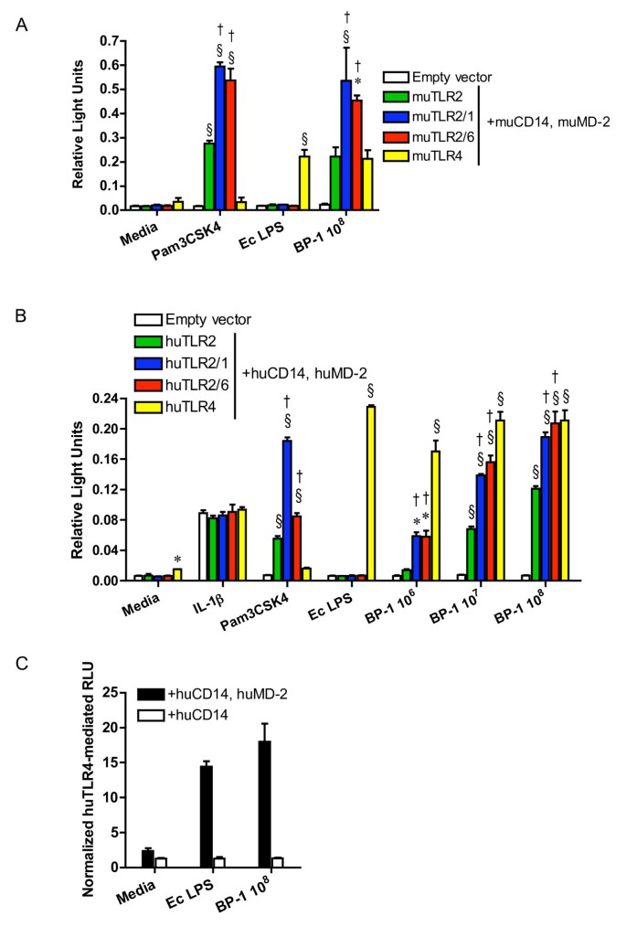
In order to identify TLRs that recognize Bp, HEK293 cells transiently transfected with plasmids expressing murine TLR2, TLR2/1, TLR2/6, or TLR4, and co-receptors CD14 and MD-2 were stimulated with a heat-killed Bp clinical isolate BP-1 (Figure 1A). Using an NF-κB-dependent luciferase reporter assay, strong _Bp_-induced NF-κB activation was detected in TLR2- and TLR4-transfected cells with augmentation of the TLR2-dependent signal in the presence of TLR1 or TLR6. To determine whether similar signaling is triggered by human receptors in response to Bp, HEK293 cells transfected with plasmids expressing human TLRs were also stimulated with Bp (Figure 1B). TLR2- and TLR4-mediated NF-κB activation was observed in a largely dose dependent fashion. Both TLR1 and TLR6 augmented the TLR2-mediated response. We also transfected a hypo-responsive variant human TLR1 plasmid in addition to TLR2 in this system and observed little amplification of the TLR2-dependent signal upon stimulation with Bp (data not shown). Because MD-2 is a necessary molecule for TLR4 signaling, human TLR4 transfection was repeated without co-transfecting MD-2. In the absence of MD-2, a TLR4-mediated signal in response to Bp was not detected (Figure 1C) although TLR2-mediated signaling was not altered (data not shown).
Figure 1
B. pseudomallei activates TLR2 and TLR4. HEK293 cells were transiently transfected with (A) murine or (B) human TLR2, TLR2/1, TLR2/6, or TLR4; co-receptors CD14 and MD-2; and NF-κB-dependent firefly ELAM luciferase and control β-actin-dependent Renilla luciferase. In (C) HEK293 cells were transiently transfected with human TLR4 and co-receptor CD14 with or without MD-2; and firefly ELAM and Renilla luciferases. Cells were stimulated with media alone, (B) IL-1β 20 ng/mL, (A & B) Pam3CSK4 100 ng/mL, E. coli 0111:B4 LPS 10 ng/mL, or heat-killed BP-1 at various concentrations in CFU/mL. NF-κB activation was measured by light emission (relative light units). In (A&B) data plotted are means ± standard deviations of triplicate conditions. In (C) the means ± standard deviations of triplicate human TLR4-mediated relative light units (normalized to mean empty vector values) are plotted from parallel experiments with or without co-transfection of MD-2. For (A & B) * indicates p < 0.05 and § indicates p = 0.001 compared with empty vector stimulated with the same ligand, and † indicates p < 0.05 compared with TLR2 stimulated with the same ligand using analysis of variance followed by the Bonferroni post-test. Other comparisons are not shown for clarity. The data in (A) represent one of two independently performed experiments. The data in (B) represent one of three independently performed experiments, but the data displayed, in contrast to the two other experiments, show the response to a hyper-responding variant TLR1 plasmid. The data in (C) represent one of three independently performed experiments.
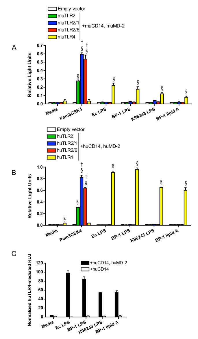
Since robust TLR4-dependent NF-κB activation was observed using heat-killed Bp, we sought to identify which Bp ligand activates TLR4 signaling. As Bp LPS was the most likely candidate, LPS was isolated and purified from the Bp clinical isolate BP-1. LPS from the Bp reference strain K96243 was acquired and also purified. Moreover, because lipid A is the specific component of LPS that activates TLR4, lipid A was isolated from BP-1. HEK293 cells transiently transfected with murine or human TLR2, TLR2/1, TR2/6, or TLR4, and co-receptors CD14 and MD-2 were stimulated with these ligands (Figure 2A &2B). In both murine- and human-transfected cells, robust TLR4-dependent NF-κB activation was identified in response to LPS from both Bp strains and in response to BP-1 lipid A without evidence of TLR2 stimulation by these ligands. Additionally, identical stimulations with Bp LPS and BP-1 lipid A of cells transfected with human TLR4 and CD14 in the absence of MD-2 were performed, and no TLR4-mediated NF-κB activation was observed (Figure 2C).
Figure 2
B. pseudomallei LPS signals via TLR4. HEK293 cells were transiently transfected with (A) murine or (B) human TLR2, TLR2/1, TLR2/6, or TLR4; co-receptors CD14 and MD-2; and NF-κB-dependent firefly ELAM luciferase and control β-actin-dependent Renilla luciferase. In (C) HEK293 cells were transiently transfected with human TLR4 and co-receptor CD14 with or without MD-2; and firefly ELAM and Renilla luciferases. Cells were stimulated with media alone, (A & B) Pam3CSK4 1000 ng/mL, E. coli 0111:B4 LPS 10 ng/mL, BP-1 LPS 10 ng/mL, K96243 LPS 10 ng/mL, or BP-1 lipid A 10 ng/mL. NF-κB activation was measured by light emission (relative light units). In (A&B) data plotted are means ± standard deviations of duplicate or triplicate conditions. In (C) the means ± standard deviations of triplicate human TLR4-mediated relative light units (normalized to mean empty vector values) are plotted from parallel experiments with or without co-transfection of MD-2. For (A & B) § indicates p < 0.001 compared with empty vector stimulated with the same ligand, and † indicates p < 0.001 compared with TLR2 stimulated with the same ligand using analysis of variance followed by the Bonferroni post-test. Other comparisons are not shown for clarity. Each graph represents one of two independently performed experiments.
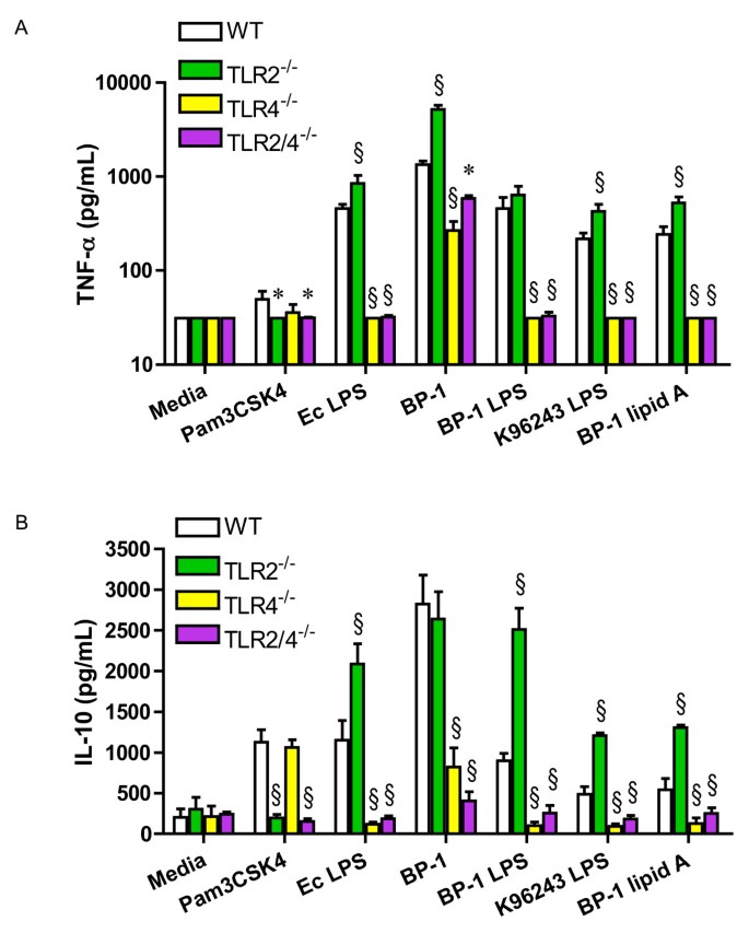
These observations in a transfected cell line prompted us to examine the cytokine response to Bp and Bp LPS in primary cells. Therefore, bone marrow-derived macrophages from wild type, TLR2-/-, TLR4-/-, and TLR2/4-/- mice were stimulated with heat-killed BP-1, BP-1 LPS, K96243 LPS, or BP-1 lipid A. Pro-inflammatory cytokine TNF-α was quantified in cell supernatants by ELISA after 24 hours of incubation (Figure 3A). Compared to wild type cells, TNF-α production by TLR4-/- cells in response to BP-1 stimulation was significantly reduced, but was completely eliminated in response to BP-1 LPS, K96243 LPS, or BP-1 lipid A. In TLR2-/- cells, however, TNF-α production after stimulation with BP-1 was either the same as or greater than that produced by wild type cells in several different experiments (3.8 fold greater in the experiment shown). TLR2-/- cells invariably produced more TNF-α than wild type cells after stimulation with BP-1 LPS, K96243 LPS, or BP-1 lipid A (1.4, 2.0, or 2.2 fold greater, respectively, in the experiment shown). Furthermore, TLR2/4-/- cells demonstrated only partial reduction in TNF-α production compared to TLR4-/- cells stimulated with BP-1. Chemokine MIP-2 was also measured in the supernatants of wild type, TLR2-/-, TLR4-/-, and TLR2/4-/- cells stimulated with the same ligands, and a similar pattern to TNF-α production was observed (data not shown). A higher concentration of Bp LPS than control Escherichia coli LPS was used in these experiments because in a separate dose response experiment stimulating wild type macrophages, both strains of Bp LPS yielded several fold less TNF-α production than similar concentrations of E. coli LPS (data not shown).
Figure 3
Absence of TLR4, but not TLR2, impairs the macrophage cytokine response to B. pseudomallei. Bone marrow harvested from wild type, TLR2-/-, TLR4-/-, or TLR2/4-/- mice was cultured in the presence of L929 cell conditioned media for 5–10 days to promote differentiation of macrophages before plating and stimulating with media alone, Pam3CSK4 1000 ng/mL, E. coli 0111:B4 LPS 100 ng/mL, heat-killed BP-1 at a bacteria to cell ratio of 100, BP-1 LPS 1000 ng/mL, K96243 LPS 1000 ng/mL, or BP-1 lipid A 1000 ng/mL. Supernatants were harvested after 24 hours and (A) TNF-α or (B) IL-10 production was measured by ELISA. Data plotted are means ± standard deviations of quadruplicate samples. * indicates p < 0.05 and § indicates p = 0.001 compared with wild type cells stimulated with the same ligand, using analysis of variance followed by the Bonferroni post-test. Other comparisons are not shown for clarity. The TNF-α data displayed are from one of six independently performed experiments stimulating various combinations of wild type, TLR2-/-, TLR4-/-, or TLR2/4-/- macrophages with these ligands, and measuring TNF-α. In one of three experiments comparing cytokine responses from TLR2-/- macrophages to wild type macrophages, production of TNF-α in response to heat-killed Bp was not significantly increased. The IL-10 data displayed are from one of five independently performed experiments stimulating various combinations of wild type, TLR2-/-, TLR4-/-, or TLR2/4-/- macrophages with these ligands, and measuring IL-10. In two of three experiments comparing cytokine responses from TLR2-/- macrophages to wild type macrophages, production of IL-10 in response to heat-killed Bp was not significantly increased.
The findings indicating augmented pro-inflammatory cytokine production in the absence of TLR2 when cells were stimulated with Bp, Bp LPS, and Bp lipid A led us to evaluate whether this was an IL-10-mediated phenomenon. IL-10 is an anti-inflammatory cytokine that may be released by cells upon TLR2 stimulation [18]. We therefore measured IL-10 in cell supernatants stimulated with heat-killed BP-1, BP-1 LPS, K96243 LPS, or BP-1 lipid A (Figure 3B). However, we did not identify any impairment in IL-10 production by TLR2-/- cells in response to these ligands. In general, the overall pattern of IL-10 production was again similar to TNF-α production, with augmentation of BP-1 LPS-, K96243 LPS-, or BP-1 lipid A-induced IL-10 release in the absence of TLR2.
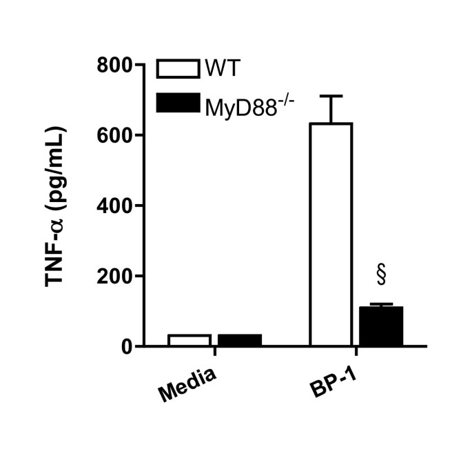
As the TLR adaptor molecule myeloid differentiation factor 88 (MyD88) mediates both TLR2 and TLR4 signaling, MyD88-/- bone marrow-derived macrophages were therefore stimulated with heat-killed BP-1 and cytokine production assayed after 24 hours (Figure 4). Compared to wild type cells, TNF-α production by MyD88-/- macrophages was markedly curtailed.
Figure 4
MyD88 regulates TNF-α production by macrophages stimulated with B. pseudomallei. Bone marrow harvested from wild type or MyD88-/- mice was cultured in the presence of L929 cell conditioned media for 5–10 days to promote differentiation of macrophages before plating and stimulating with media alone or heat-killed BP-1 at a bacteria to cell ratio of 100. Supernatants were harvested after 24 hours and TNF-α production was measured by ELISA. Data plotted are means ± standard deviations of quadruplicate samples. § indicates p < 0.001 compared with wild type cells, using the t test. The data displayed represent one of two independently performed experiments.
Discussion
In this report, we demonstrate that heat-killed Bp activates TLR2 and TLR4. We further show that Bp LPS and Bp lipid A are TLR4 ligands and we identify augmented macrophage cytokine production induced by Bp, Bp LPS or Bp lipid A in the absence of TLR2.
TLRs are innate immune receptors that recognize conserved pathogen motifs and initiate an inflammatory signaling cascade. We and others have demonstrated the essential roles of TLRs in host defense against a variety of pulmonary pathogens [5–9]. TLR2 is a promiscuous receptor that generally recognizes bacterial lipotechoic acid, peptidoglycans, or lipoproteins and functions as a heterodimer with either TLR1 or TLR6 [19–22]. LPS is the prototypical bacterial ligand for TLR4 [23] and is comprised of a core polysaccharide, polysaccharide side chains (the O-antigen) and lipid A (endotoxin), a glucosamine-based phospholipid. Lipid A usually interacts with TLR4 in addition to proteins CD14 and MD-2 to induce a pro-inflammatory signal [24]. MD-2 is a soluble molecule that is associated with the extracellular domain of TLR4 and is generally regarded as essential for LPS recognition by TLR4 [25–28]. Both TLR2 and TLR4 are therefore likely to initiate host defense signaling against Bp, a Gram-negative pathogen.
As expected, our gain-of-function studies using murine and human constructs confirm a role for TLR2, in conjunction with TLR1 or TLR6, in initiating inflammatory signaling in response to heat-killed Bp. As TLR2 forms heterodimers with TLR1 or TLR6, the signal detected after transfection of TLR2 alone is likely explained by low level endogenous expression of TLR1 in HEK293 cells [22, 29]. The role of TLR1 in facilitating TLR2-mediated NF-κB activation in response to Bp is further elucidated by our observation of minimal augmentation of the TLR2-dependent signal when we transfected a known hypo-responding variant human TLR1 plasmid [17].
We also show that heat-killed Bp reliably signals via TLR4 and that Bp LPS isolated from two separate bacterial strains activates NF-κB in a uniquely TLR4-mediated fashion. We further demonstrate that Bp LPS subcomponent lipid A is a TLR4 ligand in cells transfected with either human or murine TLR constructs. Our observations that Bp LPS and lipid A are TLR4 ligands differ from the results of Wiersinga et al. [10], who used HEK293 cells stably transfected with TLR2/CD14 or TLR4/CD14 to show that Bp LPS signals through TLR2 but not TLR4. These differences may be explained by our co-transfection of MD-2, a molecule that is not endogenously expressed by HEK293 cells [29], but that we demonstrate is essential for TLR4-dependent signaling in response to Bp, Bp LPS, or Bp lipid A. While the LPS of a several bacteria signal via TLR2 rather than TLR4 [30–33], some LPS preparations that appear to stimulate TLR2 may be contaminated with lipopeptides [34]. Alternatively, there may be strain-specific differences in Bp LPS structure that influence recognition by TLR2 and TLR4. Although our results indicating roles for TLR2 and TLR4 in this gain-of-function system used heat-killed Bp, they corroborate recent work with live Bp by others [35].
Our studies using primary murine cells provide additional evidence that Bp induces a TLR4-mediated inflammatory cytokine response and that Bp LPS and lipid A are TLR4 ligands. We demonstrate augmented _Bp_-induced TNF-α production in the absence of TLR2 alone and in the contemporaneous absence of TLR4. This differs from the TLR2-dependent TNF-α production by alveolar macrophages or whole blood stimulated with Bp by Wiersinga et al. [10] and may be attributable to the different cell types or bacterial strains used. However, our observations of enhanced cytokine production support their findings of reduced bacterial loads and improved survival in respiratory infection in TLR2-/- mice compared to wild type mice. We also observed consistent augmentation in cytokine production in TLR2-/- macrophages compared to wild type macrophages upon stimulation with Bp LPS or with ultrapure E. coli LPS, the control ligand. We have observed this phenomenon in previous studies of intracellular TNF-α production by primary bone marrow cells stimulated with Salmonella minnesota Re595 LPS, including in the absence of differential TLR7-dependent signaling between wild type and TLR2-/- cells [7]. Others have described a role for TLR2 agonists in suppressing TLR4-mediated inflammatory cytokine production [18] and a role for TLR4 in contributing to pulmonary inflammation in response to the TLR2 ligand lipoteichoic acid [36]. There are also other examples of cooperative TLR2 and TLR4 signaling in host defense [37, 38]. However, we are not aware of specific descriptions of the absence of TLR2 influencing the inflammatory response to TLR4-specific stimulation alone. The mechanisms underlying this amplifying effect of TLR2 deficiency upon TLR4-mediated cytokine production by Bp LPS are unclear. If the presence of TLR2 is indeed suppressive, our data using LPS suggests that mere expression of the receptor rather than activation may be adequate to modify TLR4-specific signaling. Other reports indicate that TLR2 stimulation may induce production of the anti-inflammatory cytokine IL-10, promoting a Th2-type response [18, 39–42]. However, our data do not support this mechanism of TLR2-mediated pro-inflammatory cytokine suppression. To the contrary, we show that Bp LPS-induced IL-10 production is augmented in the absence of TLR2, and, despite some differences largely attributable to experimental variability, generally mirrors the pattern of pro-inflammatory cytokine release observed. It may be that in the absence of TLR2, shared signaling molecules are more available to enhance activation of other TLRs, including TLR4. Further investigation will be required to elucidate the interaction of TLR2 and TLR4 in mediating recognition of Bp.
Both TLR2 and TLR4 signal via the adaptor molecule MyD88, although TLR4 may also signal in a MyD88-independent fashion via adaptor molecule TIR-domain-containing adapter-inducing interferon-β (TRIF) [43]. Our observations that pro-inflammatory cytokine production by heat-killed Bp is largely abolished in the absence of MyD88 whereas TLR2/4-/- cells demonstrate only partial impairment in TNF-α release suggest that other MyD88-dependent receptors may be important in host recognition of Bp. Specifically, TLR5 and TLR9, receptors that recognize bacterial flagellin and CpG DNA, respectively, may account for some of this residual MyD88-dependent response. TRIF-mediated TLR4 signaling may also play a role in the innate immune response to Bp infection. These additional host defense pathways require further study. Importantly, our experiments have used only heat-killed Bp and we cannot discount differential signaling induced by live Bp. Thus, additional investigations with live bacteria are warranted.
Our experiments also indicate that Bp LPS is a weaker inducer of TNF-α production in bone marrow-derived macrophages than E. coli LPS. Others have shown that Bp LPS is poorly pyrogenic but more mitogenic compared to Salmonella abortus equi [44]. Bp LPS may contribute to bacterial pathogenesis by modulating the host response and inhibiting macrophage killing [45]. Although Bp LPS is antigenically indistinguishable from the less virulent organism Burkholderia thailandensis [46], we have observed that Bp LPS may be a stronger inducer of pro-inflammatory cytokine production in macrophages than B. thailandensis LPS (unpublished data). Structural analysis of LPS preparations from these Burkholderia strains is required.
Conclusion
Bp activates TLR2 and TLR4. In the presence of MD-2, Bp LPS and lipid A are TLR4 ligands. Although the macrophage cytokine response to Bp LPS or lipid A is completely dependent on TLR4, in TLR2-/- macrophages stimulated with Bp, Bp LPS or lipid A, cytokine production is augmented. Other MyD88-dependent signaling pathways may also be important in the host response to Bp infection. These findings provide new insights into critical mechanisms of host defense in melioidosis.
Abbreviations
Bp:
Burkholderia pseudomallei
LPS:
Lipopolysaccharide
MyD88:
Myeloid differentiation factor 88
TLR:
Toll-like receptor
TRIF:
TIR-domain-containing adapter-inducing interferon-β
References
- White NJ: Melioidosis. Lancet. 2003, 361 (9370): 1715-1722. 10.1016/S0140-6736(03)13374-0.
Article CAS PubMed Google Scholar - Wiersinga WJ, van der Poll T, White NJ, Day NP, Peacock SJ: Melioidosis: insights into the pathogenicity of Burkholderia pseudomallei. Nat Rev Microbiol. 2006, 4 (4): 272-282. 10.1038/nrmicro1385.
Article CAS PubMed Google Scholar - Kanaphun P, Thirawattanasuk N, Suputtamongkol Y, Naigowit P, Dance DA, Smith MD, White NJ: Serology and carriage of Pseudomonas pseudomallei: a prospective study in 1000 hospitalized children in northeast Thailand. J Infect Dis. 1993, 167 (1): 230-233.
Article CAS PubMed Google Scholar - Akira S, Takeda K, Kaisho T: Toll-like receptors: critical proteins linking innate and acquired immunity. Nat Immunol. 2001, 2 (8): 675-680. 10.1038/90609.
Article CAS PubMed Google Scholar - Hawn TR, Smith KD, Aderem A, Skerrett SJ: Myeloid differentiation primary response gene (88)- and toll-like receptor 2-deficient mice are susceptible to infection with aerosolized Legionella pneumophila. J Infect Dis. 2006, 193 (12): 1693-1702. 10.1086/504525.
Article CAS PubMed Google Scholar - Hajjar AM, Harowicz H, Liggitt HD, Fink PJ, Wilson CB, Skerrett SJ: An essential role for non-bone marrow-derived cells in control of Pseudomonas aeruginosa pneumonia. Am J Respir Cell Mol Biol. 2005, 33 (5): 470-475. 10.1165/rcmb.2005-0199OC.
Article PubMed Central CAS PubMed Google Scholar - Skerrett SJ, Wilson CB, Liggitt HD, Hajjar AM: Redundant Toll-like receptor signaling in the pulmonary host response to Pseudomonas aeruginosa. Am J Physiol Lung Cell Mol Physiol. 2007, 292 (1): L312-22. 10.1152/ajplung.00250.2006.
Article CAS PubMed Google Scholar - Hawn TR, Verbon A, Janer M, Zhao LP, Beutler B, Aderem A: Toll-like receptor 4 polymorphisms are associated with resistance to Legionnaires' disease. Proc Natl Acad Sci U S A. 2005, 102 (7): 2487-2489. 10.1073/pnas.0409831102.
Article PubMed Central CAS PubMed Google Scholar - Dessing MC, Schouten M, Draing C, Levi M, von Aulock S, van der Poll T: Role played by Toll-like receptors 2 and 4 in lipoteichoic acid-induced lung inflammation and coagulation. J Infect Dis. 2008, 197 (2): 245-252. 10.1086/524873.
Article CAS PubMed Google Scholar - Wiersinga WJ, Wieland CW, Dessing MC, Chantratita N, Cheng AC, Limmathurotsakul D, Chierakul W, Leendertse M, Florquin S, de Vos AF, White N, Dondorp AM, Day NP, Peacock SJ, van der Poll T: Toll-Like Receptor 2 Impairs Host Defense in Gram-Negative Sepsis Caused by Burkholderia pseudomallei (Melioidosis). PLoS Med. 2007, 4 (7): e248-10.1371/journal.pmed.0040248.
Article PubMed Central PubMed Google Scholar - Westphal O, Jann K: Bacterial lipopolysaccharides: extraction with phenol–water and further applications of the procedure. Methods in Carbohydrate Chemistry. 1965, 5: 83-91.
CAS Google Scholar - Fischer W, Koch HU, Haas R: Improved preparation of lipoteichoic acids. Eur J Biochem. 1983, 133 (3): 523-530. 10.1111/j.1432-1033.1983.tb07495.x.
Article CAS PubMed Google Scholar - Folch J, Lees M, Sloane Stanley GH: A simple method for the isolation and purification of total lipides from animal tissues. J Biol Chem. 1957, 226 (1): 497-509.
CAS PubMed Google Scholar - Hirschfeld M, Ma Y, Weis JH, Vogel SN, Weis JJ: Cutting edge: repurification of lipopolysaccharide eliminates signaling through both human and murine toll-like receptor 2. J Immunol. 2000, 165 (2): 618-622.
Article CAS PubMed Google Scholar - Caroff M, Tacken A, Szabo L: Detergent-accelerated hydrolysis of bacterial endotoxins and determination of the anomeric configuration of the glycosyl phosphate present in the "isolated lipid A" fragment of the Bordetella pertussis endotoxin. Carbohydr Res. 1988, 175 (2): 273-282. 10.1016/0008-6215(88)84149-1.
Article CAS PubMed Google Scholar - Hajjar AM, Harvey MD, Shaffer SA, Goodlett DR, Sjostedt A, Edebro H, Forsman M, Bystrom M, Pelletier M, Wilson CB, Miller SI, Skerrett SJ, Ernst RK: Lack of In Vitro and In Vivo Recognition of Francisella tularensis Subspecies Lipopolysaccharide by Toll-Like Receptors. Infect Immun. 2006, 74 (12): 6730-6738. 10.1128/IAI.00934-06.
Article PubMed Central CAS PubMed Google Scholar - Hawn TR, Misch EA, Dunstan SJ, Thwaites GE, Lan NT, Quy HT, Chau TT, Rodrigues S, Nachman A, Janer M, Hien TT, Farrar JJ, Aderem A: A common human TLR1 polymorphism regulates the innate immune response to lipopeptides. Eur J Immunol. 2007, 37 (8): 2280-2289. 10.1002/eji.200737034.
Article CAS PubMed Google Scholar - Re F, Strominger JL: IL-10 released by concomitant TLR2 stimulation blocks the induction of a subset of Th1 cytokines that are specifically induced by TLR4 or TLR3 in human dendritic cells. J Immunol. 2004, 173 (12): 7548-7555.
Article CAS PubMed Google Scholar - Triantafilou M, Gamper FG, Haston RM, Mouratis MA, Morath S, Hartung T, Triantafilou K: Membrane sorting of toll-like receptor (TLR)-2/6 and TLR2/1 heterodimers at the cell surface determines heterotypic associations with CD36 and intracellular targeting. J Biol Chem. 2006, 281 (41): 31002-31011. 10.1074/jbc.M602794200.
Article CAS PubMed Google Scholar - Ozinsky A, Underhill DM, Fontenot JD, Hajjar AM, Smith KD, Wilson CB, Schroeder L, Aderem A: The repertoire for pattern recognition of pathogens by the innate immune system is defined by cooperation between toll-like receptors. Proc Natl Acad Sci U S A. 2000, 97 (25): 13766-13771. 10.1073/pnas.250476497.
Article PubMed Central CAS PubMed Google Scholar - Takeuchi O, Kawai T, Muhlradt PF, Morr M, Radolf JD, Zychlinsky A, Takeda K, Akira S: Discrimination of bacterial lipoproteins by Toll-like receptor 6. Int Immunol. 2001, 13 (7): 933-940. 10.1093/intimm/13.7.933.
Article CAS PubMed Google Scholar - Takeuchi O, Sato S, Horiuchi T, Hoshino K, Takeda K, Dong Z, Modlin RL, Akira S: Cutting edge: role of Toll-like receptor 1 in mediating immune response to microbial lipoproteins. J Immunol. 2002, 169 (1): 10-14.
Article CAS PubMed Google Scholar - Hoshino K, Takeuchi O, Kawai T, Sanjo H, Ogawa T, Takeda Y, Takeda K, Akira S: Cutting edge: Toll-like receptor 4 (TLR4)-deficient mice are hyporesponsive to lipopolysaccharide: evidence for TLR4 as the Lps gene product. J Immunol. 1999, 162 (7): 3749-3752.
CAS PubMed Google Scholar - Raetz CR, Whitfield C: Lipopolysaccharide endotoxins. Annu Rev Biochem. 2002, 71: 635-700. 10.1146/annurev.biochem.71.110601.135414.
Article PubMed Central CAS PubMed Google Scholar - Shimazu R, Akashi S, Ogata H, Nagai Y, Fukudome K, Miyake K, Kimoto M: MD-2, a molecule that confers lipopolysaccharide responsiveness on Toll-like receptor 4. J Exp Med. 1999, 189 (11): 1777-1782. 10.1084/jem.189.11.1777.
Article PubMed Central CAS PubMed Google Scholar - Nagai Y, Akashi S, Nagafuku M, Ogata M, Iwakura Y, Akira S, Kitamura T, Kosugi A, Kimoto M, Miyake K: Essential role of MD-2 in LPS responsiveness and TLR4 distribution. Nat Immunol. 2002, 3 (7): 667-672.
CAS PubMed Google Scholar - Schromm AB, Lien E, Henneke P, Chow JC, Yoshimura A, Heine H, Latz E, Monks BG, Schwartz DA, Miyake K, Golenbock DT: Molecular genetic analysis of an endotoxin nonresponder mutant cell line: a point mutation in a conserved region of MD-2 abolishes endotoxin-induced signaling. J Exp Med. 2001, 194 (1): 79-88. 10.1084/jem.194.1.79.
Article PubMed Central CAS PubMed Google Scholar - Miyake K, Ogata H, Nagai Y, Akashi S, Kimoto M: Innate recognition of lipopolysaccharide by Toll-like receptor 4/MD-2 and RP105/MD-1. J Endotoxin Res. 2000, 6 (5): 389-391.
Article CAS PubMed Google Scholar - Massari P, Visintin A, Gunawardana J, Halmen KA, King CA, Golenbock DT, Wetzler LM: Meningococcal porin PorB binds to TLR2 and requires TLR1 for signaling. J Immunol. 2006, 176 (4): 2373-2380.
Article CAS PubMed Google Scholar - Werts C, Tapping RI, Mathison JC, Chuang TH, Kravchenko V, Saint Girons I, Haake DA, Godowski PJ, Hayashi F, Ozinsky A, Underhill DM, Kirschning CJ, Wagner H, Aderem A, Tobias PS, Ulevitch RJ: Leptospiral lipopolysaccharide activates cells through a TLR2-dependent mechanism. Nat Immunol. 2001, 2 (4): 346-352. 10.1038/86354.
Article CAS PubMed Google Scholar - Martin M, Katz J, Vogel SN, Michalek SM: Differential induction of endotoxin tolerance by lipopolysaccharides derived from Porphyromonas gingivalis and Escherichia coli. J Immunol. 2001, 167 (9): 5278-5285.
Article CAS PubMed Google Scholar - Fuse ET, Tateda K, Kikuchi Y, Matsumoto T, Gondaira F, Azuma A, Kudoh S, Standiford TJ, Yamaguchi K: Role of Toll-like receptor 2 in recognition of Legionella pneumophila in a murine pneumonia model. J Med Microbiol. 2007, 56 (Pt 3): 305-312. 10.1099/jmm.0.46913-0.
Article CAS PubMed Google Scholar - Darveau RP, Pham TT, Lemley K, Reife RA, Bainbridge BW, Coats SR, Howald WN, Way SS, Hajjar AM: Porphyromonas gingivalis lipopolysaccharide contains multiple lipid A species that functionally interact with both toll-like receptors 2 and 4. Infect Immun. 2004, 72 (9): 5041-5051. 10.1128/IAI.72.9.5041-5051.2004.
Article PubMed Central CAS PubMed Google Scholar - Lee HK, Lee J, Tobias PS: Two lipoproteins extracted from Escherichia coli K-12 LCD25 lipopolysaccharide are the major components responsible for Toll-like receptor 2-mediated signaling. J Immunol. 2002, 168 (8): 4012-4017.
Article CAS PubMed Google Scholar - Hii CS, Sun GW, Goh JW, Lu J, Stevens MP, Gan YH: Interleukin-8 Induction by Burkholderia pseudomallei Can Occur without Toll-Like Receptor Signaling but Requires a Functional Type III Secretion System. J Infect Dis. 2008
Google Scholar - Knapp S, von Aulock S, Leendertse M, Haslinger I, Draing C, Golenbock DT, van der Poll T: Lipoteichoic Acid-Induced Lung Inflammation Depends on TLR2 and the Concerted Action of TLR4 and the Platelet-Activating Factor Receptor. J Immunol. 2008, 180 (5): 3478-3484.
Article CAS PubMed Google Scholar - Hirata N, Yanagawa Y, Ebihara T, Seya T, Uematsu S, Akira S, Hayashi F, Iwabuchi K, Onoe K: Selective synergy in anti-inflammatory cytokine production upon cooperated signaling via TLR4 and TLR2 in murine conventional dendritic cells. Mol Immunol. 2008
Google Scholar - Trinchieri G, Sher A: Cooperation of Toll-like receptor signals in innate immune defence. Nat Rev Immunol. 2007, 7 (3): 179-190. 10.1038/nri2038.
Article CAS PubMed Google Scholar - Redecke V, Hacker H, Datta SK, Fermin A, Pitha PM, Broide DH, Raz E: Cutting edge: activation of Toll-like receptor 2 induces a Th2 immune response and promotes experimental asthma. J Immunol. 2004, 172 (5): 2739-2743.
Article CAS PubMed Google Scholar - Agrawal S, Agrawal A, Doughty B, Gerwitz A, Blenis J, Van Dyke T, Pulendran B: Cutting edge: different Toll-like receptor agonists instruct dendritic cells to induce distinct Th responses via differential modulation of extracellular signal-regulated kinase-mitogen-activated protein kinase and c-Fos. J Immunol. 2003, 171 (10): 4984-4989.
Article CAS PubMed Google Scholar - Jotwani R, Pulendran B, Agrawal S, Cutler CW: Human dendritic cells respond to Porphyromonas gingivalis LPS by promoting a Th2 effector response in vitro. Eur J Immunol. 2003, 33 (11): 2980-2986. 10.1002/eji.200324392.
Article CAS PubMed Google Scholar - Qi H, Denning TL, Soong L: Differential induction of interleukin-10 and interleukin-12 in dendritic cells by microbial toll-like receptor activators and skewing of T-cell cytokine profiles. Infect Immun. 2003, 71 (6): 3337-3342. 10.1128/IAI.71.6.3337-3342.2003.
Article PubMed Central CAS PubMed Google Scholar - Akira S, Uematsu S, Takeuchi O: Pathogen recognition and innate immunity. Cell. 2006, 124 (4): 783-801. 10.1016/j.cell.2006.02.015.
Article CAS PubMed Google Scholar - Matsuura M, Kawahara K, Ezaki T, Nakano M: Biological activities of lipopolysaccharide of Burkholderia (Pseudomonas) pseudomallei. FEMS Microbiol Lett. 1996, 137 (1): 79-83. 10.1111/j.1574-6968.1996.tb08086.x.
Article CAS PubMed Google Scholar - Arjcharoen S, Wikraiphat C, Pudla M, Limposuwan K, Woods DE, Sirisinha S, Utaisincharoen P: Fate of a Burkholderia pseudomallei lipopolysaccharide mutant in the mouse macrophage cell line RAW 264.7: possible role for the O-antigenic polysaccharide moiety of lipopolysaccharide in internalization and intracellular survival. Infect Immun. 2007, 75 (9): 4298-4304. 10.1128/IAI.00285-07.
Article PubMed Central CAS PubMed Google Scholar - Anuntagool N, Intachote P, Wuthiekanun V, White NJ, Sirisinha S: Lipopolysaccharide from nonvirulent Ara+ Burkholderia pseudomallei isolates is immunologically indistinguishable from lipopolysaccharide from virulent Ara- clinical isolates. Clin Diagn Lab Immunol. 1998, 5 (2): 225-229.
PubMed Central CAS PubMed Google Scholar
Acknowledgements
TEW, RKE and SJS are supported by NIH award U54 AI057141. TEW is also supported by a Parker B. Francis Fellowship in Pulmonary Research.
We thank Dr. Lynn Hajjar for providing TLR plasmids and for critical review of the manuscript; Dr. Mark Wurfel for providing the hyper-responding variant human TLR1 plasmid; Dr. Chris Wilson for providing TLR- and MyD88-deficient mice; and the staff of the BSL-3 laboratories for facilitating work with Bp.
Author information
Authors and Affiliations
- Department of Medicine, University of Washington, Seattle, Washington, USA
T Eoin West, Robert K Ernst, Malinka J Jansson-Hutson & Shawn J Skerrett
Authors
- T Eoin West
- Robert K Ernst
- Malinka J Jansson-Hutson
- Shawn J Skerrett
Corresponding author
Correspondence toT Eoin West.
Additional information
Competing interests
The authors declare that they have no competing interests.
Authors' contributions
TEW, RKE and SJS designed the experiments. RKE extracted LPS and lipid A. MJJ and TEW performed the experiments. TEW and SJS analyzed the data. TEW wrote the manuscript with input from MJJ, RKE and SJS.
Authors’ original submitted files for images
Rights and permissions
This article is published under license to BioMed Central Ltd. This is an Open Access article distributed under the terms of the Creative Commons Attribution License (http://creativecommons.org/licenses/by/2.0), which permits unrestricted use, distribution, and reproduction in any medium, provided the original work is properly cited.
About this article
Cite this article
West, T.E., Ernst, R.K., Jansson-Hutson, M.J. et al. Activation of Toll-like receptors by Burkholderia pseudomallei.BMC Immunol 9, 46 (2008). https://doi.org/10.1186/1471-2172-9-46
- Received: 16 April 2008
- Accepted: 08 August 2008
- Published: 08 August 2008
- DOI: https://doi.org/10.1186/1471-2172-9-46


