v-SNARE Composition Distinguishes Synaptic Vesicle Pools (original) (raw)
. Author manuscript; available in PMC: 2012 Aug 11.
Summary
Synaptic vesicles belong to two distinct pools, a recycling pool responsible for the evoked release of neurotransmitter and a resting pool unresponsive to stimulation. The uniform appearance of synaptic vesicles has suggested that differences in location or cytoskeletal association account for these differences in function. We now find that the v-SNARE tetanus toxin-insensitive vesicle-associated membrane protein (VAMP7) differs from other synaptic vesicle proteins in its distribution to the two pools, providing the first evidence that they differ in molecular composition. We also find that both resting and recycling pools undergo spontaneous release, and when activated by deletion of the longin domain, VAMP7 influences the properties of release. Further, the endocytosis which follows evoked and spontaneous release differs in mechanism, and specific sequences confer targeting to the different vesicle pools. The results suggest that different endocytic mechanisms generate synaptic vesicles with different proteins which can endow the vesicles with distinct properties.
Introduction
The accumulation of synaptic vesicles at the nerve terminal enables the sustained release of neurotransmitter in response to persistent stimulation. However, not all synaptic vesicles contribute equally to evoked release. At most synapses, only a fraction of the synaptic vesicles present take up external tracers with stimulation, and this fraction has been termed the recycling pool (Harata et al., 2001; Rizzoli and Betz, 2005). Even after prolonged stimulation, a large proportion of synaptic vesicles at most boutons do not undergo exocytosis (Fernandez-Alfonso and Ryan, 2008), and the properties of this resting pool have remained elusive.
What accounts for the inability to release a large fraction of the synaptic vesicles at a presynaptic bouton? Resting pool vesicles may simply reside too far from the active zone, although previous work has shown that they intermingle with the recycling pool (Rizzoli and Betz, 2004). Differences in tethering to the cytoskeleton may influence vesicle mobilization by activity, and a number of proteins associated with the cytoskeleton, such as the synapsins, have been shown to influence release (Chi et al., 2001; Fenster et al., 2003; Leal-Ortiz et al., 2008; Takao-Rikitsu et al., 2004). Recent work has also suggested a role for regulation of the recycling pool by cyclin-dependent kinase 5 (cdk5) (Kim and Ryan, 2010). Consistent with a role for extrinsic factors in pool identity, synaptic vesicles within a single bouton generally appear homogeneous, and multiple synaptic vesicle proteins localize in similar proportions to recycling and resting pools (Fernandez-Alfonso and Ryan, 2008). Alternatively, intrinsic differences in molecular composition may account for the distinct behavior of recycling and resting pool vesicles. Previous work has indeed shown that synaptic vesicles recycle by multiple mechanisms (Glyvuk et al., 2010; Newell-Litwa et al., 2007; Takei et al., 1996; Zhang et al., 2009), raising the possibility that these pathways produce vesicles with different proteins.
Synaptic vesicles can recycle through an endosomal intermediate (Heuser and Reese, 1973; Hoopmann et al., 2010) as well as directly from the plasma membrane, through clathrin-dependent endocytosis (Takei et al., 1996). Synaptic vesicle formation from endosomes depends on the endosomal heterotetrameric adaptor proteins AP-3 and possibly AP-1 (Blumstein et al., 2001; Faundez et al., 1998; Glyvuk et al., 2010) rather than the related but distinct plasma membrane clathrin adaptor AP-2 (Di Paolo and De Camilli, 2006; Kim and Ryan, 2009). Although these pathways are all considered to generate the same synaptic vesicles, blocking the AP-1/3 pathway increases transmitter release at hippocampal synapses as well as at the neuromuscular junction (Polo-Parada et al., 2001; Voglmaier et al., 2006), suggesting diversion of synaptic vesicle components from a pathway that produces vesicles with a low probability of release to one that generates vesicles with a higher release probability. AP-3 (and AP-1) may therefore produce synaptic vesicles of the resting pool, and AP-2 vesicles of the recycling pool (Voglmaier and Edwards, 2007). This hypothesis predicts that since many synaptic vesicle proteins target in similar proportions to recycling and resting pools, they should use both AP-2 and AP-3 pathways. However, it also predicts that a protein preferentially dependent on one of these pathways should target more specifically to one of the pools and so differ from other synaptic vesicle proteins in its response to stimulation.
Results
To identify proteins that might depend more specifically on AP-3 for sorting to synaptic vesicles, we relied on observations made using AP-3-deficient mocha mice (Kantheti et al., 1998). Mocha mice show only a mild alteration in short-term synaptic plasticity, with no obvious reduction in the number of synaptic vesicles, or the localization of most synaptic vesicle proteins (Voglmaier et al., 2006; Vogt et al., 2000). However, a subset of synaptic vesicle proteins depend strongly on AP-3 for localization to synaptic vesicles. These include the zinc transporter ZnT3 and the tetanus toxin-insensitive vesicle-associated membrane protein (TI-VAMP or VAMP7), a v-SNARE implicated in membrane fusion (Kantheti et al., 1998; Salazar et al., 2004; Scheuber et al., 2006). We hypothesize that as proteins specifically dependent on AP-3, ZnT3 and VAMP7 may target to synaptic vesicles of the resting pool, and therefore with a low probability of release.
VAMP7-pHluorin preferentially labels synaptic vesicles unresponsive to stimulation
To assess the availability of VAMP7 for regulated exocytosis, we fused the short, lumenal C-terminus of VAMP7 to the modified green fluorescent protein ecliptic pHluorin (Alberts et al., 2006). Shifted in its pH sensitivity relative to GFP, pHluorin exhibits essentially complete fluorescence quenching at the low pH inside synaptic vesicles (Miesenböck et al., 1998; Sankaranarayanan et al., 2000). The fluorescence of pHluorin thus increases with exocytosis due to the increase in pH that accompanies exposure at the cell surface. The reacidification that rapidly follows endocytosis in turn results in fluorescence quenching, and alkalinization of the nerve terminal with a permeant weak base reveals the total pool of fluorescent protein. Transfected into hippocampal neurons, VAMP7-pHluorin colocalizes partially with the endogenous synaptic vesicle proteins SV2 and VAMP2 (Figure S1A), consistent with previous work localizing VAMP7 to synaptic vesicles as well as other endosomal membranes (Muzerelle et al., 2003; Salazar et al., 2006; Scheuber et al., 2006). Alkalinization with the permeant weak base NH4Cl increases the fluorescence of VAMP7-pHluorin (Figure 1A,B), confirming expression within an acidic, intracellular compartment, and determination of the lumenal pH using the pHluorin fusions (Mitchell and Ryan, 2004) shows no difference between vesicles containing VGLUT1 and VAMP7 at synaptic sites (Figure S1B). The surface fraction of VAMP7-pHluorin is higher than for VGLUT1 (Figure S1C), but this does not reflect over-expression (Figure S1D), and previous work has shown a similar surface fraction for endogenous as well as transfected synaptic vesicle proteins VAMP2 and synaptotagmin 1 (Dittman and Kaplan, 2006; Fernandez-Alfonso et al., 2006; Wienisch and Klingauf, 2006). Field stimulation increases the fluorescence of VAMP7-pHluorin at several boutons (Figure 1B), further supporting the localization of at least a fraction of VAMP7 to synaptic vesicles. However, identifying boutons simply based on their response to stimulation may exclude others that do not respond, even if they express VAMP7-pHluorin. On the other hand, identifying sites of intracellular VAMP7-pHluorin accumulation using NH4Cl will not discriminate between presynaptic and other sites where VAMP7 is known to localize (Coco et al., 1999).
Figure 1. VAMP7-pHluorin shows less response to electrical stimulation than VGLUT1-pHluorin.
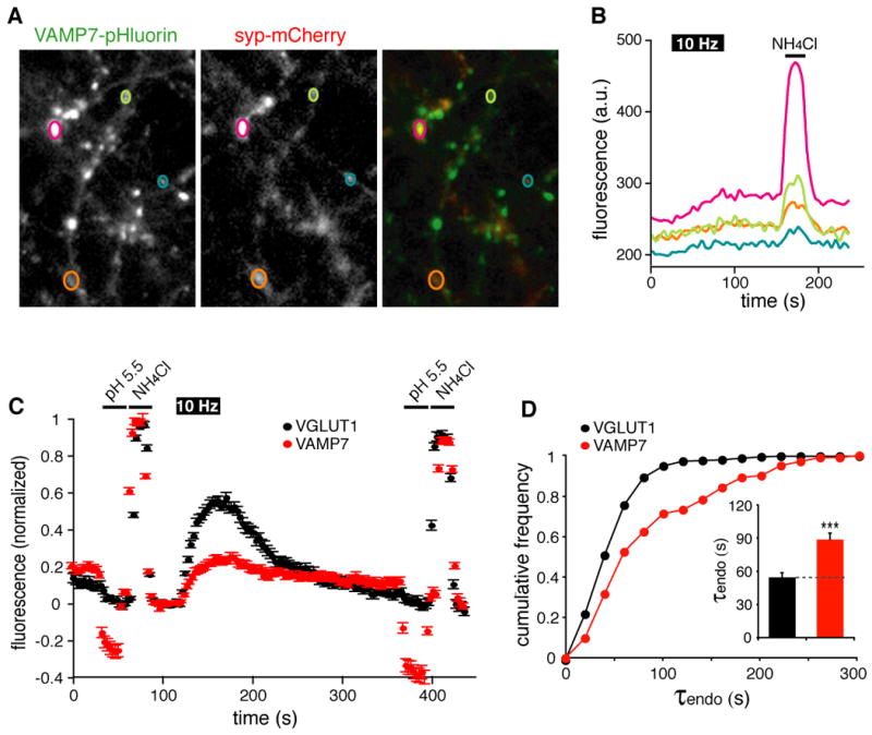
VAMP7-pHluorin-IRES2-synaptophysin(syp)-mCherry was transfected into primary hippocampal neurons, and imaged at 12-14 days in vitro. (A) Using NH4Cl (50 mM) to reveal the entire intracellular pool of pHluorin fusion, VAMP7-pHluorin and syp-mCherry partially colocalize. Synaptic boutons were selected for analysis on the sole basis of syp-mCherry fluorescence. (B) Response of VAMP7-pHluorin at individual boutons indicated in (A) to field stimulation at 10 Hz for 60 s, followed by NH4Cl. (C) Normalized to total internal pHluorin detected in NH4Cl, VAMP7 (red circles) shows more quenching at low external pH (5.5) than VGLUT1 (black circles), indicating a high surface fraction of VAMP7 (see also Figure S1C,D). In addition, VAMP7-pHluorin responds more weakly to stimulation than VGLUT1-pHluorin, and the recovery in fluorescence after stimulation is both delayed and incomplete. Since the VAMP7-pHluorin signal persistent after stimulation can be quenched by external MES buffer at pH 5.5, it reflects a delay in endocytosis rather than acidification. n=144 boutons from 3 coverslips for each construct (D) Analyzed by both cumulative frequency distribution and a direct comparison of the means (inset), the time constant for endocytosis after stimulation (τendo) is significantly slower for VAMP7-pHluorin than for VGLUT1-pHluorin. ***, p < 0.001; n = 5 coverslips containing a total of 289 boutons for VGLUT1 and 102 boutons for VAMP7. See also Figure S1.
To identify presynaptic boutons in an unbiased manner, we used an internal ribosome entry site to coexpress a monomeric form of the fluorescent Cherry protein (Shaner et al., 2004) fused to the synaptic vesicle protein synaptophysin (syp-mCherry). Syp-mCherry exhibits 78 ± 7% colocalization with endogenous SV2, 93 ± 4% with endogenous VAMP2 and 90 ± 4% with endogenous VAMP7 (Figure S2A). Identifying presynaptic boutons by mCherry fluorescence, alkalinization in NH4Cl reveals variable amounts of VAMP7-pHluorin at essentially all sites (Figure 1A,B), and after normalization to the total internal VAMP7-pHluorin, the response to stimulation is in fact quite small (Figure 1B,C). In contrast, VGLUT1-pHluorin shows a much larger response to stimulation at synapses identified in the same way (Figure 1C).
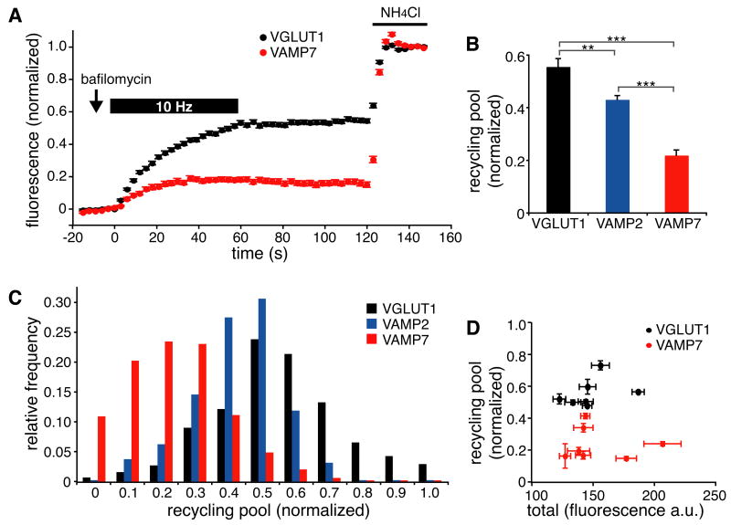
Differences in the response of VAMP7 and VGLUT1 pHluorin fusions to stimulation could reflect differences in either exocytosis or endocytosis. VAMP7 undergoes endocytosis more slowly than VGLUT1 after the stimulus (Figure 1C,D), but increased endocytosis during the stimulus might reduce the response of VAMP7-pHluorin to stimulation. To assess this possibility, we used the vacuolar H+-ATPase inhibitor bafilomycin, which prevents the reacidification and quenching of pHluorin that follows endocytosis. In the presence of bafilomycin, the response to stimulation thus reflects only exocytosis. We found that even in the presence of bafilomycin, stimulation at 10 Hz for 60 s produces a much larger increase in fluorescence of VGLUT1-pHluorin than of VAMP7-pHluorin (Figure 2A). Normalization to the total intracellular pool of pHluorin fusion revealed by NH4Cl shows that average recycling pool size is indeed much larger for VGLUT1 than for VAMP7 (Figure 2B). To compare a protein structurally similar to VAMP7, we also analyzed the v-SNARE VAMP2, and observed a much larger recycling pool for VAMP2- than VAMP7-pHluorin, consistent with a previous report (Fernandez-Alfonso and Ryan, 2008). The distribution of recycling pool size also differs markedly between VAMP7 and the other proteins, with many boutons showing little or no evoked response by VAMP7-pHluorin but very few if any boutons showing no evoked response by VGLUT1- or VAMP2-pHluorin (Figure 2C). The use of syp-mCherry expression to identify boutons in an unbiased way makes it unlikely that the distinct behavior of VAMP7 reflects expression at a subset of synapses. The hippocampal cultures contain predominantly excitatory synapses (85 ± 3%), but transfected syp-mCherry localizes to both excitatory and inhibitory synapses in the same proportions (Figure S3A,B), further excluding bias in the selection of boutons. Importantly, endogenous VAMP7 also occurs in both synapse types (Figure S3C,D). To determine whether differences in recycling pool size might simply reflect differences in expression of the two proteins, we also analyzed recycling pool size as a function of total pHluorin reporter assessed in NH4Cl. Figure 2D shows that the difference between VGLUT1 and VAMP7 in recycling pool size persists over a wide range of expression levels.
Figure 2. VAMP7-pHluorin preferentially labels synaptic vesicles unresponsive to stimulation.
(A to D) Hippocampal neurons transfected with VGLUT1-, VAMP2- or VAMP7-pHluorin were stimulated at 10 Hz for 60 s in the presence of bafilomycin to identify the recycling pool of vesicles, then treated with 50 mM NH4Cl to reveal the intracellular pool. Panel (A) shows the average traces of 142 boutons from 3 coverslips. (B) Relative to VAMP7, a substantially larger proportion of VGLUT1 and VAMP2 responds to stimulation. **, p < 0.01; ***, p < 0.001 (C) A histogram shows the distribution of recycling pool sizes for individual boutons expressing pHluorin fusions to VGLUT1, VAMP2 and VAMP7. n=9 coverslips containing a total of 438 boutons for VGLUT1, 473 for VAMP2 and 487 for VAMP7 (D) Recycling pool size shows no correlation with total pHluorin expression, and differs between VGLUT1 and VAMP7 over a range of expression levels. The data indicate mean ± SEM for individual coverslips containing 50-100 boutons each. See also Figure S2.
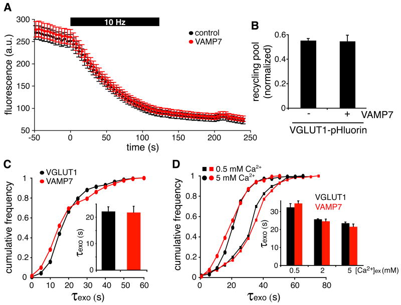
To determine whether the expression of VAMP7 might itself change recycling pool size, we used the styryl dye FM4-64 to assess release at synapses with and without VAMP7-pHluorin. Despite the reduced availability of VAMP7 for regulated exocytosis relative to VGLUT1 and other synaptic vesicle proteins including VAMP2 (Figure 2B,C), we found that the expression of VAMP7 does not affect either the rate or extent of FM4-64 destaining (Figure 3A). At boutons expressing transfected VAMP7, synaptic vesicles thus appear to cycle normally. In addition, cotransfection of untagged VAMP7 does not affect the proportion of VGLUT1 in the recycling pool (Figure 3B), and 86 ± 5% of VGLUT1-pHluorin+ boutons also express the transfected VAMP7 (Figure S2B). Further, the average time constant for exocytosis (τexo) and the distribution of τexo show no difference between VAMP7 and VGLUT1 (Figure 3C), suggesting that the VAMP7 which does respond to stimulation resides on the same synaptic vesicles expressing VGLUT1, and that the over-expression of VAMP7 does not influence their exocytosis. Consistent with this, the rates of evoked VAMP7 and VGLUT1 exocytosis show similar sensitivity to a range of external Ca++ concentrations (Figure 3D).
Figure 3. VAMP7 does not affect the evoked release of synaptic vesicles.
(A) Hippocampal neurons expressing VAMP7-pHluorin were loaded with FM4-64 by stimulation at 10 Hz for 60 s, washed for 10 min, and destained at 10 Hz for 120 s. Boutons with or without VAMP7-pHluorin show no difference in FM4-64 dye loading and unloading (τcontrol = 51.7 ± 1.0s and τvamp7 = 50.1 ± 0.9s). n = 89-90 boutons from 3 coverslips each (B) Cotransfection of untagged VAMP7 does not affect the recycling pool size of VGLUT1-pHluorin. n = 5 coverslips containing 269 boutons for without VAMP7 and 262 for with VAMP7 (C) Stimulating at 10 Hz in the presence of bafilomycin to exclude an effect of endocytosis, the time constant for exocytosis (τexo) shows no significant difference between VGLUT1- and VAMP7-pHluorin in terms of cumulative frequency distribution or a direct comparison of the means (inset). n = 6 coverslips containing 392 boutons for VGLUT1 and 296 for VAMP7 (D) The cumulative frequency distribution and mean (inset) of τexo do not differ between VGLUT1-pHluorin and VAMP7-pHluorin when stimulating in 0.5, 2 or 5 mM Ca++. n = 3 coverslips containing 120-146 boutons. Bar graphs in (B) through (D) indicate mean ± SEM. See also Figure S3.
Although a proportion of VAMP7 localizes to the recycling pool of synaptic vesicles, a much larger proportion does not. Indeed, boutons with little or no response by VAMP7 show a normal response in terms of FM dye destaining and VGLUT1 exocytosis (Figures 3A,B), indicating that vesicle composition as well as behavior varies within a single bouton. VAMP7 also localizes to endosomal and lysosomal membranes at the cell body and dendrites, but it has been found to reside only on synaptic vesicles at boutons (Muzerelle et al., 2003; Salazar et al., 2006; Scheuber et al., 2006), where we analyzed the data. We further confirm the localization of endogenous VAMP7 to membranes that behave like synaptic vesicles by both gradient fractionation and immunoisolation (Figure S4A-C). However, it remains possible that over-expression may result in mislocalization of VAMP7 to endosomes or lysosomes at presynaptic sites, and hence an apparent reduction in recycling pool size. To address this possibility, we expressed VAMP7 and VAMP2 as control fused at their lumenal, C-termini to horseradish peroxidase (HRP) (Leal-Ortiz et al., 2008). Localization of HRP within vesicles prevents diffusion of the HRP reaction product, and thus unambiguously labels the VAMP7+ or VAMP2+ population. Both VAMP7- and VAMP2-HRP indeed label only a subset of synaptic vesicles that intermingle with unlabeled vesicles (Figure 4A). In both cases, the HRP reaction product accumulates in small, round vesicles with the same diameter as unlabeled vesicles (Figure 4B,C), and not within any other compartment at or adjacent to the nerve terminal. Thus, VAMP7 localizes to membranes morphologically indistinguishable from synaptic vesicles.
Figure 4. VAMP7 localizes to synaptic vesicles.
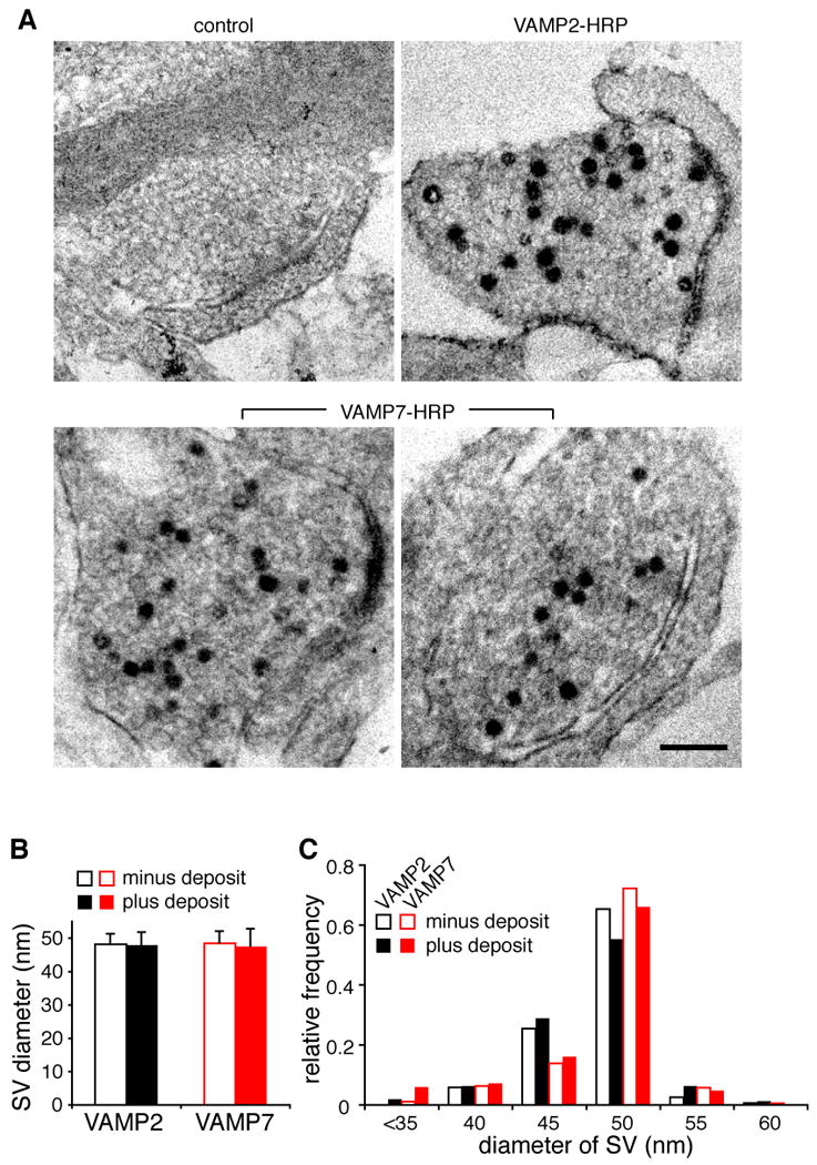
(A) Hippocampal neurons were either not transfected or transfected with VAMP2- or VAMP7-HRP, then fixed and stained for HRP. In boutons expressing VAMP2-HRP and VAMP7-HRP, a subset of synaptic vesicles contain electron-dense deposit. Size bar, 200 nm. (B) The diameter of synaptic vesicles with electron-dense deposit does not differ from that of unlabeled synaptic vesicles, for neurons transfected with either VAMP2- or VAMP7-HRP. Bar graph indicates mean ± standard deviation. (C) The distribution of synaptic vesicle size shows no difference between those with and without deposit, for either VAMP2 or 7. n = 152-172 synaptic vesicles from 10-15 synapses for each condition. See also Figure S4.
Resting and recycling pool vesicles both undergo spontaneous release
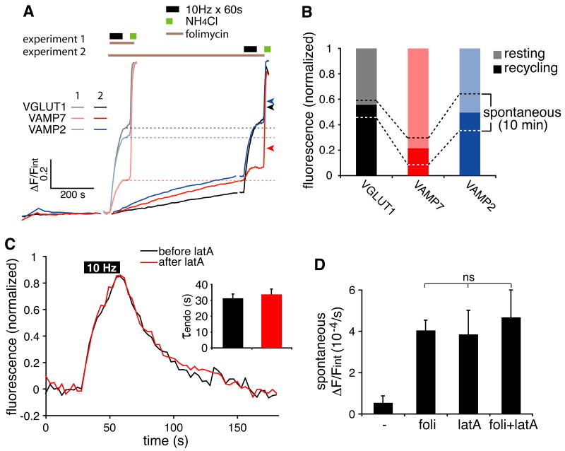
Resting pool vesicles do not apparently respond to field stimulation, but do they have the capacity for exocytosis under other circumstances? Previous work has suggested that evoked and spontaneous release may derive from distinct vesicle populations (Chung et al., 2010; Fredj and Burrone, 2009; Sara et al., 2005) (but see also (Groemer and Klingauf, 2007; Hua et al., 2010; Wilhelm et al., 2010). This predicts that as a protein enriched in the resting (and hence unresponsive) pool of synaptic vesicles, VAMP7 may undergo more spontaneous release than VGLUT1. To test this possibility, we used an optical assay for spontaneous release. Like bafilomycin, the H+-ATPase inhibitor folimycin prevents the reacidification of vesicles that have undergone exocytosis. However, folimycin is less cell permeant than bafilomycin, reducing its access to intracellular vesicles that have not undergone exocytosis (Atasoy et al., 2008). In the presence of folimycin and tetrodotoxin (TTX), the increase in fluorescence of VGLUT1- and VAMP7-pHluorin should thus reflect spontaneous release (Figure 5A). This increase in fluorescence is not due to leakage of folimycin into the neurons and alkalinization of vesicles that have not undergone exocytosis because it is greatly reduced by the high affinity, cell-permeant calcium chelator BAPTA-AM (Figure S5A), consistent with the known calcium-dependence of spontaneous release (Wasser and Kavalali, 2009). Using this assay, we find that VAMP7 undergoes substantial spontaneous exocytosis (expressed as a fraction of total intracellular stores), and the rate exceeds that of VGLUT1 (Figure 5A,B). Spontaneous release is quite heterogeneous across different boutons, but does not correlate with the level of reporter expression (Figure S5B), further excluding a role for mislocalization of over-expressed proteins. Since VAMP7 exhibits higher spontaneous but less evoked release than VGLUT1, spontaneous release cannot simply reflect the size of the recycling pool, suggesting that spontaneous release might derive from the resting pool.
Figure 5. VAMP7 undergoes spontaneous release.
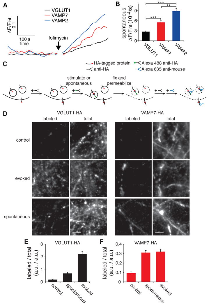
(A) To measure spontaneous release, unstimulated hippocampal neurons expressing VGLUT1- or C were treated with the cell impermeant H+-ATPase inhibitor folimycin in the presence of TTX. The rate of spontaneous fluorescence increase was normalized to total internal fluorescence determined in NH4Cl. (B) Vesicles expressing VAMP7-pHluorin or VAMP2-pHluorin undergo a higher rate of spontaneous release than those expressing VGLUT1-pHluorin. **, p < 0.01; ***, p < 0.001; n = 6 coverslips containing a total of 394 boutons for VGLUT1, 332 for VAMP2 and 419 for VAMP7 (C) Hippocampal neurons expressed HA-tagged VGLUT1 or VAMP7 were first incubated with unlabeled mouse anti-HA antibody to block protein already at the cell surface, then labeled with anti-HA conjugated to Alexa 488 by field stimulation at 10 Hz for 2 min or spontaneous uptake in 0.5 μM TTX for 20 min. For control, the spontaneous uptake of HA antibody was measured over 2 minutes (also in TTX). The cells were then fixed, permeablized and immunostained for total HA protein using a secondary antibody conjugated to Alexa 635. (D) In neurons expressing VGLUT1-HA, stimulation for two minutes evokes much more uptake of HA antibody than observed spontaneously over 20 minutes. In contrast, spontaneous exocytosis of VAMP7 approaches that observed with stimulation. Size bar, 5 μm (E,F) The extent of cell surface delivery was normalized to total reporter expression. n = 10 fields containing a total of 500 boutons from 3 coverslips of each condition. Bar graphs indicate mean ± SEM. See also Figure S5.
Since the pHluorin is large and might interfere with membrane trafficking, we have also used an alternative approach to monitor evoked and spontaneous exocytosis. There are no available antibodies that recognize the lumenal domain of either VGLUT1 or VAMP7, so we fused a short (10 residue) peptide containing the HA epitope to the lumenal domain of both proteins. Twelve days after transfection, hippocampal cultures were incubated with unlabeled anti-HA antibody to block HA-tagged protein already at the cell surface, then with HA antibody directly conjugated to Alexa 488 to detect protein newly delivered to the plasma membrane (Figure 5C). As anticipated, field stimulation at 10 Hz for 2 minutes greatly increases surface exposure of both VGLUT1- and VAMP7-HA (Figure 5D). However, incubation for 20 minutes in the absence of stimulation also enables detection of spontaneously delivered vesicles containing both VGLUT1 and VAMP7. To assess the total amount of reporter expressed at boutons, we immunostained for HA after fixation and permeabilization, using a secondary antibody conjugated to Alex 635 (Figure 5C,D). Normalized to total HA-tagged reporter, VGLUT1 shows a strong response to stimulation (Figure 5E), consistent with targeting to the recycling pool. In contrast, VAMP7-HA exhibits considerably less response to stimulation. However, both proteins show similar levels of spontaneous release, with spontaneous release of VAMP7 over 20 minutes approaching that observed after stimulation for 2 minutes (Figure 5F). The analysis by antibody labeling thus supports the preferential targeting of VAMP7 to the resting pool, which undergoes spontaneous release. Although indistinguishable from typical synaptic vesicles by morphology and standard fractionation, the VAMP7+ membranes and by inference the resting pool thus behave like constitutive secretory vesicles.
To characterize the sources of spontaneous release, we determined whether spontaneous release can occlude the effects of stimulation. Using the pHluorin-based reporters, we first measured recycling pool size by stimulation alone (experiment 1). In a separate set of coverslips from the same transfection, we measured spontaneous release for 10 minutes in the presence of folimycin and glutamate receptor antagonists (experiment 2): TTX can be difficult to wash out, so we used receptor antagonists to eliminate network activity, and the rate of spontaneous release measured this way is similar to that observed with TTX (data not shown). We then stimulated the same cells at 10 Hz for 60 s before adding NH4Cl to reveal the entire intracellular pool (Figure 6A). If spontaneous release derives solely from the recycling pool, it should reduce the subsequent evoked release so that the cumulative effect of spontaneous and evoked release (experiment 2) equals that observed for evoked release alone (experiment 1, dashed line in Figure 6A). On the other hand, if spontaneous release derives solely from the resting pool, spontaneous and evoked release should summate (arrowheads) to exceed that observed for evoked release alone. We find that in the case of all three proteins examined, the combined effect of spontaneous and evoked release exceeds the size of the recycling pool, but falls short of the fluorescence increase predicted if spontaneous and evoked release were entirely independent, indicating that spontaneous release originates from both recycling and resting pools. In addition, we were surprised to find that VAMP2 as well as VAMP7 shows more spontaneous release than VGLUT1 (Figure 5B, 6B). Since VAMP2 resembles VGLUT1 in localization to the recycling pool, the increased spontaneous release of VAMP2 further supports the origin of spontaneous release from both recycling and resting pools. In addition, it is important to note that the spontaneous release observed over 10 minutes sets only a lower bound for the full extent of spontaneous release.
Figure 6. Resting and recycling pool vesicles both undergo spontaneous release.
(A) Hippocampal neurons expressing the pHluorin constructs indicated were either stimulated at 10 Hz for 60 s in folimycin (experiment 1, weaker colors) or incubated without stimulation in folimycin, CNQX and APV for 10 minutes, followed by stimulation at 10 Hz for 60 s (experiment 2, stronger colors). In both cases, NH4Cl was used to alkalinize intracellular compartments and reveal the total pHluorin fluorescence. The dashed lines indicate the size of the recycling pool determined in experiment 1, and the arrowheads indicate the predicted size if the two pools are independent. The fluorescence reached in experiment 2 exceeds that predicted if spontaneously released vesicles come entirely from the recycling pool, but falls short of that predicted if they were entirely independent. (B) The recycling pool size derived from experiment 1 (A) is shown as a stronger color, and the resting pool as weaker. The upper dashed line indicates the cumulative fluorescence derived from both spontaneous and evoked release. The lower dashed line indicates the residual fluorescence evoked by stimulation after 10 minutes of spontaneous release. The area between upper and lower dashed lines indicates the fluorescence derived from spontaneous release over 10 minutes. Spontaneous release derives from both resting and recycling pools. n = 6 coverslips with a total of 300 boutons per group (C) In hippocampal neurons expressing VGLUT1-pHluorin, the F-actin-destabilizing agent latrunculin A (5 μM) does not interfere with the synaptic vesicle exocytosis or endocytosis evoked by 10 Hz stimulation. n = 44 boutons for each condition (D) In unstimulated hippocampal neurons, folimycin and latrunculin A both increase the fluorescence of VAMP7-pHluorin to the same extent, and without any additive effect. n = 4 coverslips containing 137 boutons for control, 218 for folimycin alone, 237 for latrunculin A and 227 for folimycin and latrunculin A. Bar graphs indicate mean ± SEM.
We also used this assay to characterize the mechanism responsible for endocytosis of spontaneously cycling vesicles. As shown previously (Holt et al., 2003; Sankaranarayanan et al., 2003), the compensatory endocytosis that follows evoked synaptic vesicle release does not depend on actin (Figure 6C). In the presence of TTX, however, actin depolymerization with latrunculin A (latA) increases the fluorescence of VAMP7 to the same extent as folimycin (Figure 6D). The increased fluorescence in latA could reflect either increased spontaneous release, or a block in endocytosis. If latA increases spontaneous release, the inhibition of vesicle reacidification that accompanies endocytosis should further promote the accumulation of VAMP7-pHluorin fluorescence. However, we find that folimycin has no additional effect in the presence of latA (Figure 6D). Actin thus appears required for the endocytosis that follows spontaneous but not evoked synaptic vesicle exocytosis.
Cytoplasmic sequences target VAMP7 and VGLUT1 to different vesicle pools
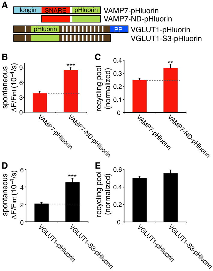
VAMP7 belongs to the longin subfamily of v-SNAREs containing an N-terminal domain that interacts with trafficking machinery as well as regulating SNARE complex formation (Burgo et al., 2009; Chaineau et al., 2008; Martinez-Arca et al., 2003; Pryor et al., 2008) (Figure 7A). Indeed, the interaction with adaptor protein AP-3 contributes to the trafficking of VAMP7 (Martinez-Arca et al., 2003). Surprisingly, deletion of the longin domain (VAMP7-ND) does not impair its targeting to synaptic boutons: 96 ± 2% colocalizes with VGLUT1-pHluorin (Figure S2B). However, the ND mutation increases the spontaneous exocytosis of VAMP7 (Figure 7B). Although loss of the longin domain might simply disinhibit SNARE complex formation, the ND mutation increases both the proportion of VAMP7 in the recycling pool (Figure 7C) and the rate of endocytosis (Figure S6A,B). In addition, over-expression of untagged VAMP7-ND does not affect the proportion of VAMP2- or VGLUT1-pHluorin in the recycling pool (Figure S6C,D). The ND mutation thus affects at least in part the trafficking of the VAMP7 protein, indicating that the longin domain targets VAMP7 toward the resting pool of synaptic vesicles. Since the longin domain interacts with AP-3 (Martinez-Arca et al., 2003), we also investigated recycling pool size in AP-3-deficient mocha mice. The loss of AP-3 disrupts the synaptic targeting of endogenous (Scheuber et al., 2006) and transfected VAMP7 (data not shown), so we used VGLUT1-pHluorin to assess recycling pool size, but found no significant change in mocha mice (46.5 ± 1.2% for mocha vs. 48.6 ± 0.9% for control). The loss of AP-3 thus has less effect on recycling pool size than the longin deletion in VAMP7, suggesting that additional factors may contribute, and recent work has indeed implicated AP-1 in synaptic vesicle recycling (Glyvuk et al., 2010; Kim and Ryan, 2009).
Figure 7. The longin domain targets VAMP7 to the resting pool.
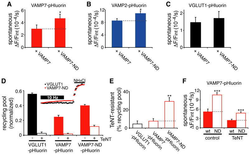
(A) Domain organization of VAMP7-pHluorin with and without (VAMP7-ND) the N-terminal longin domain, and of VGLUT1-pHluorin with and without (VGLUT1-S3) the C-terminal polyproline motifs. (B) Transfected into hippocampal neurons and imaged as described in Figure 5A, VAMP7-ND-pHluorin exhibits significantly higher spontaneous release than wild type VAMP7-pHluorin. n = 7 coverslips containing a total of 361 boutons for wild type VAMP7 and 391 boutons for VAMP7-ND (C) Deletion of the longin domain increases the proportion of VAMP7 in the recycling pool as determined by stimulation at 10 Hz for 60 s in the presence of bafilomycin, and normalization to the signal in NH4Cl. n = 6 coverslips containing 305 boutons for wild type VAMP7 and 278 boutons for VAMP7-ND (D,E) Deletion of the C-terminal polyproline motifs increases the spontaneous release of VGLUT1 (D) (n = 5-6 coverslips containing a total of 380-680 boutons), without increasing the recycling pool size (E) (p = 0.3, n = 4-5 coverslips with 140-240 boutons) **, p < 0.01; ***, p < 0.001. See also Figure S6.
We then tested the role of other sequences in targeting to different populations of synaptic vesicles. VGLUT1 contains two C-terminal polyproline motifs previously shown to interact with endophilin and other proteins (De Gois et al., 2006; Vinatier et al., 2006), some of which influence the rate of endocytosis (Voglmaier et al., 2006). We now find that deletion of these C-terminal sequences (VGLUT1-S3) substantially increases the spontaneous release of VGLUT1 (Figure 7D). However, the mutation has no effect on the recycling pool size of VGLUT1 (Figure 7E), suggesting that the polyproline motifs normally direct VGLUT1 toward a subset of vesicles with low spontaneous release that lies within either the recycling or resting pools.
Deletion of the longin domain enables VAMP7 to influence synaptic vesicle exocytosis
In addition to its role in trafficking, the longin domain of VAMP7 inhibits SNARE complex formation. Indeed, deletion of the longin domain accelerates neurite extension (Martinez-Arca et al., 2000; Martinez-Arca et al., 2001), presumably by disinhibiting the function of VAMP7 in membrane insertion. We did not observe an effect of wild type VAMP7 on synaptic vesicle exocytosis (Figures 3A,B, S6C,D), but deletion of the longin domain increases the rate of spontaneous VAMP7 exocytosis (Figure 7B). Changes in trafficking account for some of this effect, but we find that an untagged form of VAMP7-ND acts in trans to increase the spontaneous exocytosis of wild type VAMP7-pHluorin (Figure 8A). The mutant must therefore still target at least in part to the same membranes as wild type VAMP7, and affect the behavior of these vesicles, not simply trafficking of the reporter. The ND mutant also increases spontaneous release of VAMP2-pHluorin (Figure 8B), but has no effect on the spontaneous release of VGLUT1-pHluorin (Figure 8C), suggesting specificity for a subset of synaptic vesicles.
Figure 8. VAMP7-ND influences spontaneous and evoked release.
(A-C) Relative to cotransfection with wild type VAMP7, cotransfection of VAMP7-ND increases the spontaneous release of wild type VAMP7- (A) and VAMP2- (B) but not VGLUT1-pHluorin (p = 0.64) (C). n = 6 coverslips containing 317-370 boutons (D) Hippocampal neurons expressing VGLUT1-, wild type VAMP7- or VAMP7-ND-pHluorin were treated overnight with or without tetanus toxin (TeNT), and exocytosis stimulated at 10 Hz for 60 s in the presence of bafilomycin. Inset shows that TeNT essentially abolishes the response of VGLUT1, but VAMP7-ND still shows an evoked response. n = 4-6 coverslips containing 180-399 boutons per group (E) Normalized to recycling pool in the absence of tetanus toxin, the proportion of TeNT-resistant evoked release is increased for the ND mutant. (F) Even after treatment with TeNT, VAMP7-ND-pHluorin still exhibits a higher rate of spontaneous release than wild type VAMP7-pHluorin. n = 6 coverslips containing 248-366 boutons per condition; *, p < 0.05; **. p < 0.01; ***, p < 0.001 and bars indicate mean ± SEM.
VAMP2 is the dominant v-SNARE for both evoked and spontaneous synaptic vesicle exocytosis, but VAMP7 can form a SNARE complex with syntaxin 1 and SNAP-25 (Alberts et al., 2003), the dominant t-SNAREs involved in transmitter release. To assess the relative roles of VAMP2 and other v-SNAREs in the exocytosis of different synaptic vesicle pools, we selectively cleaved VAMP2 with tetanus toxin. As anticipated, tetanus toxin blocks the exocytosis of both VGLUT1 and wild type VAMP7 evoked by stimulation (Figure 8D). However, VAMP7-ND-pHluorin shows a substantial tetanus toxin-resistant response to stimulation (Figure 8D,E), suggesting that the ND mutant may itself contribute to evoked release. The increase in spontaneous release due to the ND mutation also persists after cleavage with tetanus toxin (Figure 8F), suggesting that the tetanus toxin-insensitive VAMP7 contributes to spontaneous as well as evoked release.
Discussion
The results provide the first direct evidence that synaptic vesicle recycling and resting pools differ in molecular composition. Like VGLUT1 and a number of other synaptic vesicle proteins, VAMP7 undergoes exocytosis in response to stimulation, and with properties very similar to VGLUT1. Conversely, a substantial proportion of VGLUT1 and other vesicle proteins does not respond to stimulation (Fernandez-Alfonso and Ryan, 2008), similar to VAMP7. VGLUT1, VAMP7 and other synaptic vesicle proteins thus target to both recycling and resting pools. However, we now find that the proportions differ dramatically, with higher levels of VGLUT1 and VAMP2 in the recycling pool, and of VAMP7 in the resting pool.
Previous work has suggested that synaptic vesicles undergoing spontaneous release belong to a pool distinct from those responding to stimulation (Chung et al., 2010; Fredj and Burrone, 2009; Sara et al., 2005). Although this hypothesis has proven controversial (Groemer and Klingauf, 2007; Hua et al., 2010; Wilhelm et al., 2010), it predicts that the resting pool, which is largely unresponsive to stimulation, may nonetheless undergo spontaneous release. Indeed, we now find that VAMP7, enriched on resting pool vesicles, undergoes a higher rate of spontaneous release than VGLUT1, which is enriched in the recycling pool. The spontaneous release of VAMP7 thus cannot simply reflect the size of the recycling pool, and may derive in part from the resting pool. Since the response to spontaneous release of a single vesicle (quantal size) has been used to interpret the response to evoked release, a different origin for the two events would have important implications for synaptic physiology. However, the results do not exclude a role for the recycling pool in spontaneous release. Indeed, the sequential analysis of spontaneous and evoked release at the same boutons shows that spontaneous release can occlude the effect of stimulation, indicating that spontaneous release derives from recycling as well as resting pools.
The apparent lack of response to stimulation raises the possibility that VAMP7+ resting pool vesicles may correspond to a population of membranes other than synaptic vesicles, with heterologous expression resulting in mislocalization to the recycling pool. Previous work has indeed suggested that VAMP7 may localize to only a subset of presynaptic terminals such as hippocampal mossy fibers (Coco et al., 1999; Muzerelle et al., 2003). However, recent work has demonstrated the localization of VAMP7 to synaptic vesicles (Newell-Litwa et al., 2009), and a proteomic analysis of purified synaptic vesicles from whole brain also identified VAMP7 (Takamori et al., 2006). In addition, we confirm the localization of endogenous VAMP7 to presynaptic sites by immunofluorescence and to synaptic vesicles by density gradient fractionation and immuno-isolation. Ultrastructural analysis of the VAMP7+ vesicles labeled with lumenal HRP further shows that they exhibit the typical small, round appearance of synaptic vesicles. Morphologically indistinguishable recycling and resting pool vesicles thus exhibit quantitative differences in protein composition.
What is the physiological role of the resting pool? Spontaneous release from this pool may contribute to structural changes such as process extension (Martinez-Arca et al., 2000). Recent work has also implicated spontaneous release in the regulation of synaptic strength (McKinney et al., 1999; Sutton and Schuman, 2006), suggesting additional roles in development and plasticity. Although the relatively small proportion of VGLUT1 (∼40%) localized to the resting pool might suggest that this pool does not subserve transmitter release, we have recently observed that like VAMP7, the vesicular monoamine transporter VMAT2 which fills synaptic vesicles with monoamines also shows preferential localization to the resting pool (Onoa et al., 2010). The pools may thus be specialized for the release of different transmitters, and the recent evidence for differential release of acetycholine and GABA from retinal starburst amacrine cells is consistent with this possibility (Lee et al., 2010). In addition, spontaneous release of synaptic vesicles may contribute to synapse growth (Huntwork and Littleton, 2007) or the endosomal trafficking of receptors and channels.
Preferential localization of VAMP7 to resting rather than recycling synaptic vesicles presumably reflects differences in the formation of different pools. Recycling pool vesicles are generally considered to form through clathrin- and AP2-dependent endocytosis (Di Paolo and De Camilli, 2006; Granseth et al., 2006; Kim and Ryan, 2009) whereas resting pool vesicles may use a distinct mechanism, perhaps involving AP-3 (Voglmaier and Edwards, 2007) or AP-1 (Glyvuk et al., 2010; Kim and Ryan, 2009). Indeed, we chose VAMP7 for these experiments due to its dependence on AP-3 (Scheuber et al., 2006). Consistent with a distinct endocytic pathway, we find that VAMP7 undergoes endocytosis much more slowly than other synaptic vesicle proteins, and previous work has shown that vesicles internalized after stimulation may respond more slowly to repeat stimulation (Richards et al., 2000; Richards et al., 2003). However, a delay in endocytosis may reflect inefficient targeting to an endocytic pathway as well as an intrinsically slow pathway. On the other hand, deletion of the longin domain, which interacts with AP-3, redistributes VAMP7 toward the recycling pool. The interaction of AP-3 with the longin domain thus targets VAMP7 to the resting pool of synaptic vesicles. We also find that deletion of the polyproline motifs at the C-terminus of VGLUT1 increases spontaneous exocytosis of the transporter, indicating a role for these sequences in targeting to a subset of synaptic vesicles with low rates of spontaneous release. Spontaneous recycling of VAMP7 also depends on actin, in striking contrast to evoked release (Holt et al., 2004; Sankaranarayanan et al., 2003). Different endocytic mechanisms thus appear to account for the targeting of synaptic vesicle proteins to different pools.
Since VAMP2 and 7 differ in the properties of release and belong to the family of v-SNAREs involved in membrane fusion, we further tested the possibility that like VAMP2, VAMP7 might contribute to the behavior of synaptic vesicles. Although over-expression of wild type VAMP7 has no obvious effect on synaptic vesicle exocytosis, deleting the auto-inhibitory N-terminal longin domain alone increases the spontaneous release of VAMP7 2-3 fold, and this mutation affects the behavior of vesicles containing wild type VAMP7, not simply trafficking of the VAMP7-ND reporter itself. The longin domain deletion also increases spontaneous release of VAMP7 after cleavage of VAMP2 with tetanus toxin, indicating that VAMP7 can support spontaneous release even in the absence of VAMP2. Further, the ND mutant increases evoked release even in the presence of tetanus toxin, indicating that mutant VAMP7 can also mediate the exocytosis of recycling pool vesicles in response to stimulation. When disinhibited by removal of the longin domain, VAMP7 thus has the potential to influence synaptic vesicle exocytosis. Interestingly, mocha mice show changes in asynchronous and spontaneous release, possibly due to the loss of VAMP7 (Scheuber et al., 2006).
Although differences in protein composition presumably contribute to the differences in behavior of synaptic vesicle pools, it seems unlikely that v-SNAREs are alone responsible. The differential targeting of other synaptic vesicle proteins such as the calcium sensor synaptotagmin family (Maximov et al., 2007; Xu et al., 2009) presumably contribute to the difference between pools. Recent work has indeed implicated the related calcium-sensitive C2 domain protein DOC2 specifically in spontaneous rather than evoked release (Groffen et al., 2010), although DOC2 is a soluble protein and its relationship to different vesicle pools remains unclear. By demonstrating that different synaptic vesicle pools contain different proteins, our work now provides a framework for considering their individual contribution to the behavior of these pools, and the role of these pools in synapse development, function and plasticity.
Experimental Procedures
Cell culture and immunofluorescence
Hippocampal neurons isolated from day 20 rat embryos (E20) were transfected by electroporation (Amaxa) and cultured as previously described (Li et al., 2005). Fixed cells were immunostained using antibodies to SV2 (gift of R. Kelly), VAMP2 (Synaptic Systems), DsRed (Clontech), VGLUT1 (Chemicon), GAD65 (Abcam) and VGAT (Chaudhry et al., 1998) at dilutions of 1:1000-2000 (Tan et al., 1998). A rabbit anti-VAMP7 antibody (gift of A. Peden) and a mouse anti-VAMP7 antibody (gift of V. Faundez) were used at a dilution of 1:50-100. Cy3, Cy5-conjugated secondary antibodies (Jackson ImmunoResearch) and Alexa 488-conjugated secondary antibodies (Invitrogen) were used at a dilution of 1:1000. To assess colocalization, regions of interest (ROIs) were selected in one fluorescence channel, overlaid with images from a second channel, and fluorescence in the second channel that exceeds a threshold value used to judge colocalization.
Molecular biology
VAMP7 sequences were amplified by PCR from rat brain cDNA, fused at the 3′ end of the open reading frame to the sequence encoding super-ecliptic pHluorin, then subcloned into the pCAGGS expression vector (Voglmaier et al., 2006). The mCherry cDNA (Shaner et al., 2004) was ligated to the 5′ end of the synaptophysin cDNA to generate an mCherry-synaptophysin fusion protein. VGLUT1-pHluorin (Voglmaier et al., 2006), VAMP2-pHluorin (Sankaranarayanan and Ryan, 2000) and VAMP7-pHluorin were then inserted upstream of an internal ribosome entry sequence (IRES2, Clontech) driving the translation of mCherry- synaptophysin. Sequences encoding amino acids 1-101 were deleted from VAMP7 to create VAMP7-ND. VAMP2-HRP was described before (Leal-Ortiz et al., 2008), and VAMP7-HRP was generated by replacing VAMP2 sequence with VAMP7. A 10 aa sequence containing an HA epitope tag was inserted at the C-terminus of VAMP7 to generate VAMP7-HA.
Imaging
Transfected neurons were imaged at 12-14 days in vitro (DIV) as previously described (Nemani et al., 2010; Voglmaier et al., 2006). pHluorin was imaged with 492/18 nm excitation and 535/30 nm emission filters. mCherry was imaged with 580/20 nm excitation and 630/60 nm emission filters. Images in both channels were collected every 3 seconds in experiments involving stimulation and every 15 seconds for the measurement of spontaneous release, with no evidence of focus drift (data not shown). Neurons were imaged in standard Tyrode's solution (in mM, 119 NaCl, 2.5 KCl, 2 MgCl2, 30 glucose and 25 Hepes, pH7.4) with 2 mM CaCl2 unless otherwise indicated. Low pH buffer (in mM, 119 NaCl, 2.5 KCl, 2 MgCl2, 2 CaCl2, 30 glucose, 25 MES, pH5.5) was used to quench surface pHluorin fluorescence, and NH4Cl buffer (in mM, 69 NaCl, 2.5 KCl, 2 MgCl2, 2 CaCl2, 50 NH4Cl, 30 glucose, 25 Hepes, pH 7.4) was used to reveal total pHluorin fluorescence. Glutamate receptor antagonists 6-cyano-7 nitroquinoxaline-2,3-dione (CNQX) (10 μM) and D,L-2-amino-5-phosphonovaleric acid (APV) (50 μM) were included in the Tyrode's solution during the experiments involving stimulation. Tetrodotoxin (TTX) (0.5 μM) was used for measurements of spontaneous release. Bafilomycin (0.6 μM), folimycin (0.6 μM), and latrunculin A (5 μM) were diluted from 1000× stock solutions in DMSO. Tetanus toxin (10 nM) was incubated with neurons for 16-18 hours to cleave VAMP2. BAPTA-AM (10 μM) was incubated with neurons for 1 hr to chelate intracellular calcium.
HA antibody labeling
Hippocampal neurons cotranfected with syp-mCherry and either VGLUT1-HA or VAMP7-HA were incubated with HA.11 antibody (Covance) at 1:100 dilution in Tyrode's solution for 5 min. Cells were then washed in Tyrode's solution and incubated in Alexa 488-labeled HA.11 at a dilution of 1:100 for either 2 min without stimulation (control), 2 min with 10 Hz stimulation (evoked), or 20 min without stimulation (spontaneous)—the unstimulated conditions were also maintained in 0.5 μM TTX. Cells were fixed with 4% PFA for 20 min, permeabilized with 0.02% saponin for 20 min, and stained with HA.11 (1:200) and Alexa-635 conjugated goat anti-mouse (1:500). The fluorescence of Alexa 488 and 635 was measured for boutons expressing syp-mCherry, and the ratio used to assess stimulated and spontaneous exocytosis.
Data analysis
Regions enclosing entire synaptic boutons were selected using syp-mCherry. In most experiments, fluorescence was normalized to the total intracellular fluorescence (in NH4Cl), which was determined as FNH4Cl-Finitial. pH and surface percentage were determined as previously described (Mitchell and Ryan, 2004). Briefly, VGLUT1- or VAMP7-pHluorin fluorescence at individual boutons was measured in regular Tyrode's solution, Tyrode's solution buffered to pH 5.5 (with MES), and Tyrode's containing 50 mM NH4Cl. The pH and surface fraction were calculated according to the formulas previously described (Mitchell and Ryan, 2004), assuming the pHluorin pK ∼7.1 ((Miesenböck et al., 1998; Sankaranarayanan et al., 2000). To determine the kinetics of exo- and endocytosis with 10 Hz stimulation, the change in fluorescence was normalized to the maximum change in fluorescence during stimulation (Fpoststim - Fprestim). Endocytosis kinetics were fit to a single-exponential decay (F = Fplateau + Fspan●e-kt). Exocytosis kinetics were fit to a single-exponential [F = Fmax●(1-e-kt)]. Spontaneous release was determined by subtracting the rate of fluorescence change before folimycin from the increase in fluorescence after addition of folimycin, and normalized to total intracellular fluorescence: (Kpostfoli-Kprefoli)/Finternal where K=slope (fluorescence change/time). All of the data indicate mean ± SEM, with sample number (n) referring to either coverslip or bouton number, as indicated, and nested Anova used to compare groups of data containing the indicated coverslip numbers. * indicates p<0.05, **, p<0.01 and ***, p<0.001.
Electron microscopy
Hippocampal neurons were transfected with VAMP2- or VAMP7-HRP as described above. Cells were fixed at 14 DIV with 2.5% glutaraldehyde in 0.15M cacodylate buffer, and samples were processed for EM as described previously (Leal-Ortiz et al., 2008). All sample processing and EM was performed in the Cell Sciences Imaging Facility at Stanford University.
Biochemistry
Synaptic vesicles were purified as described before (Clift-O'Grady et al., 1990). Cultured hippocampal neurons were harvested and lysed by homogenization in buffer A (in mM, 150 NaCl, 1 EGTA, 0.1 MgCl2, 10 Hepes, pH 7.4). The lysate was sedimented at 10,000g●min, followed by 1000,000g●min, and the supernatant separated by velocity sedimentation through 5-25% glycerol at 220,000g for 75 min, or by equilibrium sedimentation through 10-50% sucrose at 280,000g for 16 hr. The fractions were then immunoblotted with antibodies to synaptophysin (1:2000) and VAMP7 (1:200).
For immunoisolation, cortex from 6-week-old Spague-Dawley rats were dissected and homogenized in buffer A. The lysate was sedimented first at 30,000g●min, then 1000,000g●min. Rabbit anti-VGLUT1 serum or control rabbit serum was cross-linked to Dynal M280 (Invitrogen) magnetic beads, and the beads blocked with 5% BSA in buffer A before incubating with brain lysate. Synaptic vesicles bound to the beads were eluted with SDS sample buffer, and subjected to SDS-PAGE followed by immunoblotting with antibodies to VGLUT1 (Chemicon), SV2, synaptophysin and VAMP7 at 1:400-2000.
Supplementary Material
01
Acknowledgments
We thank A. Peden, V. Faundez and R. Kelly for antibodies to VAMP7 and SV2, J. Rothman for helpful suggestions, and T.A. Ryan and the members of the Edwards lab for discussion. This work was supported by a fellowship from the American Heart Association (to Z.H.) and a grant from NIMH (to R.H.E.).
Footnotes
Publisher's Disclaimer: This is a PDF file of an unedited manuscript that has been accepted for publication. As a service to our customers we are providing this early version of the manuscript. The manuscript will undergo copyediting, typesetting, and review of the resulting proof before it is published in its final citable form. Please note that during the production process errors may be discovered which could affect the content, and all legal disclaimers that apply to the journal pertain.
References
- Alberts P, Rudge R, Hinners I, Muzerelle A, Martinez-Arca S, Irinopoulou T, Marthiens V, Tooze S, Rathjen F, Gaspar P, Galli T. Cross talk between tetanus neurotoxin-insensitive vesicle-associated membrane protein-mediated transport and L1-mediated adhesion. Mol Biol Cell. 2003;14:4207–4220. doi: 10.1091/mbc.E03-03-0147. [DOI] [PMC free article] [PubMed] [Google Scholar]
- Alberts P, Rudge R, Irinopoulou T, Danglot L, Gauthier-Rouviere C, Galli T. Cdc42 and actin control polarized expression of TI-VAMP vesicles to neuronal growth cones and their fusion with the plasma membrane. Mol Biol Cell. 2006;17:1194–1203. doi: 10.1091/mbc.E05-07-0643. [DOI] [PMC free article] [PubMed] [Google Scholar]
- Atasoy D, Ertunc M, Moulder KL, Blackwell J, Chung C, Su J, Kavalali ET. Spontaneous and evoked glutamate release activates two populations of NMDA receptors with limited overlap. J Neurosci. 2008;28:10151–10166. doi: 10.1523/JNEUROSCI.2432-08.2008. [DOI] [PMC free article] [PubMed] [Google Scholar]
- Blumstein J, Faundez V, Nakatsu F, Saito T, Ohno H, Kelly RB. The neuronal form of adaptor protein-3 is required for synaptic vesicle formation from endosomes. J Neurosci. 2001;21:8034–8042. doi: 10.1523/JNEUROSCI.21-20-08034.2001. [DOI] [PMC free article] [PubMed] [Google Scholar]
- Burgo A, Sotirakis E, Simmler MC, Verraes A, Chamot C, Simpson JC, Lanzetti L, Proux-Gillardeaux V, Galli T. Role of Varp, a Rab21 exchange factor and TI-VAMP/VAMP7 partner, in neurite growth. EMBO Rep. 2009;10:1117–1124. doi: 10.1038/embor.2009.186. [DOI] [PMC free article] [PubMed] [Google Scholar]
- Chaineau M, Danglot L, Proux-Gillardeaux V, Galli T. Role of HRB in clathrin-dependent endocytosis. J Biol Chem. 2008;283:34365–34373. doi: 10.1074/jbc.M804587200. [DOI] [PMC free article] [PubMed] [Google Scholar]
- Chaudhry FA, Reimer RJ, Bellocchio EE, Danbolt NC, Osen KK, Edwards RH, Storm-Mathisen J. The vesicular GABA transporter VGAT localizes to synaptic vesicles in sets of glycinergic as well as GABAergic neurons. J Neurosci. 1998;18:9733–9750. doi: 10.1523/JNEUROSCI.18-23-09733.1998. [DOI] [PMC free article] [PubMed] [Google Scholar]
- Chi P, Greengard P, Ryan TA. Synapsin dispersion and reclustering during synaptic activity. Nat Neurosci. 2001;4:1187–1193. doi: 10.1038/nn756. [DOI] [PubMed] [Google Scholar]
- Chung C, Barylko B, Leitz J, Liu X, Kavalali ET. Acute dynamin inhibition dissects synaptic vesicle recycling pathways that drive spontaneous and evoked neurotransmission. J Neurosci. 2010;30:1363–1376. doi: 10.1523/JNEUROSCI.3427-09.2010. [DOI] [PMC free article] [PubMed] [Google Scholar]
- Clift-O'Grady L, Linstedt AD, Lowe AW, Grote E, Kelly RB. Biogenesis of synaptic vesicle-like structures in a pheochromocytoma cell line PC12. J Cell Biol. 1990;110:1693–1703. doi: 10.1083/jcb.110.5.1693. [DOI] [PMC free article] [PubMed] [Google Scholar]
- Coco S, Raposo G, Martinez S, Fontaine JJ, Takamori S, Zahraoui A, Jahn R, Matteoli M, Louvard D, Galli T. Subcellular localization of tetanus neurotoxin-insensitive vesicle-associated membrane protein (VAMP)/VAMP7 in neuronal cells: evidence for a novel membrane compartment. J Neurosci. 1999;19:9803–9812. doi: 10.1523/JNEUROSCI.19-22-09803.1999. [DOI] [PMC free article] [PubMed] [Google Scholar]
- De Gois S, Jeanclos E, Morris M, Grewal S, Varoqui H, Erickson JD. Identification of endophilins 1 and 3 as selective binding partners for VGLUT1 and their colocalization in neocortical glutamatergic synapses: implications for vesicular glutamate transporter trafficking and excitatory vesicle formation. Cell Mol Neurobiol. 2006;26:679–693. doi: 10.1007/s10571-006-9054-8. [DOI] [PMC free article] [PubMed] [Google Scholar]
- Di Paolo G, De Camilli P. Phosphoinositides in cell regulation and membrane dynamics. Nature. 2006;443:651–657. doi: 10.1038/nature05185. [DOI] [PubMed] [Google Scholar]
- Dittman JS, Kaplan JM. Factors regulating the abundance and localization of synaptobrevin in the plasma membrane. Proc Natl Acad Sci U S A. 2006;103:11399–11404. doi: 10.1073/pnas.0600784103. [DOI] [PMC free article] [PubMed] [Google Scholar]
- Faundez V, Horng JT, Kelly RB. A function for the AP3 coat complex in synaptic vesicle formation from endosomes. Cell. 1998;93:423–432. doi: 10.1016/s0092-8674(00)81170-8. [DOI] [PubMed] [Google Scholar]
- Fenster SD, Kessels MM, Qualmann B, Chung WJ, Nash J, Gundelfinger ED, Garner CC. Interactions between Piccolo and the actin/dynamin-binding protein Abp1 link vesicle endocytosis to presynaptic active zones. J Biol Chem. 2003;278:20268–20277. doi: 10.1074/jbc.M210792200. [DOI] [PubMed] [Google Scholar]
- Fernandez-Alfonso T, Kwan R, Ryan TA. Synaptic vesicles interchange their membrane proteins with a large surface reservoir during recycling. Neuron. 2006;51:179–186. doi: 10.1016/j.neuron.2006.06.008. [DOI] [PubMed] [Google Scholar]
- Fernandez-Alfonso T, Ryan TA. A heterogeneous “resting” pool of synaptic vesicles that is dynamically interchanged across boutons in mammalian CNS synapses. Brain Cell Biol. 2008;36:87–100. doi: 10.1007/s11068-008-9030-y. [DOI] [PMC free article] [PubMed] [Google Scholar]
- Fredj NB, Burrone J. A resting pool of vesicles is responsible for spontaneous vesicle fusion at the synapse. Nat Neurosci. 2009;12:751–758. doi: 10.1038/nn.2317. [DOI] [PMC free article] [PubMed] [Google Scholar]
- Glyvuk N, Tsytsyura Y, Geumann C, D'Hooge R, Huve J, Kratzke M, Baltes J, Boening D, Klingauf J, Schu P. AP-1/sigma1B-adaptin mediates endosomal synaptic vesicle recycling, learning and memory. EMBO J. 2010;29:1318–1330. doi: 10.1038/emboj.2010.15. [DOI] [PMC free article] [PubMed] [Google Scholar]
- Granseth B, Odermatt B, Royle SJ, Lagnado L. Clathrin-mediated endocytosis is the dominant mechanism of vesicle retrieval at hippocampal synapses. Neuron. 2006;51:773–786. doi: 10.1016/j.neuron.2006.08.029. [DOI] [PubMed] [Google Scholar]
- Groemer TW, Klingauf J. Synaptic vesicles recycling spontaneously and during activity belong to the same vesicle pool. Nat Neurosci. 2007;10:145–147. doi: 10.1038/nn1831. [DOI] [PubMed] [Google Scholar]
- Groffen AJ, Martens S, Diez Arazola R, Cornelisse LN, Lozovaya N, de Jong AP, Goriounova NA, Habets RL, Takai Y, Borst JG, et al. Doc2b is a high-affinity Ca2+ sensor for spontaneous neurotransmitter release. Science. 2010;327:1614–1618. doi: 10.1126/science.1183765. [DOI] [PMC free article] [PubMed] [Google Scholar]
- Harata N, Ryan TA, Smith SJ, Buchanan J, Tsien RW. Visualizing recycling synaptic vesicles in hippocampal neurons by FM 1-43 photoconversion. Proc Natl Acad Sci U S A. 2001;98:12748–12753. doi: 10.1073/pnas.171442798. [DOI] [PMC free article] [PubMed] [Google Scholar]
- Heuser JE, Reese TS. Evidence for recycling of synaptic vesicle membrane during transmitter release at the frog neuromuscular junction. J Cell Biol. 1973;57:315–344. doi: 10.1083/jcb.57.2.315. [DOI] [PMC free article] [PubMed] [Google Scholar]
- Holt M, Cooke A, Neef A, Lagnado L. High mobility of vesicles supports continuous exocytosis at a ribbon synapse. Curr Biol. 2004;14:173–183. doi: 10.1016/j.cub.2003.12.053. [DOI] [PubMed] [Google Scholar]
- Holt M, Cooke A, Wu MM, Lagnado L. Bulk membrane retrieval in the synaptic terminal of retinal bipolar cells. J Neurosci. 2003;23:1329–1339. doi: 10.1523/JNEUROSCI.23-04-01329.2003. [DOI] [PMC free article] [PubMed] [Google Scholar]
- Hoopmann P, Punge A, Barysch SV, Westphal V, Buckers J, Opazo F, Bethani I, Lauterbach MA, Hell SW, Rizzoli SO. Endosomal sorting of readily releasable synaptic vesicles. Proc Natl Acad Sci U S A. 2010;107:19055–19060. doi: 10.1073/pnas.1007037107. [DOI] [PMC free article] [PubMed] [Google Scholar]
- Hua Y, Sinha R, Martineau M, Kahms M, Klingauf J. A common origin of synaptic vesicles undergoing evoked and spontaneous fusion. Nat Neurosci. 2010;13:1451–1453. doi: 10.1038/nn.2695. [DOI] [PubMed] [Google Scholar]
- Huntwork S, Littleton JT. A complexin fusion clamp regulates spontaneous neurotransmitter release and synaptic growth. Nat Neurosci. 2007;10:1235–1237. doi: 10.1038/nn1980. [DOI] [PubMed] [Google Scholar]
- Kantheti P, Qiao X, Diaz ME, Peden AA, Meyer GE, Carskadon SL, Kapfhamer D, Sufalko D, Robinson MS, Noebels JL, Burmeister M. Mutation in AP-3 delta in the mocha mouse links endosomal transport to storage deficiency in platelets, microsomes and synaptic vesicles. Neuron. 1998;21:111–122. doi: 10.1016/s0896-6273(00)80519-x. [DOI] [PubMed] [Google Scholar]
- Kim SH, Ryan TA. Synaptic vesicle recycling at CNS snapses without AP-2. J Neurosci. 2009;29:3865–3874. doi: 10.1523/JNEUROSCI.5639-08.2009. [DOI] [PMC free article] [PubMed] [Google Scholar]
- Kim SH, Ryan TA. CDK5 serves as a major control point in neurotransmitter release. Neuron. 2010;67:797–809. doi: 10.1016/j.neuron.2010.08.003. [DOI] [PMC free article] [PubMed] [Google Scholar]
- Leal-Ortiz S, Waites CL, Terry-Lorenzo R, Zamorano P, Gundelfinger ED, Garner CC. Piccolo modulation of Synapsin1a dynamics regulates synaptic vesicle exocytosis. J Cell Biol. 2008;181:831–846. doi: 10.1083/jcb.200711167. [DOI] [PMC free article] [PubMed] [Google Scholar]
- Lee S, Kim K, Zhou ZJ. Role of ACh-GABA cotransmission in detecting image motion and motion direction. Neuron. 2010;68:1159–1172. doi: 10.1016/j.neuron.2010.11.031. [DOI] [PMC free article] [PubMed] [Google Scholar]
- Li H, Waites CL, Staal RG, Dobryy Y, Park J, Sulzer DL, Edwards RH. Sorting of vesicular monoamine transporter 2 to the regulated secretory pathway confers the somatodendritic exocytosis of monoamines. Neuron. 2005;48:619–633. doi: 10.1016/j.neuron.2005.09.033. [DOI] [PubMed] [Google Scholar]
- Martinez-Arca S, Alberts P, Zahraoui A, Louvard D, Galli T. Role of tetanus neurotoxin insensitive vesicle-associated membrane protein (TI-VAMP) in vesicular transport mediating neurite outgrowth. J Cell Biol. 2000;149:889–900. doi: 10.1083/jcb.149.4.889. [DOI] [PMC free article] [PubMed] [Google Scholar]
- Martinez-Arca S, Coco S, Mainguy G, Schenk U, Alberts P, Bouille P, Mezzina M, Prochiantz A, Matteoli M, Louvard D, Galli T. A common exocytotic mechanism mediates axonal and dendritic outgrowth. J Neurosci. 2001;21:3830–3838. doi: 10.1523/JNEUROSCI.21-11-03830.2001. [DOI] [PMC free article] [PubMed] [Google Scholar]
- Martinez-Arca S, Rudge R, Vacca M, Raposo G, Camonis J, Proux-Gillardeaux V, Daviet L, Formstecher E, Hamburger A, Filippini F, et al. A dual mechanism controlling the localization and function of exocytic v-SNAREs. Proc Natl Acad Sci U S A. 2003;100:9011–9016. doi: 10.1073/pnas.1431910100. [DOI] [PMC free article] [PubMed] [Google Scholar]
- Maximov A, Shin OH, Liu X, Sudhof TC. Synaptotagmin-12, a synaptic vesicle phosphoprotein that modulates spontaneous neurotransmitter release. J Cell Biol. 2007;176:113–124. doi: 10.1083/jcb.200607021. [DOI] [PMC free article] [PubMed] [Google Scholar]
- McKinney RA, Capogna M, Durr R, Gahwiler BH, Thompson SM. Miniature synaptic events maintain dendritic spines via AMPA receptor activation. Nat Neurosci. 1999;2:44–49. doi: 10.1038/4548. [DOI] [PubMed] [Google Scholar]
- Miesenböck G, De Angelis DA, Rothman JE. Visualizing secretion and synaptic transmission with pH-sensitive green fluorescent proteins. Nature. 1998;394:192–195. doi: 10.1038/28190. [DOI] [PubMed] [Google Scholar]
- Mitchell SJ, Ryan TA. Syntaxin-1A is excluded from recycling synaptic vesicles at nerve terminals. J Neurosci. 2004;24:4884–4888. doi: 10.1523/JNEUROSCI.0174-04.2004. [DOI] [PMC free article] [PubMed] [Google Scholar]
- Muzerelle A, Alberts P, Martinez-Arca S, Jeannequin O, Lafaye P, Mazie JC, Galli T, Gaspar P. Tetanus neurotoxin-insensitive vesicle-associated membrane protein localizes to a presynaptic membrane compartment in selected terminal subsets of the rat brain. Neuroscience. 2003;122:59–75. doi: 10.1016/s0306-4522(03)00567-0. [DOI] [PubMed] [Google Scholar]
- Nemani VM, Lu W, Berge V, Nakamura K, Onoa B, Lee MK, Chaudhry FA, Nicoll RA, Edwards RH. Increased expression of alpha-synuclein reduces neurotransmitter release by inhibiting synaptic vesicle reclustering after endocytosis. Neuron. 2010;65:66–79. doi: 10.1016/j.neuron.2009.12.023. [DOI] [PMC free article] [PubMed] [Google Scholar]
- Newell-Litwa K, Salazar G, Smith Y, Faundez V. Roles of BLOC-1 and adaptor protein-3 complexes in cargo sorting to synaptic vesicles. Mol Biol Cell. 2009;20:1441–1453. doi: 10.1091/mbc.E08-05-0456. [DOI] [PMC free article] [PubMed] [Google Scholar]
- Newell-Litwa K, Seong E, Burmeister M, Faundez V. Neuronal and non-neuronal functions of the AP-3 sorting machinery. J Cell Sci. 2007;120:531–541. doi: 10.1242/jcs.03365. [DOI] [PubMed] [Google Scholar]
- Onoa B, Li H, Gagnon-Bartsch JA, Elias LA, Edwards RH. Vesicular monoamine and glutamate transporters select distinct synaptic vesicle recycling pathways. J Neurosci. 2010;30:7917–7927. doi: 10.1523/JNEUROSCI.5298-09.2010. [DOI] [PMC free article] [PubMed] [Google Scholar]
- Polo-Parada L, Bose CM, Landmesser LT. Alterations in transmission, vesicle dynamics, and transmitter release machinery at NCAM-deficient neuromuscular junctions. Neuron. 2001;32:815–828. doi: 10.1016/s0896-6273(01)00521-9. [DOI] [PubMed] [Google Scholar]
- Pryor PR, Jackson L, Gray SR, Edeling MA, Thompson A, Sanderson CM, Evans PR, Owen DJ, Luzio JP. Molecular basis for the sorting of the SNARE VAMP7 into endocytic clathrin-coated vesicles by the ArfGAP Hrb. Cell. 2008;134:817–827. doi: 10.1016/j.cell.2008.07.023. [DOI] [PMC free article] [PubMed] [Google Scholar]
- Richards DA, Guatimosim C, Betz WJ. Two endocytic recycling routes selectively fill two vesicle pools in frog motor nerve terminals. Neuron. 2000;27:551–559. doi: 10.1016/s0896-6273(00)00065-9. [DOI] [PubMed] [Google Scholar]
- Richards DA, Guatimosim C, Rizzoli SO, Betz WJ. Synaptic vesicle pools at the frog neuromuscular junction. Neuron. 2003;39:529–541. doi: 10.1016/s0896-6273(03)00405-7. [DOI] [PubMed] [Google Scholar]
- Rizzoli SO, Betz WJ. The structural organization of the readily releasable pool of synaptic vesicles. Science. 2004;303:2037–2039. doi: 10.1126/science.1094682. [DOI] [PubMed] [Google Scholar]
- Rizzoli SO, Betz WJ. Synaptic vesicle pools. Nat Rev Neurosci. 2005;6:57–69. doi: 10.1038/nrn1583. [DOI] [PubMed] [Google Scholar]
- Salazar G, Craige B, Styers ML, Newell-Litwa KA, Doucette MM, Wainer BH, Falcon-Perez JM, Dell'Angelica EC, Peden AA, Werner E, Faundez V. BLOC-1 complex deficiency alters the targeting of adaptor protein complex-3 cargoes. Mol Biol Cell. 2006;17:4014–4026. doi: 10.1091/mbc.E06-02-0103. [DOI] [PMC free article] [PubMed] [Google Scholar]
- Salazar G, Love R, Werner E, Doucette MM, Cheng S, Levey A, Faundez V. The zinc transporter ZnT3 interacts with AP-3 and it is preferentially targeted to a distinct synaptic vesicle subpopulation. Mol Biol Cell. 2004;15:575–587. doi: 10.1091/mbc.E03-06-0401. [DOI] [PMC free article] [PubMed] [Google Scholar]
- Sankaranarayanan S, Atluri PP, Ryan TA. Actin has a molecular scaffolding, not propulsive, role in presynaptic function. Nat Neurosci. 2003;6:127–135. doi: 10.1038/nn1002. [DOI] [PubMed] [Google Scholar]
- Sankaranarayanan S, De Angelis D, Rothman JE, Ryan TA. The use of pHluorins for optical measurements of presynaptic activity. Biophys J. 2000;79:2199–2208. doi: 10.1016/S0006-3495(00)76468-X. [DOI] [PMC free article] [PubMed] [Google Scholar]
- Sankaranarayanan S, Ryan TA. Real-time measurements of vesicle-SNARE recycling in synapses of the central nervous system. Nat Cell Biol. 2000;2:197–204. doi: 10.1038/35008615. [DOI] [PubMed] [Google Scholar]
- Sara Y, Virmani T, Deak F, Liu X, Kavalali ET. An isolated pool of vesicles recycles at rest and drives spontaneous neurotransmission. Neuron. 2005;45:563–573. doi: 10.1016/j.neuron.2004.12.056. [DOI] [PubMed] [Google Scholar]
- Scheuber A, Rudge R, Danglot L, Raposo G, Binz T, Poncer JC, Galli T. Loss of AP-3 function affects spontaneous and evoked release at hippocampal mossy fiber synapses. Proc Natl Acad Sci U S A. 2006;103:16562–16567. doi: 10.1073/pnas.0603511103. [DOI] [PMC free article] [PubMed] [Google Scholar]
- Shaner NC, Campbell RE, Steinbach PA, Giepmans BN, Palmer AE, Tsien RY. Improved monomeric red, orange and yellow fluorescent proteins derived from Discosoma sp red fluorescent protein. Nat Biotechnol. 2004;22:1567–1572. doi: 10.1038/nbt1037. [DOI] [PubMed] [Google Scholar]
- Sutton MA, Schuman EM. Dendritic protein synthesis, synaptic plasticity, and memory. Cell. 2006;127:49–58. doi: 10.1016/j.cell.2006.09.014. [DOI] [PubMed] [Google Scholar]
- Takamori S, Holt M, Stenius K, Lemke EA, Gronborg M, Riedel D, Urlaub H, Schenck S, Brugger B, Ringler P, et al. Molecular anatomy of a trafficking organelle. Cell. 2006;127:831–846. doi: 10.1016/j.cell.2006.10.030. [DOI] [PubMed] [Google Scholar]
- Takao-Rikitsu E, Mochida S, Inoue E, Deguchi-Tawarada M, Inoue M, Ohtsuka T, Takai Y. Physical and functional interaction of the active zone proteins, CAST, RIM1, and Bassoon, in neurotransmitter release. J Cell Biol. 2004;164:301–311. doi: 10.1083/jcb.200307101. [DOI] [PMC free article] [PubMed] [Google Scholar]
- Takei K, Mundigl O, Daniell L, De Camilli P. The synaptic vesicle cycle: a single vesicle budding step involving clathrin and dynamin. J Cell Biol. 1996;133:1237–1250. doi: 10.1083/jcb.133.6.1237. [DOI] [PMC free article] [PubMed] [Google Scholar]
- Tan PK, Waites C, Liu Y, Krantz DE, Edwards RH. A leucine-based motif mediates the endocytosis of vesicular monoamine and acetylcholine transporters. J Biol Chem. 1998;273:17351–17360. doi: 10.1074/jbc.273.28.17351. [DOI] [PubMed] [Google Scholar]
- Vinatier J, Herzog E, Plamont MA, Wojcik SM, Schmidt A, Brose N, Daviet L, El Mestikawy S, Giros B. Interaction between the vesicular glutamate transporter type 1 and endophilin A1, a protein essential for endocytosis. J Neurochem. 2006;97:1111–1125. doi: 10.1111/j.1471-4159.2006.03821.x. [DOI] [PubMed] [Google Scholar]
- Voglmaier SM, Edwards RH. Do different endocytic pathways make different synaptic vesicles? Curr Opin Neurobiol. 2007;17:374–380. doi: 10.1016/j.conb.2007.04.002. [DOI] [PubMed] [Google Scholar]
- Voglmaier SM, Kam K, Yang H, Fortin DL, Hua Z, Nicoll RA, Edwards RH. Distinct endocytic pathways control the rate and extent of synaptic vesicle protein recycling. Neuron. 2006;51:71–84. doi: 10.1016/j.neuron.2006.05.027. [DOI] [PubMed] [Google Scholar]
- Vogt K, Mellor J, Tong G, Nicoll R. The actions of synaptically released zinc at hippocampal mossy fiber synapses. Neuron. 2000;26:187–196. doi: 10.1016/s0896-6273(00)81149-6. [DOI] [PubMed] [Google Scholar]
- Wasser CR, Kavalali ET. Leaky synapses: regulation of spontaneous neurotransmission in central synapses. Neuroscience. 2009;158:177–188. doi: 10.1016/j.neuroscience.2008.03.028. [DOI] [PMC free article] [PubMed] [Google Scholar]
- Wienisch M, Klingauf J. Vesicular proteins exocytosed and subsequently retrieved by compensatory endocytosis are nonidentical. Nat Neurosci. 2006;9:1019–1027. doi: 10.1038/nn1739. [DOI] [PubMed] [Google Scholar]
- Wilhelm BG, Groemer TW, Rizzoli SO. The same synaptic vesicles drive active and spontaneous release. Nat Neurosci. 2010;13:1454–1456. doi: 10.1038/nn.2690. [DOI] [PubMed] [Google Scholar]
- Xu J, Pang ZP, Shin OH, Sudhof TC. Synaptotagmin-1 functions as a Ca2+ sensor for spontaneous release. Nat Neurosci. 2009;12:759–766. doi: 10.1038/nn.2320. [DOI] [PMC free article] [PubMed] [Google Scholar]
- Zhang Q, Li Y, Tsien RW. The dynamic control of kiss-and-run and vesicular reuse probed with single nanoparticles. Science. 2009;323:1448–1453. doi: 10.1126/science.1167373. [DOI] [PMC free article] [PubMed] [Google Scholar]
Associated Data
This section collects any data citations, data availability statements, or supplementary materials included in this article.
Supplementary Materials
01
