The Drosophila Ste20 family kinase dMST functions as a tumor suppressor by restricting cell proliferation and promoting apoptosis - PubMed (original) (raw)
The Drosophila Ste20 family kinase dMST functions as a tumor suppressor by restricting cell proliferation and promoting apoptosis
Jianhang Jia et al. Genes Dev. 2003.
Abstract
In a genetic screen for mutations that restrict cell growth and organ size, we identified a new tumor suppressor gene, dMST, which encodes the Drosophila homolog of the mammalian Ste20 kinase family members MST1 and MST2. Loss-of-function mutations in dMST result in overgrown tissues containing more cells of normal size. dMST mutant cells exhibit elevated levels of Cyclin E and DIAP1, increased cell growth and proliferation, and impaired apoptosis. dMST forms a complex with Sav and Wts, two tumor suppressors also implicated in regulating both cell proliferation and apoptosis, suggesting that they act in common pathways.
Figures
Figure 1.
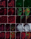
dMST mutations result in tumorous overgrowths. (A,B) Adult eyes of wild type (A) or containing dMST mutant clones (B). (B) dMST mosaic eye is enlarged and protrudes out in folds. (C,D) A wild-type wing (C) or a wing containing a dMST clone located near the wing hinge (arrow in D). (E,F) A control (E) and a dMST mosaic haltere (F). (G) A dMST mutant clone on the thorax exhibits tumorlike growth (arrow). (H) High-magnification view of wing margin containing dMST mutant clones labeled by y- (indicated by the bracket). dMST mutant cells differentiate into margin bristles of normal size. (_I,I_′) A late third instar wing disc containing dMST clones stained with a nuclear dye, 7-AAD (red in _I_′) and anti-Myc antibody (green in _I,I_′). dMST mutant clones lack hs-Myc-GFP expression, whereas the twin spots exhibit enhanced hs-Myc-GFP expression. (_J_–_K_′) High-magnification view of dMST mutant clones and twin spots labeled with 7-AAD (red in _J,J_′), which labels the nuclei, or anti-Armadillo (Arm) antibody (red in _K,K_′), which stains cell membrane. dMST mutant cells are recognized by the lack of Myc-GFP expression (green). dMST mutant cells are of the normal size compared with wild-type cells.
Figure 2.
dMST controls both cell proliferation and apoptosis. (A_–_D) BrdU incorporation (A,B) or phospho-H3 staining (C,D) of wild-type eye discs (A,C) or dMST mosaic eye discs (B,D). Arrowheads indicate the second mitotic wave (SMW). All eye discs are oriented with anterior to the left. (B,D) dMST mosaic discs exhibit extensive BrdU incorporation and increased phospho-H3-positive cells posterior to the SMW. (E) Cyclin E expression in wild type. (_F_–_F_″) Cyclin E expression in dMST mosaic discs. dMST mutant cells are labeled by the lack of Myc-GFP expression (green). dMST mutant cells posterior to the SMW accumulate high levels of Cyclin E cell autonomously (arrows). (G,H) Phalloidin staining of wild type (G) or dMST mosaic (H) pupal eye disc (46 h APF). (G) The wild-type pupal eye disc contains a single layer of interommatidial cells. (H) In contrast, the dMST mutant disc contains multiple layers of interommatidial cells. (_I_–_I_″) A dMST mosaic pupal eye disc (38 h APF) doubly stained to show TUNEL (red) and Myc-GFP (green) expression. dMST mutant cells are labeled by the lack of green staining. The TUNEL staining is confined to wild-type cells. (J) A late third eye disc expressing GMR-hid and stained with an antibody against an activated form of the Drosophila caspase Drice (Drice-Act), which labels dying cells (Yoo et al. 2002). Expression of GMR-hid induces precocious cell death. (_K,K_′) A dMST mosaic eye disc expressing GMR-hid stained with antibodies against Drice-Act (red) and Myc (green). dMST mutant cells are recognized by the lack of green staining. Ectopic cell death induced by GMR-hid is blocked in dMST mutant cells. (L,M) Adult eyes derived from _GMR-hid_-expressing discs (L) or dMST mosaic discs expressing GMR-hid (M). (N_–_Q) Diap1 protein accumulation (red in _N_–_O_′) or diap1-lacZ expression (red in _P_–_Q_′) in wild-type (N,P) or dMST mosaic (_O,O_′,_Q,Q_′) eye discs. dMST mutant cells, which lack Myc-GFP expression (green), exhibit high levels of DIAP1 protein (_O,O_′) and diap1-lacZ expression (_Q,Q_′).
Figure 3.
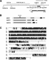
dMST encodes the homolog of the mammalian Ste20-like kinases MST1 and MST2. (A) The genomic region of the dMST locus. The positions of two of the P-element insertions used for mapping—l(2)k06323 and _l(2)k00705_—are indicated. (B) Functional domains of dMST, and human MST1 (hMST1) and MST2 (hMST2). The kinase domains are shaded in gray. The black and hatched boxes indicate the dimerization and kinase inhibitory domains, respectively. The arrows indicate the positions of dMSTBF33 and dMSTJM1 mutations. (C) Sequence alignment among dMST, hMST1, and hMST2. The kinase domain, kinase inhibitory domain, and dimerization domain are underlined by a single solid line, a broken line, and a double solid line, respectively. The asterisk indicates K71, which is changed to R in dMSTnK>R mutant. The arrowhead marks the boundary between dMSTn and dMSTc.
Figure 5.
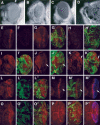
Genetic interaction among dMST, sav, and wts. (A_–_D) Adult eyes of wild type (A), GMR-gal4/UAS-dMSTn (B), GMR-Wts (C), and GMR-gal4/UAS-dMSTn;GMR-Wts (D). (E,F) Drice-Act staining in eye discs expressing GMR-gal4/UAS-dMSTn (E) or GMR-gal4/UAS-dMSTnKR (F). (_G_–_H_′) Drice-Act staining (red) in eye discs expressing GMR-gal4/UAS-dMSTn and containing sav (_G,G_′) or wts (_H,H_′) mutant clones. sav or wts mutant cells are labeled by the lack of Myc-GFP staining (green). (_I_–_J_′) DIAP1 expression (red) in sav mosaic eye disc (_I,I_′) or sav mosaic eye disc expressing GMR-gal4/UAS-dMSTn (_J,J_′). sav mutant cells are labeled by the lack of Myc-GFP staining (green). dMSTn blocks the up-regulation of DIAP in sav mutant cells (arrow). (_K,K_′) DIAP1 expression in wts mosaic disc expressing GMR-gal4/UAS-dMSTn. wts mutant cells (labeled by the lack of green staining) exhibit high levels of DIAP1 even though they express GMR-gal4/UAS-dMSTn (arrow). (_L_–_P_″) Cyclin E staining (red) in sav (_L_–_L_″), wts (_O_–_O_″) mosaic discs, or sav mosaic disc expressing GMR-gal4/UAS-dMSTn (_M_–_M_″), or mosaic disc expressing GMR-gal4/UAS-dMSTn (_P_–_P_″). sav and wts mutant cells are labeled by the lack of Myc-GFP staining (green). dMSTn expression is indicated by Flag staining (blue). sav mutant cells accumulate high levels of Cyclin E cell autonomously (_L_–_L_″), which is suppressed by dMSTn (arrows in _M_–_M_″). (_P_–_P_″) In contrast, wts mutant cells accumulate high levels of Cyclin E even though they express dMSTn.
Figure 4.
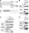
dMST interacts with Sav and Wts. (A) Schematic drawing of Sav and dMST constructs used for transfection. The WW and coiled-coil (C-C) domains of Sav are indicated by shaded and black boxes, respectively. The Sav constructs contain two copies of HA tag, whereas the dMST constructs have a Flag tag at the N terminus. (B_–_D) S2 cells were transfected in indicated DNA constructs. (Top and middle panels) Cell extracts were immunoprecipitated (IP) with indicated antibodies, followed by Western blot (WB) with corresponding antibodies, as indicated. (Bottom panels) Cell lysates were also directly subjected to Western blot with indicated antibodies to determine the expression levels of individual constructs. The Wts construct contains six copies of Myc tag at the N terminus. The asterisks indicate the positions of proteins expressed from the transfected constructs (bottom panels) or coimmunoprecipitated proteins (top panels). The arrows indicate immunoglobin G. Of note, dMSTf and dMSTc appear to be much larger than their predicted sizes.
Similar articles
- The Drosophila Mst ortholog, hippo, restricts growth and cell proliferation and promotes apoptosis.
Harvey KF, Pfleger CM, Hariharan IK. Harvey KF, et al. Cell. 2003 Aug 22;114(4):457-67. doi: 10.1016/s0092-8674(03)00557-9. Cell. 2003. PMID: 12941274 - Hippo promotes proliferation arrest and apoptosis in the Salvador/Warts pathway.
Udan RS, Kango-Singh M, Nolo R, Tao C, Halder G. Udan RS, et al. Nat Cell Biol. 2003 Oct;5(10):914-20. doi: 10.1038/ncb1050. Epub 2003 Sep 21. Nat Cell Biol. 2003. PMID: 14502294 - The DeMSTification of mammalian Ste20 kinases.
Radu M, Chernoff J. Radu M, et al. Curr Biol. 2009 May 26;19(10):R421-5. doi: 10.1016/j.cub.2009.04.022. Curr Biol. 2009. PMID: 19467213 Review. - Filling out the Hippo pathway.
Saucedo LJ, Edgar BA. Saucedo LJ, et al. Nat Rev Mol Cell Biol. 2007 Aug;8(8):613-21. doi: 10.1038/nrm2221. Nat Rev Mol Cell Biol. 2007. PMID: 17622252 Review.
Cited by
- The Hippo pathway regulates homeostatic growth of stem cell niche precursors in the Drosophila ovary.
Sarikaya DP, Extavour CG. Sarikaya DP, et al. PLoS Genet. 2015 Feb 2;11(2):e1004962. doi: 10.1371/journal.pgen.1004962. eCollection 2015 Feb. PLoS Genet. 2015. PMID: 25643260 Free PMC article. - Salvador-Warts-Hippo pathway in a developmental checkpoint monitoring helix-loop-helix proteins.
Wang LH, Baker NE. Wang LH, et al. Dev Cell. 2015 Jan 26;32(2):191-202. doi: 10.1016/j.devcel.2014.12.002. Epub 2015 Jan 8. Dev Cell. 2015. PMID: 25579975 Free PMC article. - Salt-inducible kinases regulate growth through the Hippo signalling pathway in Drosophila.
Wehr MC, Holder MV, Gailite I, Saunders RE, Maile TM, Ciirdaeva E, Instrell R, Jiang M, Howell M, Rossner MJ, Tapon N. Wehr MC, et al. Nat Cell Biol. 2013 Jan;15(1):61-71. doi: 10.1038/ncb2658. Nat Cell Biol. 2013. PMID: 23263283 Free PMC article. - Disease implication of hyper-Hippo signalling.
Wang SP, Wang LH. Wang SP, et al. Open Biol. 2016 Oct;6(10):160119. doi: 10.1098/rsob.160119. Open Biol. 2016. PMID: 27805903 Free PMC article. Review. - YAP antagonizes TEAD-mediated AR signaling and prostate cancer growth.
Li X, Zhuo S, Cho YS, Liu Y, Yang Y, Zhu J, Jiang J. Li X, et al. EMBO J. 2023 Feb 15;42(4):e112184. doi: 10.15252/embj.2022112184. Epub 2023 Jan 2. EMBO J. 2023. PMID: 36588499 Free PMC article.
References
- Abrams J.M. 1999. An emerging blueprint for apoptosis in Drosophila. Trends Cell. Biol. 9: 435-440. - PubMed
- Amanai K. and Jiang, J. 2001. Distinct roles of central missing and dispatched in sending the Hedgehog signal. Development 128: 5119-5127. - PubMed
- Basler K. and Struhl, G. 1994. Compartment boundaries and the control of Drosophila limb pattern by hedgehog protein. Nature 368: 208-214. - PubMed
- Cheung W.L., Ajiro, K., Samejima, K., Kloc, M., Cheung, P., Mizzen, C.A., Beeser, A., Etkin, L.D., Chernoff, J., Earnshaw, W.C., et al. 2003. Apoptotic phosphorylation of histone H2B is mediated by mammalian sterile 20 kinase. Cell 113: 507-517. - PubMed
- Creasy C.L. and Chernoff, J. 1995a. Cloning and characterization of a human protein kinase with homology to Ste20. J. Biol. Chem. 270: 21695-21700. - PubMed
Publication types
MeSH terms
Substances
LinkOut - more resources
Full Text Sources
Other Literature Sources
Molecular Biology Databases
Research Materials
Miscellaneous


