Early hematopoietic lineage restrictions directed by Ikaros - PubMed (original) (raw)
Early hematopoietic lineage restrictions directed by Ikaros
Toshimi Yoshida et al. Nat Immunol. 2006 Apr.
Abstract
Ikaros is expressed in early hematopoietic progenitors and is required for lymphoid differentiation. In the absence of Ikaros, there is a lack of markers defining fate restriction along lympho-myeloid pathways, but it is unclear whether formation of specific progenitors or expression of their markers is affected. Here we use a reporter based on Ikaros regulatory elements to separate early progenitors in wild-type and Ikaros-null mice. We found previously undetected Ikaros-null lympho-myeloid progenitors lacking the receptor tyrosine kinase Flt3 that were capable of myeloid but not lymphoid differentiation. In contrast, lack of Ikaros in the common myeloid progenitor resulted in increased formation of erythro-megakaryocytes at the expense of myeloid progenitors. Using this approach, we identify previously unknown pivotal functions for Ikaros in distinct fate 'decisions' in the early hematopoietic hierarchy.
Figures
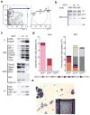
Figure 1
Molecular and functional subdivision of the HSC compartment using the Ikaros-GFP reporter. (a) Analysis of BM HSCs for Ikaros-GFP expression. Lineage-negative BM cells were examined for expression of Sca-1, c-Kit (LSK-left plot) and Ikaros-GFP (right histogram). The percentage of the LSKs is indicated (left) as well as the percentage of LSK progenitors falling within the two -GFPneg—lo and -GFP+ gates (right). Sorted LSK -GFPneg—lo and -GFP+ subsets were used for subsequent studies. (b) Semi-quantitative RT-PCR analysis of endogenous Zfpn1a1 (Ikaros) expression with primers spanning exons 3–8. Ikaros isoforms are indicated on the right side of the panel. Analysis is representative of two independent studies. (c) Semi-quantitative RT-PCR analysis for lineage-specific genes in LSK -GFP subsets. C, normalization and identity controls; GM, granulocyte-macrophage; EMk, erythro-megakaryocyte; Ly, lymphoid. Data is representative of at least four independent studies. (d) Myelo-erythroid differentiation potential of 100 sorted cells from LSK -GFP subpopulations revealed by colony assays performed in the presence of Epo or mixed cytokines (Mix). The percentage of colonies obtained from each subset is shown. Colonies were scored from day 2–4 for the Epo cultures, and from day 5–17 for the Mix cultures. CFU-E, colony-forming unit erythroid; BFU-E, burst forming unit erythroid; EMk, erythoid-megakaryocyte; Mk, megakaryocyte; G, granulocyte; GM, granulocyte-macrophage; M, macrophage; Mc, mast cells; Mix, mixed colonies contain more than three lineages usually G, M, E and Mk. The colony assays shown are from one representative out of more than five independent experiments with similar results. (e) Microscopic examination of LSK -GFP colonies. A 50x magnification of a representative mix-lineage colony from the LSK -GFPneg—lo cultures (upper left) and a GM colony from the LSK -GFP+ cultures (upper right) visualized by fluorescence microscopy. A 600x magnified image of a mix-colony from the LSK -GFPneg—lo culture (lower left) and a GM colony from the LSK -GFP+ culture (lower right) stained by May-Giemsa and visualized by bright field microscopy. (f) Lymphoid activities of LSK -GFPneg—lo and -GFP+ subsets. 200–500 sorted cells were co-cultured on OP9-GFP (top) and OP9-DL1 (middle and bottom) stromal cells. Differentiating cells were analyzed by expression of lymphoid markers and their percentages are shown in each quadrant. One representative out of three independent studies is shown. (g) Flt3 expression in LSK -GFP subsets. The shaded and open histograms represent cells stained by Flt3 and isotype control antibodies respectively. Data shown is representative of two independent studies (and Supplementary Fig. 4c online).
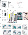
Figure 2
Molecular and functional subdivision of erythro-myeloid progenitors by the Ikaros-GFP reporter. (a) Analysis of Ikaros-GFP expression in erythro-myeloid progenitors. Lineage-depleted BM cells were first analyzed for expression of c-Kit and lack of Sca-1 expression (LK-left) and then for Ikaros-GFP activity (right). Three gates were set on the LK Ikaros-GFP histogram, -GFP−, -GFPint, -GFPhi, and the number of cells falling into each gate is shown (and Supplementary Fig. 1). The LK -GFP−, -GFPint, and -GFPhi subsets were sorted and used in subsequent studies. (b) Semi-quantitative RT-PCR analysis of endogenous Zfpn1a1 (Ikaros) expression as shown in Fig. 1b. (c) Semi-quantitative RT-PCR analysis for expression of lineage-specific genes in the LK -GFP−, -GFPint and -GFPhi subsets as shown in Fig. 1c. (c) The erythro-myeloid differentiation potential of 250 cells sorted from the LK -GFP subsets was determined under Epo and mixed cytokine (Mix) cultures as described in Fig. 1d. The colony nomenclature used is described in Fig. 1d except for m.BFU-E which represents mature BFU-E. Data shown is representative of at least two independent studies performed with LK -GFP subsets. (d) Microscopic examination of LK -GFP colonies. A 600x magnified image of an EMk colony from an LK -GFP− culture (upper left), a Mix colony from an LK -GFPint culture (upper right) and an M colony from an LK -GFPhi culture (lower left) all stained by May-Giemsa and visualized by bright field microscopy. A 50x magnified image of an M colony from an LK -GFPhi culture (lower right) visualized by fluorescence microscopy.
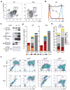
Figure 3
Molecular and functional subdivision of the Ikaros-null LSK with the Ikaros-GFP reporter. (a) Analysis of Ikaros-null BM LSKs (left) for Ikaros-GFP expression (middle) as described in Fig. 1a (Supplementary Fig. 1, online). Expression of Flt3 in wild-type (blue line) and Ikaros-null (red line) LSKs is shown (right) together with an isotype control for staining (dotted line). Flt3 expression is representative of at least two independent experiments. The Ikaros-null LSK -GFPneg—lo and -GFPhi subsets were sorted and used for subsequent studies. (b) Semi-quantitative RT-PCR analysis of lineage-specific genes in Ikaros null LSK -GFPneg—lo and -GFP+ subsets (as described in Fig. 1c), is representative of at least two independent experiments. (c) Myeloid differentiation potential of 100 LSK -GFPneg—lo and -GFP+ sorted cells from wild-type (WT) and Ikaros-null (Null) mice cultured under mixed cytokine (Mix) conditions. The percentage of cells yielding colonies and the type of colony are presented. Colonies were scored from day 5–17 as described in Fig. 1d. Data is representative of three independent experiments. (d) Microscopic examination of Ikaros-null LSK colonies. A 50x magnification of a representative mix-lineage colony (upper left) from the Ikaros-null LSK -GFPneg—lo exhibiting clusters of GFP− (erythroid) and GFP+ (myeloid) cells, and GM colonies (lower left) from the Ikaros-null LSK -GFP+ cultures. A 600x magnification of these colonies stained by May-Giemsa is shown under bright field (upper and lower right). (e) FACS analysis of mix colonies from wild-type (WT) and Ikaros-null (Null) LSK -GFPneg—lo and -GFP+ populations at day 12 of culture. Cells were harvested and stained for Mac-1 (left) revealing myeloid and for CD41 (right) revealing Mk differentiation. Data is representative of two independent studies. (f) T lymphoid activity of the Ikaros-null LSK -GFP+ subset. 250–500 cells were sorted and co-cultured on OP9-DL1 stromal cells. Cells were labeled with antibodies against CD44 and CD25 or CD4 and CD8 after 28 days of co-culture and subjected to FACS analysis. The percentage of cells going through specific stages of T cell differentiation is indicated in each quadrant. Data is representative of two independent studies.
Figure 4
Clonal analysis of wild-type and Ikaros-null LMPPs. (a) Single wild-type and Ikaros-null LMPPs were initially expanded (6 days) in the presence of SCF, Flt3L and IL-7 (+IL-7) or SCF and Flt3L (-IL-7). A fraction of the expanding clones (18 wild type and 32 mutants) were subsequently examined for their potential under culture conditions that favor B-myeloid and T cell differentiation. The differentiation outcome in the secondary cultures was determined by expression of lymphoid and myeloid surface markers (i.e. B220, CD19 and Mac-1 or Thy-1.2, CD44 and CD25) and by morphology using May-Giemsa staining. The percentage of wild type and Ikaros-null clones undergoing differentiation, lympho-myeloid (MyLy: T+M or B+M or B+T+M-black), lymphoid (Ly: T or B or T+B-medium gray) or myeloid (My-light gray), is shown in a bar graph representing the total number of LMPP clones under investigation. The percent of clones that displayed no growth (NG) after secondary culture is shown in white. (b) A total of 216 wild-type and 800 Ikaros-null clones were initially expanded under SCF, Flt3L and IL-7 conditions in three independent experiments. From these clones, 57 wild-type and 107 Ikaros-null were subsequently split and analyzed for lymphoid (T and B) and Myeloid (My) differentiation potential. The average percentage of clones with T or My differentiation potential is shown. Note that clones with multi-lineage potential are included in this score.
Figure 5
Molecular and functional analysis of Ikaros-null erythro-myeloid progenitors. (a) The CMP, GMP and MEP distribution in Ikaros-null BM LKs. Lineage negative BM cells (not transgenic for Ikaros-GFP) from wild-type and Ikaros-null mice were labeled with antibodies against c-Kit, Sca-1, CD34 and FcγRII/III and electronically gated to reveal CMP (CD34intFcgRII/IIIlo), GMP (CD34+FcgRII/IIIhi), and MEP (CD34−FcgRII/IIIlo) within the LK compartment. The percentage of LK progenitors inside each gate is shown. Data is representative of at least three independent experiments. (b) Relative Ikaros-GFP reporter expression in the wild-type and Ikaros-null LK compartment. LK progenitors from wild-type (blue line) or Ikaros-null (red line) BM, transgenic for Ikaros-GFP, were analyzed for reporter expression. The percentage of Ikaros-null LK -GFP subsets is shown. Data is representative of at least three independent experiments. (c) Semi-quantitative RT-PCR analysis of LK -GFP−, -GFPint and -GFPhi cells from Ikaros-null mice for lineage-specific markers. Experiments were performed as described in Fig. 1c. (d) Differentiation potential of Ikaros-null erythro-myeloid progenitors. 250 LK GFP−, GFPint and GFPhi cells sorted from wild-type and Ikaros-null BM were assayed under mixed cytokine (Mix) conditions (as described in Fig. 1d). One representative out of three independent experiments with similar results is shown. (e) Erythro-myeloid colony assays on unfractionated wild-type and Ikaros-null BM under mixed cytokine conditions. 2.5×10⁴ nucleated cells were plated and colonies were scored from days 5–17. Data is representative of at least two independent experiments. (f) Analysis of day 12 mix cultures with wild-type and Ikaros-null LK -GFPint and -GFPhi cells for expression of lineage-specific markers. Cells were harvested and analyzed for myeloid (Mac-1-left panel) or Mk (CD41-right) markers by FACS.
Comment in
- Hematopoiesis flies high with Ikaros.
Smale ST, Dorshkind K. Smale ST, et al. Nat Immunol. 2006 Apr;7(4):367-9. doi: 10.1038/ni0406-367. Nat Immunol. 2006. PMID: 16550201 No abstract available.
Similar articles
- Genome-wide lineage-specific transcriptional networks underscore Ikaros-dependent lymphoid priming in hematopoietic stem cells.
Ng SY, Yoshida T, Zhang J, Georgopoulos K. Ng SY, et al. Immunity. 2009 Apr 17;30(4):493-507. doi: 10.1016/j.immuni.2009.01.014. Epub 2009 Apr 2. Immunity. 2009. PMID: 19345118 Free PMC article. - Ikaros isoform x is selectively expressed in myeloid differentiation.
Payne KJ, Huang G, Sahakian E, Zhu JY, Barteneva NS, Barsky LW, Payne MA, Crooks GM. Payne KJ, et al. J Immunol. 2003 Mar 15;170(6):3091-8. doi: 10.4049/jimmunol.170.6.3091. J Immunol. 2003. PMID: 12626565 - Hematopoiesis flies high with Ikaros.
Smale ST, Dorshkind K. Smale ST, et al. Nat Immunol. 2006 Apr;7(4):367-9. doi: 10.1038/ni0406-367. Nat Immunol. 2006. PMID: 16550201 No abstract available. - Awakening lineage potential by Ikaros-mediated transcriptional priming.
Yoshida T, Ng SY, Georgopoulos K. Yoshida T, et al. Curr Opin Immunol. 2010 Apr;22(2):154-60. doi: 10.1016/j.coi.2010.02.011. Epub 2010 Mar 17. Curr Opin Immunol. 2010. PMID: 20299195 Free PMC article. Review. - Ikaros and chromatin regulation in early hematopoiesis.
Ng SY, Yoshida T, Georgopoulos K. Ng SY, et al. Curr Opin Immunol. 2007 Apr;19(2):116-22. doi: 10.1016/j.coi.2007.02.014. Epub 2007 Feb 20. Curr Opin Immunol. 2007. PMID: 17307348 Review.
Cited by
- A new lymphoid-primed progenitor marked by Dach1 downregulation identified with single cell multi-omics.
Amann-Zalcenstein D, Tian L, Schreuder J, Tomei S, Lin DS, Fairfax KA, Bolden JE, McKenzie MD, Jarratt A, Hilton A, Jackson JT, Di Rago L, McCormack MP, de Graaf CA, Stonehouse O, Taoudi S, Alexander WS, Nutt SL, Ritchie ME, Ng AP, Naik SH. Amann-Zalcenstein D, et al. Nat Immunol. 2020 Dec;21(12):1574-1584. doi: 10.1038/s41590-020-0799-x. Epub 2020 Oct 19. Nat Immunol. 2020. PMID: 33077975 - A lineage-specific requirement for YY1 Polycomb Group protein function in early T cell development.
Assumpção ALFV, Fu G, Singh DK, Lu Z, Kuehnl AM, Welch R, Ong IM, Wen R, Pan X. Assumpção ALFV, et al. Development. 2021 Apr 1;148(7):dev197319. doi: 10.1242/dev.197319. Epub 2021 Apr 15. Development. 2021. PMID: 33766932 Free PMC article. - Regulation of murine natural killer cell commitment.
Huntington ND, Nutt SL, Carotta S. Huntington ND, et al. Front Immunol. 2013 Jan 30;4:14. doi: 10.3389/fimmu.2013.00014. eCollection 2013. Front Immunol. 2013. PMID: 23386852 Free PMC article. - Cellular signaling and epigenetic regulation of gene expression in leukemia.
Gowda C, Song C, Ding Y, Iyer S, Dhanyamraju PK, McGrath M, Bamme Y, Soliman M, Kane S, Payne JL, Dovat S. Gowda C, et al. Adv Biol Regul. 2020 Jan;75:100665. doi: 10.1016/j.jbior.2019.100665. Epub 2019 Oct 5. Adv Biol Regul. 2020. PMID: 31623972 Free PMC article. Review. - Chromatin restriction by the nucleosome remodeler Mi-2β and functional interplay with lineage-specific transcription regulators control B-cell differentiation.
Yoshida T, Hu Y, Zhang Z, Emmanuel AO, Galani K, Muhire B, Snippert HJ, Williams CJ, Tolstorukov MY, Gounari F, Georgopoulos K. Yoshida T, et al. Genes Dev. 2019 Jul 1;33(13-14):763-781. doi: 10.1101/gad.321901.118. Epub 2019 May 23. Genes Dev. 2019. PMID: 31123064 Free PMC article.
References
- Morrison SJ, Uchida N, Weissman IL. The biology of hematopoietic stem cells. Annu Rev Cell Dev Biol. 1995;11:35–71. - PubMed
- Kondo M, et al. Biology of Hematopoieitc stem cells and progenitors: implications for clinical application. Annu Rev Immunol. 2003;21:759–806. - PubMed
- Lemischka IR. Clonal, in vivo behavior of the totipotent hematopoietic stem cell. Semin Immunol. 1991;3:349–355. - PubMed
- Kondo M, Weissman IL, Akashi K. Identification of clonogenic common lymphoid progenitors in mouse bone marrow. Cell. 1997;91:661–672. - PubMed
- Akashi K, Traver D, Miyamoto T, Weissman IL. A clonogenic common myeloid progenitor that gives rise to all myeloid lineages. Nature. 2000;404:193–197. - PubMed
Publication types
MeSH terms
Substances
Grants and funding
- R37 AI033062/AI/NIAID NIH HHS/United States
- 5R37 R01 AI33062/AI/NIAID NIH HHS/United States
- R01 AI033062/AI/NIAID NIH HHS/United States
- R01 AI042254/AI/NIAID NIH HHS/United States
- 5T32 AI07529/AI/NIAID NIH HHS/United States
LinkOut - more resources
Full Text Sources
Other Literature Sources
Medical
Molecular Biology Databases
Miscellaneous


