Crystal structures of leucyl/phenylalanyl-tRNA-protein transferase and its complex with an aminoacyl-tRNA analog - PubMed (original) (raw)
Crystal structures of leucyl/phenylalanyl-tRNA-protein transferase and its complex with an aminoacyl-tRNA analog
Kyoko Suto et al. EMBO J. 2006.
Abstract
Eubacterial leucyl/phenylalanyl-tRNA protein transferase (L/F-transferase), encoded by the aat gene, conjugates leucine or phenylalanine to the N-terminal Arg or Lys residue of proteins, using Leu-tRNA(Leu) or Phe-tRNA(Phe) as a substrate. The resulting N-terminal Leu or Phe acts as a degradation signal for the ClpS-ClpAP-mediated N-end rule protein degradation pathway. Here, we present the crystal structures of Escherichia coli L/F-transferase and its complex with an aminoacyl-tRNA analog, puromycin. The C-terminal domain of L/F-transferase consists of the GCN5-related N-acetyltransferase fold, commonly observed in the acetyltransferase superfamily. The p-methoxybenzyl group of puromycin, corresponding to the side chain of Leu or Phe of Leu-tRNA(Leu) or Phe-tRNA(Phe), is accommodated in a highly hydrophobic pocket, with a shape and size suitable for hydrophobic amino-acid residues lacking a branched beta-carbon, such as leucine and phenylalanine. Structure-based mutagenesis of L/F-transferase revealed its substrate specificity. Furthermore, we present a model of the L/F-transferase complex with tRNA and substrate proteins bearing an N-terminal Arg or Lys.
Figures
Figure 1
Overall architecture of E. coli L/F-transferase. (A) Stereo view of the E. coli L/F-transferase structure. The NH2-terminal domain (residues 2–62) and the COOH-terminal domain (residues 63–232) are colored blue and green, respectively. The puromycin bound to the hydrophobic pocket is colored yellow. (B) Topology diagram of L/F-transferase. The rimmed elements in the COOH-terminal domain (α3–α5) and (β5–β12) are common to the GNAT superfamily fold. The α-helices and β-strands in the COOH-terminal domains are colored red and yellow, respectively. (C) Comparison of the structures of E. coli L/F-transferase (left), W. viridescens FemX (_wv_FemX; middle, PDB accession number 1P4N; Biarrotte-Sorin et al, 2004) and S. aureus FemA (_sa_FemA; PDB accession number 1LRZ; Benson et al, 2002). The COOH-terminal domain of L/F-transferase is topologically similar to the domain 2′s of _wv_FemX and _sa_FemA. The conserved α-helices and β-strands in L/F-transferase, _wv_FemX and _sa_FemA, are colored red and yellow, respectively.
Figure 2
Sequence alignment of the L/F-transferases from E. coli (Aat_ E. coli), Xylella fastidosa (Aat_X-fast; accession number ZP_00683190.1), Pseudomonas aeruginosa (Aat_P. aeru; accession number ZP_00204849.1), Synechocystis (Aat_Synech; accession number NP_440931.1), and Mesorhizobium loti (Aat_M. loti; accession number NP_102051.1). The sequence of R-transferase from Plasmodium falciparum (ATEL_P. fa; accession number NP_473045.1). The secondary structure elements of E. coli L/F-transferase are indicated above the alignment. The α-helices and β-strands in the NH2-terminal domain are colored blue, and those of the COOH-terminal domain are colored red and yellow, respectively, as shown in Figure 1B.
Figure 3
Recognition of the puromycin by E. coli L/F-transferase. (A) Chemical structure of puromycin (left) and that of the 3′-ends of Leu-tRNALeu and Phe-tRNAPhe (middle and right, respectively). The amino-acid moiety and the base moiety are colored pink and blue, respectively. (B) ∣_Fo−Fc_∣ omit map of puromycin (contour level 3.0σ). (C) Recognition of the _p_-methoxybenzyl group and the puromycin base by the hydrophobic pocket, as shown by a surface model. (D) Ribbon model of (C). The hydrophobic amino acid involved in the recognition of the _p_-methoxybenzyl group and the base moiety of puromycin are colored green and blue, respectively. (E) The C-shaped edge of the hydrophobic pocket is composed of continuous amino-acid residues (Gly155-Glu156-Ser157-Met158; colored yellow and highlighted). The α-, β- and γ-carbons of puromycin are also shown.
Figure 4
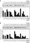
In vitro activity assays for various mutants of L/F-transferase. (A) The activity of [14C]Leu incorporation into α-casein from [14C]Leu-tRNALeu by mutant L/F-transferases (see Materials and methods). The upper gel shows the [14C]Leu-labeled α-casein. The lower graph shows the quantification of the relative intensity of the labeled product of the upper gel, where the incorporation of [14C]Leu into α-casein by wild-type L/F-transferase was taken as 1.0. The bars on the graph indicate the s.d. of more than three independent experiments. (B) The activity of [14C]Phe incorporation into α-casein from [14C]Phe-tRNAPhe by mutant L/F-transferases, as in (A).
Figure 5
Recognition of the aminoacyl-moiety by L/F-transferase. Recognition of the side chains of Leu (A) and Phe (B) by L/F-transferase. The α- and β-carbons of Leu and Phe were superimposed onto those of puromycin (colored orange). Discrimination of Ile (C) and Val (D) by L/F-transferase. The α- and β-carbons of Ile and Val were superimposed as in (A) and (B). The steric hindrance between the branched methyl groups at the β-carbons of Ile and Val. The C-shaped edge of the hydrophobic pocket is colored yellow. The chemical structures of each amino acid are depicted in parentheses.
Figure 6
Model of aminoacyl-tRNA binding to L/F-transferase. (A) Two views of a ribbon diagram of the docking model of L/F-transferase and tRNA. The tRNA backbone is shown as a green line (upper two panels). Two views of the L/F-transferase-tRNA complex model, showing the surface colored according to its calculated electrostatic potential (lower panel; blue, positively charged +8KT; red, negatively charged –8KT). The electrostatic surface model was calculated by the program APBS (Baker et al, 2001). The upper view displays the top side of the complex of front views. (B) Electrostatic potentials of _wv_FemX (upper) and _sa_FemA (lower). The marked regions on the diagrams show the positively charged regions corresponding to α2 of L/F-transferase and predicted as RNA binding regions. (C) The simultaneous tRNA binding to L/F-transferase (colored green) and EF-Tu (colored pink). The tRNA phosphate backbones are shown as a green line and a pink line for the L/F-transferase-tRNA complex and the EE-Tu-tRNA complex, respectively. The EF-Tu and Cys-tRNACys complex structure was from PDB 1B23 (Nissen et al, 1999). The 3′-terminus of Cys-tRNACys was manually modeled to accommodate the puromycin binding pocket of L/F-transferase.
Similar articles
- The crystal structure of leucyl/phenylalanyl-tRNA-protein transferase from Escherichia coli.
Dong X, Kato-Murayama M, Muramatsu T, Mori H, Shirouzu M, Bessho Y, Yokoyama S. Dong X, et al. Protein Sci. 2007 Mar;16(3):528-34. doi: 10.1110/ps.062616107. Epub 2007 Jan 22. Protein Sci. 2007. PMID: 17242373 Free PMC article. - Probing the leucyl/phenylalanyl tRNA protein transferase active site with tRNA substrate analogues.
Fung AW, Ebhardt HA, Krishnakumar KS, Moore J, Xu Z, Strazewski P, Fahlman RP. Fung AW, et al. Protein Pept Lett. 2014 Jul;21(7):603-14. doi: 10.2174/0929866521666140212110639. Protein Pept Lett. 2014. PMID: 24521222 - An alternative mechanism for the catalysis of peptide bond formation by L/F transferase: substrate binding and orientation.
Fung AW, Ebhardt HA, Abeysundara H, Moore J, Xu Z, Fahlman RP. Fung AW, et al. J Mol Biol. 2011 Jun 17;409(4):617-29. doi: 10.1016/j.jmb.2011.04.033. Epub 2011 Apr 20. J Mol Biol. 2011. PMID: 21530538 - The molecular basis for the post-translational addition of amino acids by L/F transferase in the N-end rule pathway.
Fung AW, Fahlman RP. Fung AW, et al. Curr Protein Pept Sci. 2015;16(2):163-80. Curr Protein Pept Sci. 2015. PMID: 25692952 Review. - Perspectives and Insights into the Competition for Aminoacyl-tRNAs between the Translational Machinery and for tRNA Dependent Non-Ribosomal Peptide Bond Formation.
Fung AW, Payoe R, Fahlman RP. Fung AW, et al. Life (Basel). 2015 Dec 31;6(1):2. doi: 10.3390/life6010002. Life (Basel). 2015. PMID: 26729173 Free PMC article. Review.
Cited by
- Idiosyncratic features in tRNAs participating in bacterial cell wall synthesis.
Villet R, Fonvielle M, Busca P, Chemama M, Maillard AP, Hugonnet JE, Dubost L, Marie A, Josseaume N, Mesnage S, Mayer C, Valéry JM, Ethève-Quelquejeu M, Arthur M. Villet R, et al. Nucleic Acids Res. 2007;35(20):6870-83. doi: 10.1093/nar/gkm778. Epub 2007 Oct 11. Nucleic Acids Res. 2007. PMID: 17932062 Free PMC article. - Unraveling the biochemistry and provenance of pupylation: a prokaryotic analog of ubiquitination.
Iyer LM, Burroughs AM, Aravind L. Iyer LM, et al. Biol Direct. 2008 Nov 3;3:45. doi: 10.1186/1745-6150-3-45. Biol Direct. 2008. PMID: 18980670 Free PMC article. - The determination of tRNALeu recognition nucleotides for Escherichia coli L/F transferase.
Fung AW, Leung CC, Fahlman RP. Fung AW, et al. RNA. 2014 Aug;20(8):1210-22. doi: 10.1261/rna.044529.114. Epub 2014 Jun 16. RNA. 2014. PMID: 24935875 Free PMC article. - Amidoligases with ATP-grasp, glutamine synthetase-like and acetyltransferase-like domains: synthesis of novel metabolites and peptide modifications of proteins.
Iyer LM, Abhiman S, Maxwell Burroughs A, Aravind L. Iyer LM, et al. Mol Biosyst. 2009 Dec;5(12):1636-60. doi: 10.1039/b917682a. Epub 2009 Oct 13. Mol Biosyst. 2009. PMID: 20023723 Free PMC article. - New biochemistry in the Rhodanese-phosphatase superfamily: emerging roles in diverse metabolic processes, nucleic acid modifications, and biological conflicts.
Burroughs AM, Aravind L. Burroughs AM, et al. NAR Genom Bioinform. 2023 Mar 23;5(1):lqad029. doi: 10.1093/nargab/lqad029. eCollection 2023 Mar. NAR Genom Bioinform. 2023. PMID: 36968430 Free PMC article.
References
- Abramochkin G, Shrader TE (1996) Aminoacyl-tRNA recognition by the leucyl/phenylalanyl-tRNA-protein transferase. J Biol Chem 271: 22901–22907 - PubMed
- Benson TE, Prince DB, Mutchler VT, Curry KA, Ho AM, Sarver RW, Hagadorn JC, Choi GH, Garlick RL (2002) X-ray crystal structure of Staphylococcus aureus FemA. Structure 10: 1107–1115 - PubMed
- Biarrotte-Sorin S, Maillard AP, Delettre J, Sougakoff W, Arthur M, Mayer C (2004) Crystal structures of Weissella viridescens FemX and its complex with UDP-MurNAc-pentapeptide: insights into FemABX family substrates recognition. Structure 12: 257–267 - PubMed
- Brunger AT, Adams PD, Clore GM, DeLano WL, Gros P, Grosse-Kunstleve RW, Jiang JS, Kuszewski J, Nilges M, Pannu NS, Read RJ, Rice LM, Simonson T, Warren GL (1998) Crystallography & NMR system: a new software suite for macromolecular structure determination. Acta Crystallogr D 54: 905–921 - PubMed
Publication types
MeSH terms
Substances
LinkOut - more resources
Full Text Sources
Molecular Biology Databases
Research Materials
Miscellaneous


