T-cell recognition of human melanoma antigens - PubMed (original) (raw)
Review
T-cell recognition of human melanoma antigens
Y Kawakami et al. J Immunother Emphasis Tumor Immunol. 1993 Aug.
Abstract
The adoptive transfer of tumor-infiltrating lymphocytes (TILs) with interleukin-2 (IL-2) has antitumor activity in some patients with metastatic melanoma. We have analyzed molecular mechanisms of TIL recognition of human melanoma. Some cultured TILs specifically lysed autologous and some allogeneic melanomas sharing a variety of class I major histocompatibility complex (MHC) molecules. HLA-A2-restricted melanoma-specific TILs lysed many HLA-A2+ melanoma cell lines from different patients but failed to lyse HLA-A2- melanoma and HLA-A2+ nonmelanoma cell lines. However, these TILs were capable of lysing many naturally HLA-A2- melanomas after introduction of the HLA-A2.1 gene by vaccinia virus. These results indicate that shared melanoma antigens (Ag) are expressed in melanomas regardless of their human leukocyte antigen types. In order to identify these shared melanoma Ags, we have tested some known proteins expressed in melanoma. Expression of tyrosinase or HMB45 Ag correlated with lysis of TILs. We are also attempting to isolate antigenic peptides by high performance liquid chromatography separation and genes encoding melanoma Ag by cDNA expression cloning. The T-cell component of the antimelanoma response was also analyzed by determining the genetic structure of the T-cell receptor (TCR) used by melanoma TILs. However, we did not observe common TCR variable region usage by different melanoma TILs. We could establish melanoma cell clones and lines resistant to TIL lysis due to the absence of or defects in the expression of Ag, MHC, or beta 2-microglobulin molecules. These data indicate multiple mechanisms for melanoma escape from T-cell immunosurveillance.(ABSTRACT TRUNCATED AT 250 WORDS)
Figures
FIG. 1
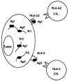
T-cell recognition of melanoma antigenic peptides in the context of different class I MHCs. Common (Pr1) or different (Pr2, 3) melanoma Ag proteins may provide Ag peptides (Ag1, Ag2) to different MHCs, and these Ag peptides may be recognized by T-cells in the context of different MHCs. If such common Ag proteins exist, we may be able to use them to immunize patients expressing a variety of HLA types.
Similar articles
- Common expression of melanoma tumor-associated antigens recognized by human tumor infiltrating lymphocytes: analysis by human lymphocyte antigen restriction.
Hom SS, Topalian SL, Simonis T, Mancini M, Rosenberg SA. Hom SS, et al. J Immunother (1991). 1991 Jun;10(3):153-64. J Immunother (1991). 1991. PMID: 1868040 - T cell receptor (TCR) structure of autologous melanoma-reactive cytotoxic T lymphocyte (CTL) clones: tumor-infiltrating lymphocytes overexpress in vivo the TCR beta chain sequence used by an HLA-A2-restricted and melanocyte-lineage-specific CTL clone.
Sensi M, Salvi S, Castelli C, Maccalli C, Mazzocchi A, Mortarini R, Nicolini G, Herlyn M, Parmiani G, Anichini A. Sensi M, et al. J Exp Med. 1993 Oct 1;178(4):1231-46. doi: 10.1084/jem.178.4.1231. J Exp Med. 1993. PMID: 8376931 Free PMC article. - Shared human melanoma antigens. Recognition by tumor-infiltrating lymphocytes in HLA-A2.1-transfected melanomas.
Kawakami Y, Zakut R, Topalian SL, Stötter H, Rosenberg SA. Kawakami Y, et al. J Immunol. 1992 Jan 15;148(2):638-43. J Immunol. 1992. PMID: 1729379 - Recognition of shared melanoma antigens by human tumor-infiltrating lymphocytes.
Topalian SL, Hom SS, Kawakami Y, Mancini M, Schwartzentruber DJ, Zakut R, Rosenberg SA. Topalian SL, et al. J Immunother (1991). 1992 Oct;12(3):203-6. doi: 10.1097/00002371-199210000-00013. J Immunother (1991). 1992. PMID: 1445813 Review. - Lymphocyte-melanoma interaction: role of surface molecules.
Becker JC, Bröcker EB. Becker JC, et al. Recent Results Cancer Res. 1995;139:205-14. doi: 10.1007/978-3-642-78771-3_15. Recent Results Cancer Res. 1995. PMID: 7597291 Review.
Cited by
- New dimensions in vaccinology: A new insight.
Tomar D, Chattree V, Tripathi V, Khan AA, Bakshi AR, Rao DN. Tomar D, et al. Indian J Clin Biochem. 2005 Jan;20(1):213-30. doi: 10.1007/BF02893073. Indian J Clin Biochem. 2005. PMID: 23105525 Free PMC article. - Cloning of the gene coding for a shared human melanoma antigen recognized by autologous T cells infiltrating into tumor.
Kawakami Y, Eliyahu S, Delgado CH, Robbins PF, Rivoltini L, Topalian SL, Miki T, Rosenberg SA. Kawakami Y, et al. Proc Natl Acad Sci U S A. 1994 Apr 26;91(9):3515-9. doi: 10.1073/pnas.91.9.3515. Proc Natl Acad Sci U S A. 1994. PMID: 8170938 Free PMC article. - Identification of a human melanoma antigen recognized by tumor-infiltrating lymphocytes associated with in vivo tumor rejection.
Kawakami Y, Eliyahu S, Delgado CH, Robbins PF, Sakaguchi K, Appella E, Yannelli JR, Adema GJ, Miki T, Rosenberg SA. Kawakami Y, et al. Proc Natl Acad Sci U S A. 1994 Jul 5;91(14):6458-62. doi: 10.1073/pnas.91.14.6458. Proc Natl Acad Sci U S A. 1994. PMID: 8022805 Free PMC article. - CCL5 mediates breast cancer metastasis and prognosis through CCR5/Treg cells.
Qiu J, Xu L, Zeng X, Wu H, Liang F, Lv Q, Du Z. Qiu J, et al. Front Oncol. 2022 Aug 10;12:972383. doi: 10.3389/fonc.2022.972383. eCollection 2022. Front Oncol. 2022. PMID: 36033472 Free PMC article. - Why do tumor-infiltrating lymphocytes have variable efficacy in the treatment of solid tumors?
Li B. Li B. Front Immunol. 2022 Oct 21;13:973881. doi: 10.3389/fimmu.2022.973881. eCollection 2022. Front Immunol. 2022. PMID: 36341370 Free PMC article. Review.
References
- Rosenberg SA, Packard BS, Aebersold PM, et al. Use of tumor-infiltrating lymphocytes and interleukin-2 in the immunotherapy of patients with metastatic melanoma: a preliminary report. N Engl J Med. 1988;319:1676–80. - PubMed
- Pockaj BA, Sherry R, Wei J, et al. Localization of 111 indium-labeled tumor infiltrating lymphocytes to tumor in patients receiving adoptive immunotherapy: augmentation with cyclophosphamide and association with response. (submitted) - PubMed
- Topalian SL, Solomon D, Rosenberg SA. Tumor specific cytolysis by lymphocytes infiltrating human melanomas. J Immunol. 1989;142:3714–25. - PubMed
- Schwartzentruber DJ, Topalian SL, Mancini M, Rosenberg SA. Specific release of granulocyte-macrophage colony-stimulating factor, tumor necrosis factor-α, and IFN-γ by human tumor-infiltrating lymphocytes after autologous tumor stimulation. J Immunol. 1991;146:3674–81. - PubMed
Publication types
MeSH terms
Substances
LinkOut - more resources
Full Text Sources
Other Literature Sources
Medical
Research Materials
