Anatomy and Physiology of Human Ear (original) (raw)
Last Updated : 25 May, 2026
The human ear is a specialised sensory organ responsible for hearing and maintaining body balance. It detects sound waves from the environment and converts them into nerve impulses that are interpreted by the brain. In addition to hearing, the ear also helps maintain equilibrium and body posture.
Anatomy of the Human Ear
The ear is structurally complex and can be divided into three main parts, and each part performs specific functions in hearing and balance.
1. Outer Ear
The outer ear is the visible part of the ear and is responsible for collecting sound waves.
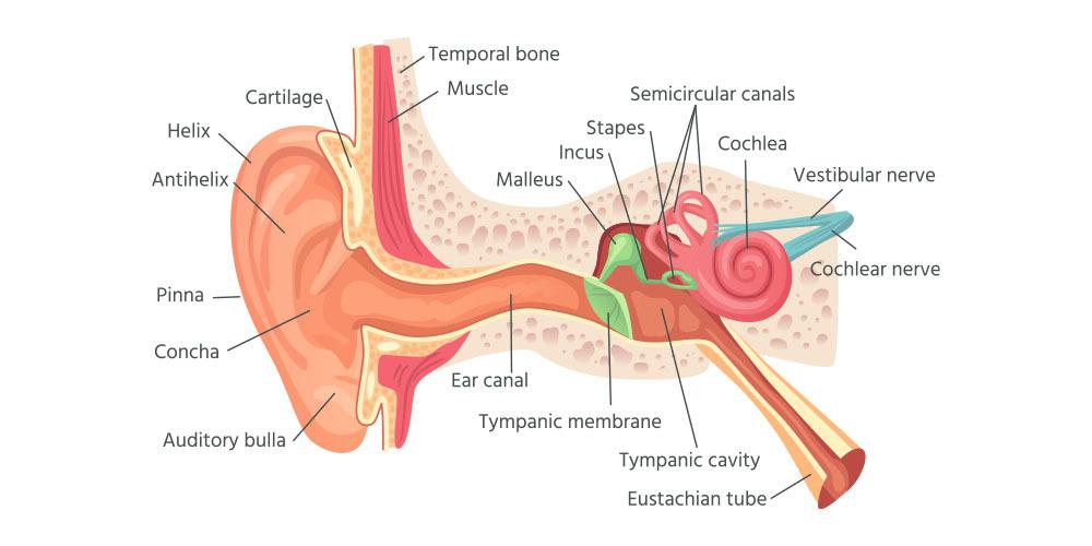
**Components of the Outer Ear:
****(a) Pinna (Auricle)**
- The pinna is the external flap-like structure of the ear.
- Made of elastic cartilage covered by skin.
- Helps collect and direct sound waves into the ear canal.
- Collects sound waves.
- Helps determine the direction of sound.
****(b) External Auditory Canal**
- A tube-like passage leading inward from the pinna.
- Lined with hairs and wax-secreting glands.
- Carries sound waves to the tympanic membrane.
- Earwax protects against dust and microbes.
****(c) Tympanic Membrane (Eardrum)**
- Thin membrane separating the outer and middle ear.
- Vibrates when sound waves strike it.
- Converts sound waves into mechanical vibrations.
2. Middle Ear
The middle ear is an air-filled chamber located between the outer and inner ear.
**Components of the Middle Ear:
****(a) Ear Ossicles**
- The middle ear contains three tiny bones called auditory ossicles: Malleus (Hammer), Incus (Anvil), and Stapes (Stirrup).
- The stapes is the smallest bone in the human body.
- Amplify and transmit sound vibrations to the inner ear.
****(b) Eustachian Tube**
- Connects the middle ear to the pharynx.
- Maintains equal air pressure on both sides of the eardrum.
- Helps proper vibration of the tympanic membrane.
3. Inner Ear
The inner ear is the most delicate and complex part of the ear. It contains structures responsible for hearing and balance.
**Components of the Inner Ear:
****(a) Cochlea:**
- Spiral-shaped, coiled structure and filled with fluid.
- Inside the cochlea lies the Organ of Corti, which contains sensory hair cells.
- Converts mechanical vibrations into nerve impulses.
****(b) Vestibular Apparatus**
- It includes semicircular canals, utricle, and saccule.
- Maintains body balance and equilibrium.
Physiology of Hearing
The process of hearing involves several steps.
**Step 1: Collection of Sound Waves
The pinna collects sound waves and directs them through the auditory canal toward the eardrum.
**Step 2: Vibration of the Tympanic Membrane
Sound waves strike the tympanic membrane (eardrum), causing it to vibrate.
**Step 3: Amplification by Ear Ossicles
Vibrations are transmitted through the malleus, incus, and stapes, which amplify the sound vibrations.
**Step 4: Transmission to the Inner Ear
The stapes transfers vibrations to the oval window of the cochlea, producing pressure waves in the cochlear fluid.
**Step 5: Stimulation of Hair Cells
Movement of the cochlear fluid bends the hair cells present in the Organ of Corti, generating nerve impulses.
**Step 6: Transmission to the Brain
The auditory nerve carries these nerve impulses to the brain, where they are interpreted as sound.
Functions of the Human Ear
- Hearing.
- Maintenance of balance.
- Coordination of body posture.
- Detection of sound direction.
Disorders of the Ear
- **Deafness: Loss of hearing ability due to damage to ear structures or the auditory nerve.
- **Otitis: Inflammation or infection of the ear.
- **Vertigo: Sensation of dizziness due to balance disorders.
Importance of the Human Ear
- Enables communication through hearing.
- Helps maintain equilibrium.
- Assists in environmental awareness.