NMR Metabolomic Profiling Reveals New Roles of SUMOylation in DNA Damage Response (original) (raw)
. Author manuscript; available in PMC: 2011 Oct 1.
Published in final edited form as: J Proteome Res. 2010 Oct 1;9(10):5382–5388. doi: 10.1021/pr100614a
Abstract
Post-translational modifications by the Small Ubiquitin-like Modifier (SUMO) family of proteins have been established as critical events in the cellular response to a wide range of DNA damaging reagents and radiation; however, the detailed mechanism of SUMOylation in DNA damage response is not well understood. In this study, we used nuclear magnetic resonance (NMR) spectroscopy based metabolomics approach to examine the effect of an inhibitor of SUMO-mediated protein-protein interactions on MCF7 breast cancer cell response to radiation. Metabolomics is sensitive to changes in cellular functions, and thus provides complementary information to other biological studies. The peptide inhibitor (SUMO interaction motif mimic, SIM) and a control peptide were stably expressed in MCF-7 cell line. Metabolite profiles of the cell lines before and after radiation were analyzed using solution NMR methods. Various statistical methods were used to isolate significant changes. Differences in the amounts of glutamine, aspartate, malate, alanine, glutamate and NADH between the SIM-expressing and control cells suggest a role for SUMOylation in regulating mitochondrial function. This is also further verified following the metabolism of 13C-labeled glutamine. The inability of the cells expressing the SIM peptide to increase production of the antioxidants carnosine and glutathione after radiation damage suggests an important role of SUMOylation in regulating the levels of antioxidants that protect cells from free radicals and reactive oxygen species generated by radiation. This study reveals previously unknown roles of SUMOylation in DNA damage response.
Introduction
DNA damage response mechanisms are critical to the protection of genome integrity as well as to cancer therapy, because both radiation and chemotherapy kill cancer cells by inducing DNA damage that leads to genomic instability and/or cell death. Post-translational modifications play a central role in prompt cellular response to DNA damage. For example, upon DNA damage, response proteins localize to sites of DNA damage within seconds.1 Such a fast response cannot be the result of changes in expression levels of repair proteins, but rather is due to mechanisms regulated by post-translational modifications, such as phosphorylation2 and acetylation.3 Post-translational modifications are also involved in signaling, cell cycle checkpoints and apoptosis associated with DNA damage.4
Post-translational modifications with the Small Ubiquitin-like Modifier (SUMO) family of proteins have recently been established as a critical post-translational modification required in cellular response to a wide range of DNA damaging reagents.5-8 For over a decade, it has been known that cells defective in SUMOylation have increased sensitivity to a wide variety of DNA damaging reagents as well as to radiation.7, 8 Like ubiquitination, SUMOylation results in covalent attachment of one or a chain of SUMO proteins to other cellular proteins. In most cases, ubiquitin-like proteins serve as platforms for interactions with other proteins.9, 10 The SUMO proteins form a ubiquitin-like fold in their central regions, which contain an α-helix packed on a β-sheet.11-13 However, the amino acid residues at the protein surfaces of SUMO and ubiquitin are very different, resulting in completely different surface properties between them. The unique surface composition of the SUMO proteins dictates its specific protein-protein interaction networks, and consequently its post-modification effects, and thus SUMOylation does not directly target proteins for degradation by the Proteosome. Instead, SUMOylation regulates protein localization and protein-protein interactions in most cases.14
Previously, we have identified the SUMO-binding (or interacting) motif (SBM, SIM) 15, 16 that mediates SUMO-dependent protein-protein interactions for nearly all known SUMO-dependent down-stream effects. In a recent study, we found that expression of a peptide containing the SIM sequence, which inhibits SUMO-mediated protein-protein interactions, significantly enhances the susceptibility of cancer cells to radiation and chemotherapeutic drugs that cause DNA damage.17 In particular, the SIM peptide inhibits the non-homologous end joining (NHEJ) pathway, which is the major pathway for the repair of DNA double-strand breaks (DSB), suggesting that the SUMO:SIM mediated protein-protein interactions are important in such processes. Other recent studies have indicated that SUMOylation is also important for the homologous recombination DNA repair pathway.18-20 All of these studies suggest that SUMOylation is important for the repair of DNA damage. Nevertheless, it remains unclear whether SUMOylation is important for other aspects of cellular response to DNA damaging agents and radiation.
We have previously created MCF7 cell lines that stably express either a SIM peptide or a control peptide in order to study the role of SUMO:SIM-meditated protein-protein interactions in DNA damage response.17 We have shown that the SIM peptide did not confer noticeable toxicity to cells, but significantly increased cell sensitivity to DNA damaging chemotherapeutic drugs and radiation.17 In order to gain insights into cellular pathways that are responsible for the increased sensitivity to DNA damaging agents, we carried out NMR based metabolomics analysis to compare the cell lines expressing the SIM and control peptide. Metabolomics refers to the global profiling of metabolites in cells.21, 22 Unlike other systems biology approaches, metabolomics directly reflects acute cellular status. Metabolomics permits the quantification of non-protein small molecules, including Krebs cycle intermediates, free amino acids, cytosolic components, which can be end-products, intermediates or ligands triggering biological pathways. Thus metabolomics provide complimentary information to other biological approaches.
In this study, we employed NMR based metabolomics to identify other potential pathways in which SUMOylation plays in DNA damage response. We identified small, but reproducible differences in the concentrations of several metabolites between the MCF7 cell line expressing the SIM peptide and that expressing the control peptide. The differences in metabolite concentrations indicate that the SIM peptide alters mitochondria functions and the production of glutathione, which is important for removing reactive oxygen species. Altered mitochondria function can result in increased production of free radicals from electron transfer chains.23, 24 In addition, we found a significant effect of the SIM peptide inhibiting the production of carnosine upon radiation. This molecule is a naturally occurring dipeptide antioxidant and a free radical scavenger.25, 26 Free radicals, such as hydroxyl radicals which arise from the absorption of ionizing radiation by water, are the major cause of DNA damage by ionizing radiation.27, 28 Thus, results from this study indicate that SUMO:SIM-mediated protein-protein interactions play a previously unappreciated role in DNA damage response – protection of DNA from free radicals through mitochondria-dependent and -independent mechanisms.
Experimental Procedures
Cell lines and cultures
Cell lines stably expressing the SIM mimic peptide and those with the control peptide were grown following typical culturing techniques for MCF-7 cells. Cultures were maintained in DMEM (Mediatech, Inc., Manassas, VA) supplemented with sodium pyruvate, 2 mM glutamine, 10% v/v fetal bovine serum (Omega Scientific, Tarzana, CA), non-essential amino acids (Irvine Scientific, Santa Ana, CA), penicillin-streptomycin and 1 mg/ml G418 (Invitrogen, Carlsbad, CA) in a humidified 37 °C incubator with 5% CO2. To obtain approximately 1×10⁷ cells per sample, cells were grown in one T175 culture flask per cell line per sample to 85-90% confluency. Three 100 mm petri dishes can be used instead of a filter cap culture flask to achieve the same cell number. Two culture flasks were prepared identically for the SIM-expressing cells and for the control peptide-expressing cells; one flask for each cell line was exposed to 4 grays of cesium-137 gamma radiation. Cultures were returned to the 37 °C incubator for 2 hours to allow repair and recovery pathways to commence. This experimental set was consecutively repeated with subsequent passages for a total of three trials.
Sample preparation for NMR spectroscopy
Mechanical harvesting was used to lift cells into the medium. The Sarstedt cell scrapers recommended for these procedures are comprised of soft rubber (Sarstedt, Inc., Newton, NC), minimizing cell lysis during the cell detachment procedure. Cells and medium were transferred into a 50 ml conical tube and centrifuged at 1000 rpm for 5 minutes. Media were removed by aspiration and the cells washed twice with sterile PBS (phosphate buffered saline; Irvine Scientific, Santa Ana, CA); any residual media will lead to skewing of metabolite concentrations. In order to aid identification of water-soluble compounds and hydrophobic compounds, a chemical extraction protocol was utilized for metabolite preparation from cells. This type of dual phase extraction has the added benefit of removing proteins from the cell sample;29, 30 this will produce cleaner chemical shift data and it will prevent enzymatic reactions from occurring that could alter the metabolomic profile. The wet mass of the harvested sample was used to calculate the appropriate amount of solvent; samples were resuspended in 7.4 ml/g cold methanol and 2.22 ml/g cold water. Cells underwent sonication in an ice-bath at 25% duty cycle for 1 minute on and 1 minute off, repeated three times. The homogenate was transferred to a borosilicate glass culture tube, then 3.7 ml/g cold chloroform was added. The tube opening was covered with aluminum foil and sealed with parafilm to prevent evaporation. The suspension was vortexed and kept at 4 °C overnight. To this monophasic solution, 3.7 ml/g cold chloroform and 3.7 ml/g cold water were added, and the solutions were vortexed and centrifuged for 5 minutes. The upper hydrophilic layer and the lower lipophilic layer were carefully transferred to separate clean tubes and dried in a speed-vac concentrator.
1H NMR spectroscopy
Hydrophilic samples dried from the methanol-water fractions were each resuspended in 400 μl 100% D2O containing 6.4 μM DSS (Cambridge Isotope Laboratories, MA). The internal standard 4,4-dimethyl-4-silapentane-1-sulfonic acid served as a chemical shift reference and a concentration standard. One-dimensional spectra of polar samples were acquired at 25 °C on a Bruker Avance 600 spectrometer using a sequence with water suppression method WATERGATE W5;31 256 transients and 32 K data points were collected with a spectral width of 16 ppm. Dried lipophilic samples were saved for later analysis.
NMR Data and Statistical Analysis
Spectra were imported into the Chenomx NMR Suite Processor (version 6.0, Chenomx Inc., Edmonton, Canada) for Fourier transformation, a cubic-spline-based baseline adjustment and phasing. Chemical shifts were referenced to the methyl protons of DSS. The integral of the DSS methyl proton peak was set within Chenomx to represent 6.4 μM DSS. The Chenomx NMR Suite Profiler was used to identify metabolites by fitting compound signatures from the provided NMR spectral library. Metabolite concentrations were calculated using Chenomx by determining heights of compound signatures that best fit the sample spectra. The table of identified and calculated concentrations were exported and saved in an Excel worksheet. The concentrations were converted from micromolar units to nanomoles per gram by multiplying the NMR sample volume and dividing the wet mass of the sample. For each set of samples (each cell line, with or without radiation treatment), we calculated in Excel the fold change for both cell lines (i.e. log2(after radiation/before radiation)); significant changes were determined using a Student's t-test with a critical α=0.05.
Results
NMR profiling of metabolites in MCF7 cells
In order to compare the SIM and control expression cell lines both before and after radiation, each cell line was cultured in two sets of flasks. One set was harvested and followed by metabolite extraction, and the other set was irradiated with 4 grays gamma radiation and allowed to recover for 2 hours before harvesting for metabolite extraction. The two hour recovery was used, because we have previously shown that DNA damage in the cells expressing the control peptide was nearly fully recovered after two hours, but not in cells expressing the SIM peptide.17 The hydrophilic and hydrophobic metabolites were obtained and separated using a methanol/chloroform extraction from cultured MCF7 cells expressing either the SIM or control peptide. NMR spectra of the hydrophilic fractions were collected. Figure 1 shows representative spectra of the two cell lines with assignments of the metabolites indicated. Chenomx NMR Suite software was utilized to identify the compounds visible in the spectra. Most compounds can be identified based on their standard NMR spectra in various metabolite databases including the Human Metabolome Database,32 Madison Metabolomics Consortium Database,33 and Biological Magnetic Resonance Data Bank.34, 35 The compounds identified in the MCF-7 cells include amino acids, glycolysis products, TCA intermediates and choline derivatives.
Figure 1.
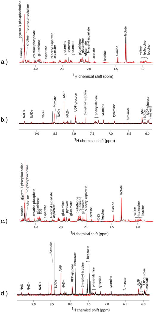
Effect of SUMOylation on the metabolic profile of MCF-7 cells. Proton NMR spectra of the hydrophilic metabolites from cells not exposed to radiation (A & B)) and two hours after exposure to 4 Gy ionizing radiation (C & D). Spectra in black represent typical results from the cells expressing the control peptide, while those in red represent the cells expressing the SIM peptide.
In order to draw meaningful conclusions, differences in metabolite concentration resulting from slight variations in culturing and sample preparation conditions and cell passage numbers need to be eliminated. Thus, the entire process of cell culturing, metabolite extraction and NMR data collection was repeated multiple times. Variations were observed in the metabolite NMR spectra for cells from the same origin but cultured at different times. Possible reasons for such variations could be differences in culture confluency at time of harvest. The largest variants were N,N-dimethylformamide (DMF; 8, 3 and 2.9 ppm) peaks. These were found in our samples inconsistently as a result of contamination from the speed vacuum used to dry the solvent after water/ethanol extraction; the same speed vacuum was also used by others to concentrate samples dissolved in DMF, concurrently with our samples.
The SIM peptide alters mitochondria activities
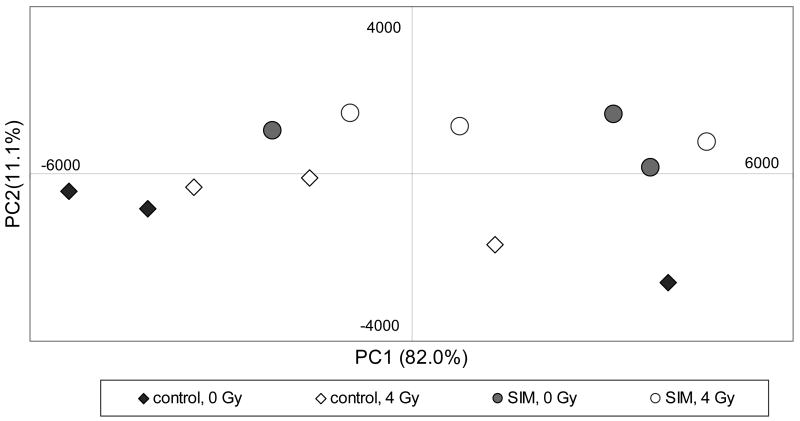
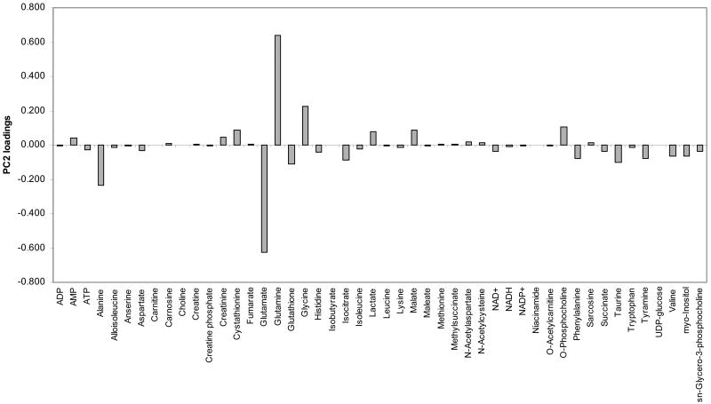
In order to analyze whether the two groups of spectra from cells expressing the SIM or control peptide are significantly different, we employed principal components analysis (PCA) to identify the compounds that are statistically different between the two groups of three repeats. The concentrations of each sample were used as input to calculate the principal components and scores using StatistiXL software (version 1.8; StatistiXL, Nedlands, Western Australia). There is a clear distinction along the PC2 axis between the SIM-expressing cells and the control cells regardless of radiation treatment (Figure 2), and inspection of the PC2 loadings shows glutamine, glutamate, alanine and glycine as key features (Figure 3). These metabolites are directly linked to mitochondrial function. Therefore, PCA analysis indicates that the SIM peptide leads to alteration in mitochondrial function. As we previously demonstrated using fluorescence microscopy, the SIM peptide is localized to the nucleus, and not in the mitochondria.17 Thus, these results suggest that the SIM:SUMO-mediated protein-protein interactions are likely involved in regulating the production and balance of mitochondria components.
Figure 2.
Principal components analysis of metabolic differences between cell lines. PCA scores of components 1 and 2 from control (diamonds) and SIM-expressing cells (circles), with 4 Gy radiation treatment (open) or without (filled). The data from three sets of experiments are shown.
Figure 3.
Loadings for principal component 2 of the data from the three sets of experiments using the SIM or control expressing cell lines, with and without radiation.
In order to further probe and confirm the effect of the SIM peptide on mitochondria regulation, we compared glutamine uptake and metabolism between the two different cell lines. Glutamine is an important component of the cell culture media, and thus isotope 13C-labeled glutamine was used to follow its metabolism. In this experiment, the SIM and control cells were cultured similarly as before in media, supplemented with unlabeled glutamine, to 85-90% confluency. Prior to the dual phase extraction, the media were replaced with DMEM supplemented with uniformly 13C-labeled glutamine (Isotec Inc., Miamisburg, OH) and the cells were allowed to incubate for an additional 2 hours at 37 °C. The metabolites that were synthesized from the labeled glutamine within 2 hours were visible by 13C-HSQC (Figure 4). Glutamine and glutamate comprise the majority of the labeled metabolites, and concentrations of these two amino acids were similar in both cell lines. In contrast, the levels of malate and aspartate were consistently lower in the SIM-expressing cells than the control cells (Figure 4). Due to the fact that conversion of glutamine to malate and aspartate requires mitochondria (Figure 5), this result suggests that the SIM-expressing cells have mitochondrial dysfunction (most likely reduced activity) relative to control cells. Altered mitochondria activity can lead to increased production of free radicals and ROS from normal respiratory activity, and thus increased sensitivity to radiation and DNA damaging chemotherapeutic drugs.23, 24
Figure 4.
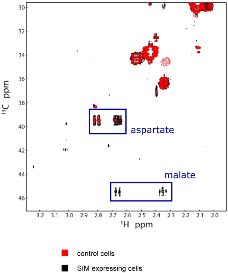
13C HSQC of cell metabolites after incubation with uniformly labeled 13C-glutamine. Extracts from control cells (black) and SIM cells (red).
Figure 5.
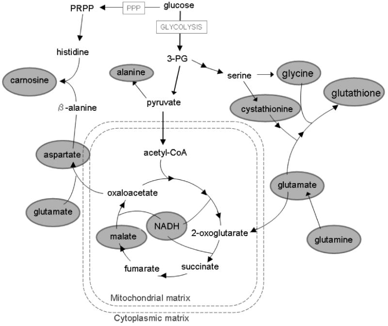
Diagram of metabolic pathways involved in radiation response, observed by NMR measurements. Circled metabolites indicate compounds with significant value changes between controls and SIM cells (p<0.05).
The role of SIM peptide in eliminating free radicals
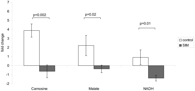
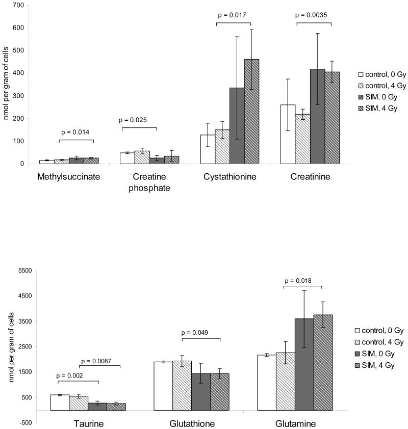
In order to compare how the SIM-expressing cells differ from the control cells in radiation response, we calculated the fold-change in concentrations of detectable metabolites after radiation for each cell line, with or without radiation. The cells with and without radiation were seeded at the same time and cultured together and were processed at the same time using identical procedures. Therefore, the ratios only reflect the changes due to radiation, and any differences due to cell passages or experimental conditions are minimized. Despite the varying levels of metabolites in cells cultured at different times, the ratios of some metabolites before and after radiation are very consistent with small variations from time to time. A Student's t-test revealed that the changes in carnosine, malate, and NADH between the cells expressing the functional SIM peptide and those with the mutant peptide were significantly different (p<0.5; Figure 6). Upon closer inspection of the average concentrations of these compounds, the levels of carnosine and malate increased considerably after radiation exposure in the cells expressing the control peptide. However, their levels did not increase, but decreased slightly after radiation in SIM (inhibitor)-expressing cells (Figure 6). A Student's t-test of the measured concentrations also revealed other metabolites that showed significant differences between the different cell types. Creatine-phosphate and taurine were observed at higher levels in control cells compared to the SIM-expressing cells when both groups were not treated with radiation (Figure 7). Methylsuccinate, cystathionine, creatinine, taurine, glutathione and glutamine levels were significantly different in the control cells expressing the control peptide compared to the SIM-peptide cells, after exposure to 4 grays of radiation treatment (Figure 7).
Figure 6.
Changes in cells as a result of radiation treatment. For controls (white bars) and SIM cells (gray bars), change was calculated as the log2 ratio of after radiation values to no radiation values. Bar graphs are average fold change ± standard deviation (n=3).
Figure 7.
Effect of inhibiting SUMOylation recognition on individual metabolites. Average measured amount of compounds from control cells with no treatment (white), controls with 4 Gy radiation (white with stripes), SIM cells without treatment (gray), and SIM cells after 4 Gy radiation (gray with stripes). Error bars are standard deviation. Brackets indicate which comparisons are significant according to Student's test p-values.
Discussion
This study highlights the complementary information provided by quantitative metabolomics studies. Although SUMOylation is established as being important in DNA damage response, the mechanisms of how SUMOylation is involved are not well understood, and most studies to date have focused on DNA repair pathways. Metabolomics studies described here have suggested previously unknown functions of SUMOylation in DNA damage response—regulating mitochondria function and the production of free radical reducers and scavengers, such as carnosine. Mitochondrial dysfunction or dysregulation can lead to increased sensitivity to radiation and DNA damaging reagents due to increased production of free radicals from the electron transfer chains.23, 24 The notable differences in glutathione and carnosine suggest that the SIM peptide inhibited the production of small molecules that are protective to cells from free radical induced damage to DNA and proteins. The major DNA damage by radiation is due to free radicals generated during radiation.27, 28 Both glutathione and carnosine are free radical scavengers, and thus increased levels of these compounds in control cell line should reduce the continuous damage of DNA by free radicals. The mechanism by which SUMOylation regulates mitochondrial function and the production of glutathione and carnosine remains to be elucidated by future molecular and cellular biology studies, for example investigating expression levels and regulation of relevant enzymes. The information provided in the current study promises to stimulate future studies in this area.
We have also demonstrated for the first time a direct way to assess mitochondria function in cancer cells using NMR and uniformly 13C-labeled glutamine. NMR measurements can directly probe the uptake and metabolism of glutamine and obtain information on mitochondrial function. The SIM-expressing cells do not show apparent differences in growth from the control cells, indicating that the SIM peptide, alone, is not toxic to cells. This is likely due to the fact that the SIM peptide did not have sufficient affinity to SUMO to inhibit all SUMO:SIM-mediated protein-protein interactions in cells. Nonetheless, comparison of the metabolite concentrations indicated differences in mitochondria functions between the SIM-expression and control cell lines. Moreover, metabolism of the 13C-labeled glutamine further suggested that the SIM peptide altered normal mitochondria function.
This study suggests that SIM-mimetics can be potential sensitizers in cancer therapy. Because of SUMOylation's role in tumor growth36 and metastasis;37 such an approach is likely to have other benefits. In particular, hypoxia, a common condition associated with aggressive tumors, can upregulate SUMO-1 levels by as much as 100-fold,38, 39 and hypoxic tumors are more resistant to chemo- and radiation therapies.40-45 In such cases, the SIM peptide should be particularly useful to inhibit the functions of increased levels of SUMO. Currently, small molecular mimmetics of SIM have not been reported. The findings described here justify efforts to develop such mimmetics that will be more stable than peptides and can be delivered in vivo in a more straightforward manner than peptides.
Acknowledgments
This work is funded by NIH Grants R01GM074748 and R01GM086171 to Y Chen, KE is the recipient of NIH Postdoctoral Training Grant to Promote Diversity in Health-Related Research Y-J Li is a recipient of the NIH National Research Service Award (F32CA134180).
References
- 1.Bekker-Jensen S, Lukas C, Melander F, Bartek J, Lukas J. Dynamic assembly and sustained retention of 53BP1 at the sites of DNA damage are controlled by Mdc1/NFBD1. J Cell Biol. 2005;170(2):201–11. doi: 10.1083/jcb.200503043. [DOI] [PMC free article] [PubMed] [Google Scholar]
- 2.Stucki M, Jackson SP. gammaH2AX and MDC1: anchoring the DNA-damage-response machinery to broken chromosomes. DNA Repair (Amst) 2006;5(5):534–43. doi: 10.1016/j.dnarep.2006.01.012. [DOI] [PubMed] [Google Scholar]
- 3.Bird AW, Yu DY, Pray-Grant MG, Qiu Q, Harmon KE, Megee PC, Grant PA, Smith MM, Christman MF. Acetylation of histone H4 by Esa1 is required for DNA double-strand break repair. Nature. 2002;419(6905):411–5. doi: 10.1038/nature01035. [DOI] [PubMed] [Google Scholar]
- 4.Huen MS, Chen J. Assembly of checkpoint and repair machineries at DNA damage sites. Trends Biochem Sci. 35(2):101–8. doi: 10.1016/j.tibs.2009.09.001. [DOI] [PMC free article] [PubMed] [Google Scholar]
- 5.Andrews EA, Palecek J, Sergeant J, Taylor E, Lehmann AR, Watts FZ. Nse2, a component of the Smc5-6 complex, is a SUMO ligase required for the response to DNA damage. Mol Cell Biol. 2005;25(1):185–96. doi: 10.1128/MCB.25.1.185-196.2005. [DOI] [PMC free article] [PubMed] [Google Scholar]
- 6.Zhao X, Blobel G. A SUMO ligase is part of a nuclear multiprotein complex that affects DNA repair and chromosomal organization. Proc Natl Acad Sci U S A. 2005 doi: 10.1073/pnas.0500537102. [DOI] [PMC free article] [PubMed] [Google Scholar]
- 7.Seufert W, Futcher B, Jentsch S. Role of a ubiquitin-conjugating enzyme in degradation of S- and M-phase cyclins. Nature. 1995;373(6509):78–81. doi: 10.1038/373078a0. [DOI] [PubMed] [Google Scholar]
- 8.Shayeghi M, Doe CL, Tavassoli M, Watts FZ. Characterisation of Schizosaccharomyces pombe rad31, a UBA-related gene required for DNA damage tolerance. Nucleic Acids Res. 1997;25(6):1162–9. doi: 10.1093/nar/25.6.1162. [DOI] [PMC free article] [PubMed] [Google Scholar]
- 9.Pichler A, Knipscheer P, Oberhofer E, van Dijk WJ, Korner R, Olsen JV, Jentsch S, Melchior F, Sixma TK. SUMO modification of the ubiquitin-conjugating enzyme E2-25K. Nat Struct Mol Biol. 2005;12(3):264–9. doi: 10.1038/nsmb903. [DOI] [PubMed] [Google Scholar]
- 10.Macauley MS, Errington WJ, Okon M, Scharpf M, Mackereth CD, Schulman BA, McIntosh LP. Structural and dynamic independence of isopeptide-linked RanGAP1 and SUMO-1. J Biol Chem. 2004;279(47):49131–7. doi: 10.1074/jbc.M408705200. [DOI] [PubMed] [Google Scholar]
- 11.Bayer P, Arndt A, Metzger S, Mahajan R, Melchior F, Jaenicke R, Becker J. Structure determination of the small ubiquitin-related modifier SUMO-1. J Mol Biol. 1998;280(2):275–86. doi: 10.1006/jmbi.1998.1839. [DOI] [PubMed] [Google Scholar]
- 12.Jin C, Shiyanova T, Shen Z, Liao X. Heteronuclear nuclear magnetic resonance assignments, structure and dynamics of SUMO-1, a human ubiquitin-like protein. Int J Biol Macromol. 2001;28(3):227–34. doi: 10.1016/s0141-8130(00)00169-0. [DOI] [PubMed] [Google Scholar]
- 13.Huang WC, Ko TP, Li SS, Wang AH. Crystal structures of the human SUMO-2 protein at 1.6 A and 1.2 A resolution: implication on the functional differences of SUMO proteins. Eur J Biochem. 2004;271(20):4114–22. doi: 10.1111/j.1432-1033.2004.04349.x. [DOI] [PubMed] [Google Scholar]
- 14.Meulmeester E, Melchior F. Cell biology: SUMO. Nature. 2008;452(7188):709–11. doi: 10.1038/452709a. [DOI] [PubMed] [Google Scholar]
- 15.Song J, Durrin LK, Wilkinson TA, Krontiris TG, Chen Y. Identification of a SUMO-binding motif that recognizes SUMO-modified proteins. Proc Natl Acad Sci U S A. 2004;101(40):14373–14378. doi: 10.1073/pnas.0403498101. [DOI] [PMC free article] [PubMed] [Google Scholar]
- 16.Song J, Zhang Z, Hu W, Chen Y. Small ubiquitin-like modifier (SUMO) recognition of a SUMO binding motif: a reversal of the bound orientation. J Biol Chem. 2005;280(48):40122–9. doi: 10.1074/jbc.M507059200. [DOI] [PubMed] [Google Scholar]
- 17.Li YJ, Stark JM, Chen DJ, Ann DK, Chen Y. Role of SUMO:SIM-mediated protein-protein interaction in non-homologous end joining. Oncogene. doi: 10.1038/onc.2010.108. [DOI] [PMC free article] [PubMed] [Google Scholar]
- 18.Bergink S, Jentsch S. Principles of ubiquitin and SUMO modifications in DNA repair. Nature. 2009;458(7237):461–7. doi: 10.1038/nature07963. [DOI] [PubMed] [Google Scholar]
- 19.Galanty Y, Belotserkovskaya R, Coates J, Polo S, Miller KM, Jackson SP. Mammalian SUMO E3-ligases PIAS1 and PIAS4 promote responses to DNA double-strand breaks. Nature. 2009;462(7275):935–9. doi: 10.1038/nature08657. [DOI] [PMC free article] [PubMed] [Google Scholar]
- 20.Morris JR, Boutell C, Keppler M, Densham R, Weekes D, Alamshah A, Butler L, Galanty Y, Pangon L, Kiuchi T, Ng T, Solomon E. The SUMO modification pathway is involved in the BRCA1 response to genotoxic stress. Nature. 2009;462(7275):886–90. doi: 10.1038/nature08593. [DOI] [PubMed] [Google Scholar]
- 21.Daviss B. Growing Pains for Metabolomics. The Scientist. 2005;19(8):25–28. [Google Scholar]
- 22.Nicholson JK, Lindon JC, Holmes E. ‘Metabonomics’: understanding the metabolic responses of living systems to pathophysiological stimuli via multivariate statistical analysis of biological NMR spectroscopic data. Xenobiotica. 1999;29(11):1181–1189. doi: 10.1080/004982599238047. [DOI] [PubMed] [Google Scholar]
- 23.Walker DW, Hajek P, Muffat J, Knoepfle D, Cornelison S, Attardi G, Benzer S. Hypersensitivity to oxygen and shortened lifespan in a Drosophila mitochondrial complex II mutant. Proc Natl Acad Sci U S A. 2006;103(44):16382–7. doi: 10.1073/pnas.0607918103. [DOI] [PMC free article] [PubMed] [Google Scholar]
- 24.Leeuwenburgh C, Heinecke JW. Oxidative stress and antioxidants in exercise. Curr Med Chem. 2001;8(7):829–38. doi: 10.2174/0929867013372896. [DOI] [PubMed] [Google Scholar]
- 25.La Mendola D, Sortino S, Vecchio G, Rizzarelli E. Synthesis of New Carnosine Derivatives of β-Cyclodextrin and Their Hydroxyl Radical Scavenger Ability. Helvetica Chimica Acta. 2002;85(6):1633–1643. [Google Scholar]
- 26.Boldyrev AA, Stvolinsky SL, Fedorova TN, Suslina ZA. Carnosine As a Natural Antioxidant and Geroprotector: From Molecular Mechanisms to Clinical Trials. Rejuvenation Research. 2010;13(2-3):156–158. doi: 10.1089/rej.2009.0923. [DOI] [PubMed] [Google Scholar]
- 27.Dainton FS. Radiation chemistry. IV. A review of the evidence for the production of free radicals in water consequent on the absorption of ionizing radiations. Br J Radiol. 1951;24(284):428–33. doi: 10.1259/0007-1285-24-284-428. [DOI] [PubMed] [Google Scholar]
- 28.Breen AP, Murphy JA. Reactions of oxyl radicals with DNA. Free Radical Biology and Medicine. 1995;18(6):1033–1077. doi: 10.1016/0891-5849(94)00209-3. [DOI] [PubMed] [Google Scholar]
- 29.Bligh EG, Dyer WJ. A rapid method of total lipid extraction and purification. Can J Biochem Physiol. 1959;37(8):911–7. doi: 10.1139/o59-099. [DOI] [PubMed] [Google Scholar]
- 30.Wu H, Southam AD, Hines A, Viant MR. High-throughput tissue extraction protocol for NMR- and MS-based metabolomics. Analytical Biochemistry. 2008;372(2):204–212. doi: 10.1016/j.ab.2007.10.002. [DOI] [PubMed] [Google Scholar]
- 31.Liu M, Mao Xa, Ye C, Huang H, Nicholson JK, Lindon JC. Improved WATERGATE Pulse Sequences for Solvent Suppression in NMR Spectroscopy. Journal of Magnetic Resonance. 1998;132(1):125–129. [Google Scholar]
- 32.Wishart DS, Knox C, Guo AC, Eisner R, Young N, Gautam B, Hau DD, Psychogios N, Dong E, Bouatra S, Mandal R, Sinelnikov I, Xia J, Jia L, Cruz JA, Lim E, Sobsey CA, Shrivastava S, Huang P, Liu P, Fang L, Peng J, Fradette R, Cheng D, Tzur D, Clements M, Lewis A, De Souza A, Zuniga A, Dawe M, Xiong Y, Clive D, Greiner R, Nazyrova A, Shaykhutdinov R, Li L, Vogel HJ, Forsythe I. HMDB: a knowledgebase for the human metabolome. Nucl Acids Res. 2009;37(suppl_1):D603–610. doi: 10.1093/nar/gkn810. [DOI] [PMC free article] [PubMed] [Google Scholar]
- 33.Cui Q, Lewis IA, Hegeman AD, Anderson ME, Li J, Schulte CF, Westler WM, Eghbalnia HR, Sussman MR, Markley JL. Metabolite identification via the Madison Metabolomics Consortium Database. Nat Biotech. 2008;26(2):162–164. doi: 10.1038/nbt0208-162. [DOI] [PubMed] [Google Scholar]
- 34.Ulrich EL, Akutsu H, Doreleijers JF, Harano Y, Ioannidis YE, Lin J, Livny M, Mading S, Maziuk D, Miller Z, Nakatani E, Schulte CF, Tolmie DE, Kent Wenger R, Yao H, Markley JL. BioMagResBank. Nucl Acids Res. 2008;36(suppl_1):D402–408. doi: 10.1093/nar/gkm957. [DOI] [PMC free article] [PubMed] [Google Scholar]
- 35.Robinette SL, Zhang F, Bruschweiler-Li L, Bruschweiler R. Web Server Based Complex Mixture Analysis by NMR. Analytical Chemistry. 2008;80(10):3606–3611. doi: 10.1021/ac702530t. [DOI] [PubMed] [Google Scholar]
- 36.Mo YY, Yu Y, Theodosiou E, Rachel Ee PL, Beck WT. A role for Ubc9 in tumorigenesis. Oncogene. 2005;24(16):2677–83. doi: 10.1038/sj.onc.1208210. [DOI] [PubMed] [Google Scholar]
- 37.Kim JH, Choi HJ, Kim B, Kim MH, Lee JM, Kim IS, Lee MH, Choi SJ, Kim KI, Kim SI, Chung CH, Baek SH. Roles of sumoylation of a reptin chromatin-remodelling complex in cancer metastasis. Nat Cell Biol. 2006;8(6):631–9. doi: 10.1038/ncb1415. [DOI] [PubMed] [Google Scholar]
- 38.Comerford KM, Leonard MO, Karhausen J, Carey R, Colgan SP, Taylor CT. Small ubiquitin-related modifier-1 modification mediates resolution of CREB-dependent responses to hypoxia. Proc Natl Acad Sci U S A. 2003;100(3):986–91. doi: 10.1073/pnas.0337412100. [DOI] [PMC free article] [PubMed] [Google Scholar]
- 39.Shao R, Zhang FP, Tian F, Anders Friberg P, Wang X, Sjoland H, Billig H. Increase of SUMO-1 expression in response to hypoxia: direct interaction with HIF-1alpha in adult mouse brain and heart in vivo. FEBS Lett. 2004;569(1-3):293–300. doi: 10.1016/j.febslet.2004.05.079. [DOI] [PubMed] [Google Scholar]
- 40.Harrison L, Blackwell K. Hypoxia and anemia: factors in decreased sensitivity to radiation therapy and chemotherapy? Oncologist. 2004;9 5:31–40. doi: 10.1634/theoncologist.9-90005-31. [DOI] [PubMed] [Google Scholar]
- 41.Xu RH, Pelicano H, Zhou Y, Carew JS, Feng L, Bhalla KN, Keating MJ, Huang P. Inhibition of glycolysis in cancer cells: a novel strategy to overcome drug resistance associated with mitochondrial respiratory defect and hypoxia. Cancer Res. 2005;65(2):613–21. [PubMed] [Google Scholar]
- 42.Unruh A, Ressel A, Mohamed HG, Johnson RS, Nadrowitz R, Richter E, Katschinski DM, Wenger RH. The hypoxia-inducible factor-1 alpha is a negative factor for tumor therapy. Oncogene. 2003;22(21):3213–20. doi: 10.1038/sj.onc.1206385. [DOI] [PubMed] [Google Scholar]
- 43.Brown JM. Tumor microenvironment and the response to anticancer therapy. Cancer Biol Ther. 2002;1(5):453–8. doi: 10.4161/cbt.1.5.157. [DOI] [PubMed] [Google Scholar]
- 44.Moeller BJ, Cao Y, Li CY, Dewhirst MW. Radiation activates HIF-1 to regulate vascular radiosensitivity in tumors: role of reoxygenation, free radicals, and stress granules. Cancer Cell. 2004;5(5):429–41. doi: 10.1016/s1535-6108(04)00115-1. [DOI] [PubMed] [Google Scholar]
- 45.Erler JT, Cawthorne CJ, Williams KJ, Koritzinsky M, Wouters BG, Wilson C, Miller C, Demonacos C, Stratford IJ, Dive C. Hypoxia-mediated down-regulation of Bid and Bax in tumors occurs via hypoxia-inducible factor 1-dependent and -independent mechanisms and contributes to drug resistance. Mol Cell Biol. 2004;24(7):2875–89. doi: 10.1128/MCB.24.7.2875-2889.2004. [DOI] [PMC free article] [PubMed] [Google Scholar]
