Nodal homolog (original) (raw)
From Wikipedia, the free encyclopedia
Mammalian protein found in Homo sapiens
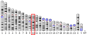
Nodal homolog is a secretory protein that in humans is encoded by the NODAL gene[5][6] which is located on chromosome 10q22.1.[7] It belongs to the transforming growth factor beta superfamily (TGF-β superfamily). Like many other members of this superfamily it is involved in cell differentiation in early embryogenesis, playing a key role in signal transfer from the primitive node, in the anterior primitive streak, to lateral plate mesoderm (LPM).[8][9]
Nodal signaling is important very early in development for mesoderm and endoderm formation and subsequent organization of left-right axial structures.[10][11][12] In addition, Nodal seems to have important functions in neural patterning, stem cell maintenance[7][12] and many other developmental processes, including left/right handedness.[11][13]
Nodal induction of gastrulation
[edit]
The primitive node serves as the primary organizer while producing Nodal, which works as the signaling molecule for early embryonic development and gastrulation. Following the formation of the primitive node, secretion of Nodal induces local cell migration.[14] Secondary signals such as DKK1 enable migration through upregulating or downregulating cell adhesion molecules, thereby allowing movement and association with like cells.[15]
First, cranially or anteriorly, anterior visceral endoderm (AVE) develops as the first wave of Nodal induces migration of visceral endoderm relative to the primitive node. AVE begins secreting inhibitory factors such as Lefty quickly following Nodal expression and works to inhibit Nodal and establish anterior-posterior axis patterning.[15]
As the primitive node extends cranially, epiblast cells exposed to high concentrations of nodal begin initial movement into the primitive streak and become endoderm, while epiblast cells exposed to intermediate concentrations of nodal become mesoderm, and cells that are not stimulated by nodal become ectoderm. This process results in transition from the single layer epiblast into three germ layers of progenitor cells for all other adult body systems. Simultaneous action of cilia on the primitive node surface pushes increased concentrations to the left side of the embryo, establishing the left-right concentration gradient preceding asymmetrical organogenesis in later development due to downstream signaling cascades. Absence of Nodal leads to failed gastrulation and nonviability.[14][15]
Nodal can bind type I and type II serine/threonine kinase receptors, with Cripto-1 acting as its co-receptor.[16] Signaling through SMAD 2/3 and subsequent translocation of SMAD 4 to the nucleus promotes the expression of genes involved in proliferation and differentiation.[7] Nodal also further activates its own expression via a positive feedback loop.[12][16] It is tightly regulated by inhibitors Lefty A, Lefty B, Cerberus, and Tomoregulin-1, which can interfere with Nodal receptor binding.[9][12]
Species specific Nodal ligands
[edit]
Nodal is a widely distributed cytokine.[17] The presence of Nodal is not limited to vertebrates, it is also known to be conserved in other deuterostomes (cephalochordates, tunicates and echinoderms) and protostomes such as snails, but neither the nematode C. elegans (another protosome) nor the fruit fly Drosophila (an arthropod) have a copy of nodal.[18][19] Although mouse and human only have one nodal gene, the zebrafish contain three nodal paralogs: squint, cyclops and southpaw, and the frog five (xnr1,2,3,5 and 6). Even though the zebrafish Nodal homologs are very similar, they have specialized to perform different roles; for instance, Squint and Cyclops are important for mesoendoderm formation, whereas the Southpaw has a major role in asymmetric heart morphogenesis and visceral left-right asymmetry.[20] Another example of protein speciation is the case of the frog where Xnr1 and Xnr2 regulate movements in gastrulation in contrast to Xnr5 and Xnr6 that are involved in mesoderm induction.[21] In mouse, Nodal has been implicated in left-right asymmetry, neural pattering and mesoderm induction (see nodal signaling).
Nodal signaling regulates mesoderm formation in a species-specific manner. Thus, in Xenopus, Xnr controls dorso-ventral mesoderm formation along the marginal zone. In zebrafish, Squint and Cyclops are responsible for animal-vegetal mesoderm formation. In chicken and mouse, Vg1 and Nodal respectively promote primitive streak formation in the epiblast.[12] In chick development, Nodal is expressed in Koller's sickle.[22] Studies have shown that a nodal knockout in mouse causes the absence of the primitive streak and failure in the formation of mesoderm, leading to developmental arrest just after gastrulation.[23][24][25]
Compared to mesoderm specification, endoderm specification requires a higher expression of Nodal. Here, Nodal stimulates mixer homeoproteins, which can interact with SMADs in order to up-regulate endoderm specific genes and repress mesoderm specific genes.[12]
Left-right asymmetry (LR asymmetry) of visceral organs in vertebrates is also established through nodal signaling. Whereas Nodal is initially symmetrically expressed in the embryo, after gastrulation, Nodal becomes asymmetrically restricted to the left side of the organism.[7][12] It is highly conserved among deuterostomes.[26][27] An ortholog of Nodal was found in snails and was shown to be involved in left-right asymmetry as well in 2008.[27]
In order to enable anterior neural tissue development, Nodal signaling needs to be repressed after inducing mesendoderm and LR asymmetry.[12][16]
Recent research on mouse and human embryonic stem cells (hESCs) indicates that Nodal seems to be involved in the maintenance of stem cell self-renewal and pluripotent potentials.[7][12][28][29] Thus, overexpression of Nodal in hESCs lead to the repression of cell differentiation.[12] On the contrary, inhibition of Nodal and Activin signaling enabled the differentiation of hESCs.[7]
- ^ a b c GRCh38: Ensembl release 89: ENSG00000156574 – Ensembl, May 2017
- ^ a b c GRCm38: Ensembl release 89: ENSMUSG00000037171 – Ensembl, May 2017
- ^ "Human PubMed Reference:". National Center for Biotechnology Information, U.S. National Library of Medicine.
- ^ "Mouse PubMed Reference:". National Center for Biotechnology Information, U.S. National Library of Medicine.
- ^ Gebbia M, Ferrero GB, Pilia G, Bassi MT, Aylsworth A, Penman-Splitt M, et al. (November 1997). "X-linked situs abnormalities result from mutations in ZIC3". Nature Genetics. 17 (3): 305–308. doi:10.1038/ng1197-305. PMID 9354794. S2CID 22916101.
- ^ "NODAL - Nodal homolog precursor - Homo sapiens (Human) - NODAL gene & protein". www.uniprot.org. Archived from the original on 30 March 2022. Retrieved 30 March 2022.
- ^ a b c d e f Strizzi L, Postovit LM, Margaryan NV, Seftor EA, Abbott DE, Seftor RE, et al. (2008). "Emerging roles of nodal and Cripto-1: from embryogenesis to breast cancer progression". Breast Disease. 29: 91–103. doi:10.3233/bd-2008-29110. PMC 3175751. PMID 19029628.
- ^ Kawasumi A, Nakamura T, Iwai N, Yashiro K, Saijoh Y, Belo JA, et al. (May 2011). "Left-right asymmetry in the level of active Nodal protein produced in the node is translated into left-right asymmetry in the lateral plate of mouse embryos". Developmental Biology. 353 (2): 321–330. doi:10.1016/j.ydbio.2011.03.009. PMC 4134472. PMID 21419113.
- ^ a b Branford WW, Yost HJ (May 2004). "Nodal signaling: CrypticLefty mechanism of antagonism decoded". Current Biology. 14 (9): R341 – R343. Bibcode:2004CBio...14.R341B. doi:10.1016/j.cub.2004.04.020. PMID 15120085.
- ^ "Entrez Gene: NODAL nodal homolog (mouse)".
- ^ a b Dougan ST, Warga RM, Kane DA, Schier AF, Talbot WS (May 2003). "The role of the zebrafish nodal-related genes squint and cyclops in patterning of mesendoderm". Development. 130 (9): 1837–1851. doi:10.1242/dev.00400. PMID 12642489.
- ^ a b c d e f g h i j Shen MM (March 2007). "Nodal signaling: developmental roles and regulation". Development. 134 (6): 1023–1034. doi:10.1242/dev.000166. PMID 17287255.
- ^ Brandler WM, Morris AP, Evans DM, Scerri TS, Kemp JP, Timpson NJ, et al. (September 2013). "Common variants in left/right asymmetry genes and pathways are associated with relative hand skill". PLOS Genetics. 9 (9): e1003751. doi:10.1371/journal.pgen.1003751. PMC 3772043. PMID 24068947.
- ^ a b Robertson EJ (August 2014). "Dose-dependent Nodal/Smad signals pattern the early mouse embryo". Seminars in Cell & Developmental Biology. RNA biogenesis & TGFβ signalling in embryonic development. 32: 73–79. doi:10.1016/j.semcdb.2014.03.028. PMID 24704361. Archived from the original on 2023-04-08. Retrieved 2024-04-16.
- ^ a b c Kumar A, Lualdi M, Lyozin GT, Sharma P, Loncarek J, Fu XY, et al. (April 2015). "Nodal signaling from the visceral endoderm is required to maintain Nodal gene expression in the epiblast and drive DVE/AVE migration". Developmental Biology. 400 (1): 1–9. doi:10.1016/j.ydbio.2014.12.016. PMC 4806383. PMID 25536399.
- ^ a b c Schier AF (Aug 2003). "Nodal signaling in vertebrate development". Annual Review of Cell and Developmental Biology. 19: 589–621. doi:10.1146/annurev.cellbio.19.041603.094522. PMID 14570583.
- ^ Chen, Hsu-Hsin, Geijsen, Neils (2006). "Signaling germline commitment". In Simón, Carlos, Pellicer, Antonio (eds.). Stem cells in human reproduction: basic science and therapeutic potential. CRC Press. p. 74. ISBN 978-0-415-39777-3.
- ^ Chea HK, Wright CV, Swalla BJ (October 2005). "Nodal signaling and the evolution of deuterostome gastrulation". Developmental Dynamics. 234 (2): 269–278. doi:10.1002/dvdy.20549. PMID 16127715. S2CID 24982316.
- ^ Schier AF (November 2009). "Nodal morphogens". Cold Spring Harbor Perspectives in Biology. 1 (5): a003459. doi:10.1101/cshperspect.a003459. PMC 2773646. PMID 20066122.
- ^ Baker K, Holtzman NG, Burdine RD (September 2008). "Direct and indirect roles for Nodal signaling in two axis conversions during asymmetric morphogenesis of the zebrafish heart". Proceedings of the National Academy of Sciences of the United States of America. 105 (37): 13924–13929. Bibcode:2008PNAS..10513924B. doi:10.1073/pnas.0802159105. PMC 2544555. PMID 18784369.
- ^ Luxardi G, Marchal L, Thomé V, Kodjabachian L (February 2010). "Distinct Xenopus Nodal ligands sequentially induce mesendoderm and control gastrulation movements in parallel to the Wnt/PCP pathway". Development. 137 (3): 417–426. doi:10.1242/dev.039735. PMID 20056679.
- ^ Schnell S (18 December 2007). Multiscale Modeling of Developmental Systems. Academic Press. ISBN 978-0-08-055653-6. Retrieved 7 December 2013.
- ^ Conlon FL, Lyons KM, Takaesu N, Barth KS, Kispert A, Herrmann B, et al. (July 1994). "A primary requirement for nodal in the formation and maintenance of the primitive streak in the mouse". Development. 120 (7): 1919–1928. doi:10.1242/dev.120.7.1919. PMID 7924997.
- ^ Zhou X, Sasaki H, Lowe L, Hogan BL, Kuehn MR (February 1993). "Nodal is a novel TGF-beta-like gene expressed in the mouse node during gastrulation". Nature. 361 (6412): 543–547. Bibcode:1993Natur.361..543Z. doi:10.1038/361543a0. PMID 8429908. S2CID 4318909. Archived from the original on 2022-01-28. Retrieved 2019-09-11.
- ^ Reissmann E, Jörnvall H, Blokzijl A, Andersson O, Chang C, Minchiotti G, et al. (August 2001). "The orphan receptor ALK7 and the Activin receptor ALK4 mediate signaling by Nodal proteins during vertebrate development". Genes & Development. 15 (15): 2010–2022. doi:10.1101/gad.201801. PMC 312747. PMID 11485994.
- ^ Hamada H, Meno C, Watanabe D, Saijoh Y (February 2002). "Establishment of vertebrate left-right asymmetry". Nature Reviews. Genetics. 3 (2): 103–113. doi:10.1038/nrg732. PMID 11836504. S2CID 20557143.
- ^ a b Grande C, Patel NH (February 2009). "Nodal signalling is involved in left-right asymmetry in snails". Nature. 457 (7232): 1007–1011. Bibcode:2009Natur.457.1007G. doi:10.1038/nature07603. PMC 2661027. PMID 19098895.
- ^ Chng Z, Vallier L, Pedersen R (2011). "Activin/nodal signaling and pluripotency". Vitamins and Hormones. 85: 39–58. doi:10.1016/B978-0-12-385961-7.00003-2. ISBN 978-0-12-385961-7. PMID 21353875.
- ^ Fei T, Chen YG (April 2010). "Regulation of embryonic stem cell self-renewal and differentiation by TGF-beta family signaling". Science China. Life Sciences. 53 (4): 497–503. doi:10.1007/s11427-010-0096-2. PMID 20596917. S2CID 9074927.
Postovit LM, Seftor EA, Seftor RE, Hendrix MJ (April 2007). "Targeting Nodal in malignant melanoma cells". Expert Opinion on Therapeutic Targets. 11 (4): 497–505. doi:10.1517/14728222.11.4.497. PMID 17373879. S2CID 43180068.
Yan YT, Liu JJ, Luo Y, E C, Haltiwanger RS, Abate-Shen C, et al. (July 2002). "Dual roles of Cripto as a ligand and coreceptor in the nodal signaling pathway". Molecular and Cellular Biology. 22 (13): 4439–4449. doi:10.1128/MCB.22.13.4439-4449.2002. PMC 133918. PMID 12052855.
Roberts HJ, Hu S, Qiu Q, Leung PC, Caniggia I, Gruslin A, et al. (May 2003). "Identification of novel isoforms of activin receptor-like kinase 7 (ALK7) generated by alternative splicing and expression of ALK7 and its ligand, Nodal, in human placenta". Biology of Reproduction. 68 (5): 1719–1726. doi:10.1095/biolreprod.102.013045. PMID 12606401.
Munir S, Xu G, Wu Y, Yang B, Lala PK, Peng C (July 2004). "Nodal and ALK7 inhibit proliferation and induce apoptosis in human trophoblast cells". The Journal of Biological Chemistry. 279 (30): 31277–31286. doi:10.1074/jbc.M400641200. PMID 15150278.
Haffner C, Frauli M, Topp S, Irmler M, Hofmann K, Regula JT, et al. (August 2004). "Nicalin and its binding partner Nomo are novel Nodal signaling antagonists". The EMBO Journal. 23 (15): 3041–3050. doi:10.1038/sj.emboj.7600307. PMC 514924. PMID 15257293.
Besser D (October 2004). "Expression of nodal, lefty-a, and lefty-B in undifferentiated human embryonic stem cells requires activation of Smad2/3". The Journal of Biological Chemistry. 279 (43): 45076–45084. doi:10.1074/jbc.M404979200. PMID 15308665.
Bamforth SD, Bragança J, Farthing CR, Schneider JE, Broadbent C, Michell AC, et al. (November 2004). "Cited2 controls left-right patterning and heart development through a Nodal-Pitx2c pathway". Nature Genetics. 36 (11): 1189–1196. doi:10.1038/ng1446. PMID 15475956.
Vallier L, Reynolds D, Pedersen RA (November 2004). "Nodal inhibits differentiation of human embryonic stem cells along the neuroectodermal default pathway". Developmental Biology. 275 (2): 403–421. doi:10.1016/j.ydbio.2004.08.031. PMID 15501227.
Hart AH, Willson TA, Wong M, Parker K, Robb L (August 2005). "Transcriptional regulation of the homeobox gene Mixl1 by TGF-beta and FoxH1". Biochemical and Biophysical Research Communications. 333 (4): 1361–1369. doi:10.1016/j.bbrc.2005.06.044. PMID 15982639.
Vallier L, Alexander M, Pedersen RA (October 2005). "Activin/Nodal and FGF pathways cooperate to maintain pluripotency of human embryonic stem cells". Journal of Cell Science. 118 (Pt 19): 4495–4509. doi:10.1242/jcs.02553. PMID 16179608.
Strizzi L, Postovit LM, Margaryan NV, Seftor EA, Abbott DE, Seftor RE, et al. (2008). "Emerging roles of nodal and Cripto-1: from embryogenesis to breast cancer progression". Breast Disease. 29: 91–103. doi:10.3233/BD-2008-29110. PMC 3175751. PMID 19029628.
Branford WW, Yost HJ (May 2004). "Nodal signaling: CrypticLefty mechanism of antagonism decoded". Current Biology. 14 (9): R341 – R343. Bibcode:2004CBio...14.R341B. doi:10.1016/j.cub.2004.04.020. PMID 15120085.
Shen MM (March 2007). "Nodal signaling: developmental roles and regulation". Development. 134 (6): 1023–1034. doi:10.1242/dev.000166. PMID 17287255.
Schier AF (Aug 2003). "Nodal signaling in vertebrate development". Annual Review of Cell and Developmental Biology. 19: 589–621. doi:10.1146/annurev.cellbio.19.041603.094522. PMID 14570583.
Chng Z, Vallier L, Pedersen R (2011). "Activin/nodal signaling and pluripotency". Vitamins and Hormones. 85: 39–58. doi:10.1016/B978-0-12-385961-7.00003-2. ISBN 978-0-12-385961-7. PMID 21353875.
Fei T, Chen YG (April 2010). "Regulation of embryonic stem cell self-renewal and differentiation by TGF-beta family signaling". Science China. Life Sciences. 53 (4): 497–503. doi:10.1007/s11427-010-0096-2. PMID 20596917. S2CID 9074927.
nodal+protein at the U.S. National Library of Medicine Medical Subject Headings (MeSH)


