Cell the Unit of Life Class 11 Notes CBSE Biology Chapter 8 (original) (raw)
Last Updated : 23 Jul, 2025
**Class 11 Biology NCERT Notes for Chapter 8 Cell The Unit of Life: Cells are the basic units of life and serve as the building blocks of all living organisms. They differ in structure, composition, and function, but have similarities. The human body is made up of more than trillions of cells. They give structure to the body, extract nutrients from food, convert those nutrients into energy, and perform specific functions.
**NCERT Notes for Chapter 8 Cell The Unit of Life further explains thatcells also contain the body's genetic material and can make copies of themselves. A bacterium or yeast is often a distinct, complete organism as it is single-celled or unicellular. Other cells acquire special functions as they mature. Understanding the structure and function of cells is crucial for comprehending the complexity of biological systems.
Table of Content
- Discovery of Cell
- Cell Theory
- Overview of Cell
- Cell Structure and Function
- Structure of Cell Wall
- Functions of Cell Wall
- Endomembrane System
- Mitochondria
- Conclusion - Cell the Unit of Life Class 11 Notes
- FAQs on Cell Structure and Function
Discovery of Cell
Without the development of the microscope, the discovery of the cell would not have been possible. Interested in learning more about the microscopic world, scientist Robert Hooke perfected the existing compound microscope design in 1665. He placed a piece of cork under the microscope. To him, the cork appeared to be made up of tiny pores, which he called "cells."
Shortly after Hooke's discovery, Dutch scientist Antonie van Leeuwenhoek discovered tinier hidden organisms, bacteria, and protozoa. He was a master microscope maker and perfected the design of a simple microscope, which had only one lens, allowing it to magnify an object anywhere from two to three hundred times its original size.
Cell Theory
In biology, the cell theory is a scientific theory that has three principles: First part was formulated by Theodor Schwann in 1839 which states that organisms are made up of cells.
Based on a conclusion made by Schwann and Matthias Schleiden, after comparing their observations of plant and animal cells, they put forward the second part of the cell theory. It states that cells are the basic structural/organizational unit of all organisms. The third part, which was described by Rudolf Virchow in 1858, asserts that cells come from pre-existing cells.
Overview of Cell
Cells are divided into two broad categories: Prokaryotic and Eukaryotic. The unicellular organisms of the kingdom Bacteria and Archaea are classified as Prokaryotes. Whereas, animal cells, plant cells, fungi, and protists are eukaryotes. All cells share four common components:
- a plasma membrane, an outer covering that separates the cell’s interior from its surrounding environment;
- Cytoplasm, consisting of a jelly-like region within the cell in which other cellular components are found;
- DNA, the genetic material of the cell; and
- ribosomes, particles that synthesize proteins. However, eukaryotes differ from prokaryotes in several ways.
**Also read: Difference between Prokaryotic and Eukaryotic Cells
**Prokaryotic Cell
A **prokaryotic cell is a single-celled or unicellular organism that has neither a true nucleus nor membrane-bound organelles. Organisms belonging to the Bacteria and Archaea domains are based on the prokaryotic cell. Prokaryotic cells are covered with a cell membrane which acts as an extra layer of protection. It also helps the cell maintain its shape and prevents dehydration. These cells contain both free DNA and ribosomes. Although ribosomes are organelles, they are not attached to the plasma membrane.
**Eukaryotic Cell
Any cell or organism with a clearly defined nucleus is called a **eukaryotic cell. A eukaryotic cell has a nuclear membrane that surrounds the nucleus and contains well-defined chromosomes. Eukaryotic cells also contain organelles, including mitochondria, Golgi apparatus, endoplasmic reticulum, and lysosomes. Eukaryotic cells are larger and more complex than prokaryotic cells, which are found in the archaeal and bacterial domains.

Prokaryotic and Eukaryotic Cells
Cell Structure and Function
The cell structure consists of individual elements with specific functions. These components include the cell membrane, cell wall, cytoplasm, nucleus, and cell organelles. Let's study them in detail:
**Cell Membrane
The cell membrane, also called the **plasma membrane, is the membrane present in all cells that separate the cell interior from the outside environment. In bacterial and plant cells, the cell wall is attached to the outer surface of the cell membrane.

**Structure of Cell Membrane
The main components of the plasma membrane are phospholipids, proteins, and carbohydrate groups related to certain lipids and proteins.
- **Phospholipids: The membrane is made up in part of molecules called phospholipids, which spontaneously assemble into a bilayer with hydrophilic (water-loving) heads on the outside or exterior of the bilayer and hydrophobic (water-hating) tails are on the interior or inside of the bilayer. These interactions with water allow for the formation of plasma membranes.
- **Proteins: Proteins lie between the lipids that make up the membrane, and these transmembrane proteins allow the passage of molecules that otherwise would not be able to enter the cell, creating channels, pores, or gates. The cell thus controls the flow of these molecules as they enter and exit. Cell membrane proteins play a role in many other functions, such as cell signaling, cell recognition, and enzyme activity.
- **Carbohydrates: Carbohydrates are also found in the plasma membrane. In particular, most membrane carbohydrates are part of the glycoproteins that are formed when a carbohydrate binds to a protein. Glycoproteins play a role in cell-cell interactions, including cell adhesion, the process by which cells stick together.
**Fluid Mosaic Model
In 1972, **Singer and Nicolson proposed a flowing mosaic pattern. According to this model, the near-liquid nature of lipids allows lateral movement of proteins across the bilayer. This ability to move within the membrane is measured by its fluidity. The plasma membrane consists of lipids arranged in bilayers and within the membrane, with the pole head pointing outward and the hydrophilic tails pointing inward. This protects the non-polar tail of the saturated hydrocarbons from the aquatic environment. The cell membrane is comprised of 52% proteins and 40% lipids. Peripheral proteins reside on the surface of the membrane while integral proteins are partially or fully buried within the membrane.
**Functions of Cell Membrane
Some of the activities performed by the cell membrane are described below:
- **Physical barrier: The plasma membrane separates the cytoplasm from the extracellular fluid outside the cell, protecting all cellular components from the external environment and allowing separate activities inside and outside the cell.
- **Structural Support: Protects the cytoskeleton, a network of protein fibers within the cell that holds all parts of the cell together and gives the cell its shape.
- **Selective Permeability: Plasma membranes are selectively permeable (or semipermeable), meaning that only molecules such as water, oxygen, and carbon dioxide can pass through them with ease. But the membrane can control how fast certain molecules, such as sodium, potassium, and other polar molecules, can move in and out of the cell.
- **Endocytosis and exocytosis: Endocytosis allows a cell to take up large amounts of molecules or even whole bacteria from the extracellular fluid. Exocytosis occurs when the cell releases these materials. The cell membrane plays an important role in both processes.
- **Cell signaling: Another important function of the membrane is to facilitate communication and signaling between cells. It does this by using different proteins and carbohydrates in the membrane.
**What is Cell Wall?
Cell walls are the hard, inanimate objects that make up the plasma membrane's outer covering in fungi and plants. The majority of plant cells, as well as those of fungi, bacteria, algae, and some archaea, have this outer layer right near the cell membrane. Animal cells, however, do not have a cell wall.
Structure of Cell Wall
Different organisms have different cell walls with different compositions. Strong fibers made of cellulose, glucose, and polymer make up the majority of the cell wall in plants. Sugars and peptidoglycan, a polymer of amino acids, make up the bacterial cell wall. Chitin, glucans, and proteins make up the bulk of fungal cell walls. The plant's cell wall has multiple layers and is divided into these three sections:
- **Middle lamella: Polysaccharides known as pectins are found in this layer of the outer cell wall. Pectins facilitate cell adhesion by assisting the adherence of the cell walls of surrounding cells.
- **Primary cell wall: In developing plant cells, this layer develops between the middle lamella and plasma membrane. Hemicellulose fibers and pectin polysaccharides form a gel-like matrix that is predominantly made up of cellulose microfibrils. The basic cell wall offers the durability and adaptability required for cell development.
- **Secondary cell wall: In some plant cells, this layer develops between the primary cell wall and plasma membrane. Some secondary cell walls furthermore include lignin in addition to cellulose and hemicellulose. Lignin helps plant vascular tissue cells transmit water and reinforces the cell wall.

Cell Wall
Functions of Cell Wall
A cell's cell wall serves a variety of vital roles, including support, structure, and protection.
- **Support: The cell wall offers support and mechanical strength. It also regulates the way that cells grow.
- **Control cell growth: To divide and expand, cells must enter the cell cycle, which is signaled by the cell wall.
- **Control diffusion: Because the cell wall is permeable, some molecules, such as proteins, can enter the cell while blocking the entry of others.
- **Communication: Plant cell walls have pores or channels known as plasmodesmata that allow chemicals and communication impulses to move between individual plant cells. These channels or pores allow cells to communicate with one another.
- **Defense: The cell wall acts as a barrier against pathogens such as plant viruses. It also aids in stopping water loss.
- **Storage: Carbohydrates are kept in the cell wall for usage throughout plant growth, particularly in seeds.
Endomembrane System
The endoplasmic reticulum (ER), Golgi Complex, Lysosomes, and Vacuoles are components of the endomembrane system.
Endoplasmic Reticulum (ER)
Endoplasmic Reticulum is a network or reticulum of minute tubular structures that are dispersed throughout the cytoplasm. The two forms of ER, known as rough ER and smooth ER, can be distinguished by differences in certain morphological and functional properties.
- **Rough Endoplasmic Reticulum: RER refers to the endoplasmic reticulum that has ribosomes on its surface.
- **Smooth Endoplasmic Reticulum: These are known as smooth endoplasmic reticulum (SER) when there are no ribosomes present.

**Functions of Endoplasmic Reticulum
- RER is typically seen in cells that are actively producing and secreting proteins. They extend far and are contiguous with the nucleus's outer membrane.
- The principal location for lipid synthesis is the smooth endoplasmic reticulum. Steroid hormones that resemble lipids are produced in animal cells by the SER.
**Golgi Apparatus
A Golgi body, often referred to as a Golgi apparatus, is an organelle found in cells that aid in the processing and packaging of proteins and lipid molecules, particularly proteins intended for cell export. The Camillo Golgi-named Golgi body consists of a collection of membranes.
**Structure of Golgi Apparatus
They are composed of several flat, disc-shaped sacs or cisternae that range in diameter from 0.5 to 1.0 m. These are stacked parallel to one another. There are different amounts of cisternae in a Golgi complex. The developing face of the Golgi cisternae is convex, and the mature face is concave, and they are concentrated close to the nucleus. The cis and trans faces of the organelle are entirely separate but connected.
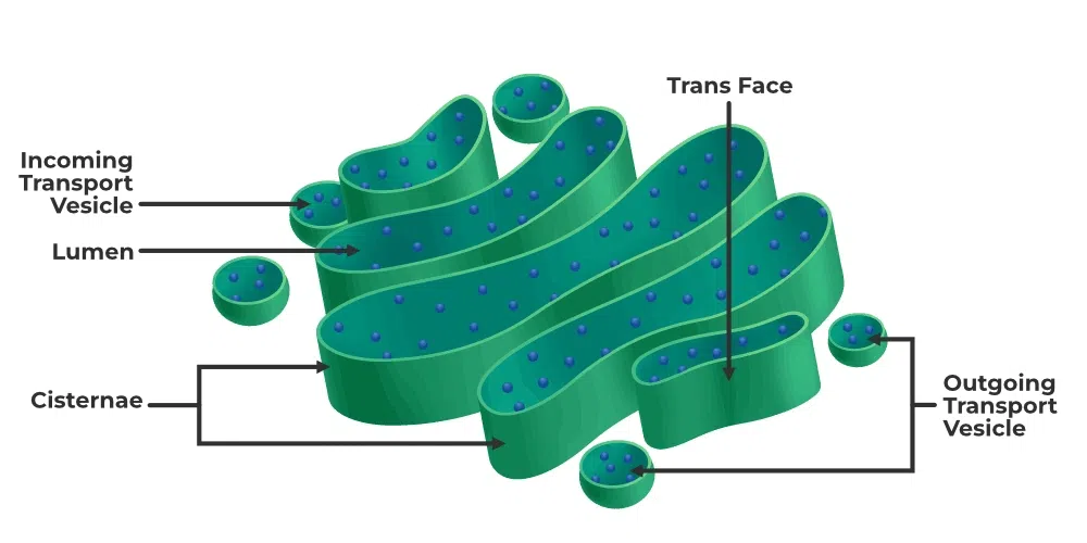
**Functions of Golgi Apparatus
- The primary site of glycoprotein and glycolipid synthesis is the Golgi apparatus.
- Its primary function is to bundle chemicals for delivery to intracellular destinations or for secretion outside the cell.
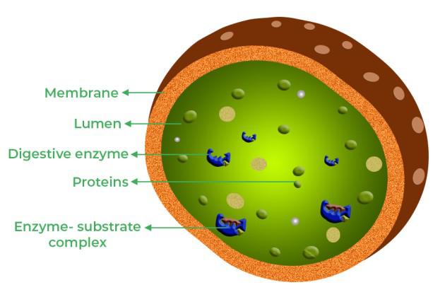
**Lysosomes
Lysosomes are membrane-bound vesicles created during the Golgi apparatus packing process. It has been discovered that the isolated lysosomal vesicles are extremely rich in practically all varieties of hydrolytic enzymes that are most active at the acidic pH.
**Functions of Lysosome
Lysosomal vesicles contain hydrolytic enzymes that can break down lipids, proteins, carbohydrates, and nucleic acids.
**Vacuoles
The membrane-bound compartment in the cytoplasm is known as the **vacuole. Water, sap, excretory material, and other substances are present. Tonoplast is the given name of the isolated membrane that surrounds the vacuole. In plant cells, vacuoles possess the capacity to consume up to 90% of the entire cell volume.
**Functions of Vacoule
- In plants, the tonoplast helps with the movement of various ions and other substances up concentration gradients and into the vacuole.
- The contractile vacuole in amoebas is crucial for excretion and osmoregulation. Food vacuoles are created by enveloping the food particles in numerous cells, just like in protists.
**Mitochondria
The rod-shaped organelles known as **mitochondria serve as the cell's power house by generating adenosine triphosphate (ATP) from oxygen and nutrients.
Also Read: Why is Mitochondria known as Power house of the Cell?
**Structure of Mitochondria
Typically, mitochondria are cylindrical or sausage-shaped, measuring 1.0–4.1 m in length and 0.2–1.0 m in diameter. Each mitochondrion is a structure that is double-membrane bound, with the outer membrane and the inner membrane partitioning the lumen into the outer compartment and the inner compartment, respectively. The matrix, a dense, uniform material, fills the interior compartment. The organelle's continuous limiting boundary is formed by the outer membrane. The cristae are a series of infoldings that the inner membrane forms as it moves toward the matrix.
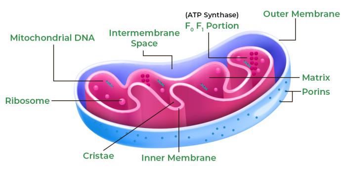
**Functions of Mitochondria
Mitochondria carry out the following functions:
- The locations of aerobic respiration are mitochondria.
- Mitochondria also provide cellular energy in the form of ATP.
- The matrix also contains a single circular DNA molecule, a few RNA molecules, ribosomes, and the elements necessary for protein synthesis.
- Contributes to the process of apoptosis, or programmed cell death.
- Aids in the liver cells' ability to detoxify ammonia.
**Plastids
Plastids, which are found in plant cells and euglenoids, are double-membrane bound organelles with their own DNA and ribosomes.
**Structure & Functions of Plastid
Due to their dimensions, these can be viewed easily under a microscope. They carry unique pigments, that provide the plants with distinctive colors. Plastids can be divided into chloroplasts, chromoplasts, and leucoplasts according to the kind of pigment they carry.
- **Chloroplasts: Similar to mitochondria, chloroplasts are a double membrane-bound structure. The area known as the stroma is the part of the chloroplast that is limited by its own inner membrane. The stroma contains a variety of thylakoids, which are structured flattened membranous sacs. The stroma lamellae, which connect the thylakoids of the various grana, are flat membrane tubules as well. A region known as the lumen is enclosed by the membrane of the thylakoids. The pigments chlorophyll and carotenoids found in chloroplasts are in charge of capturing the light energy required for photosynthesis.
- **Chromoplasts: Carotene, xanthophylls, and other fat-soluble carotenoid pigments are found in chromoplasts. As a result, the plant portion has a yellow, orange, or red coloring.
- **Leucoplasts: These nutrient-storing, colorless plastids come in a variety of sizes and forms. They are known as Amyloplasts, which store carbohydrates like potatoes, Elaioplasts, which store oils and fats, and Aleuroplasts, which store proteins when they are specialized for bulk storage of starch, lipid, or protein.

**Ribosomes
The place where protein synthesis occurs in cells is called a **ribosome, an intercellular structure consisting of both RNA and protein.
In the absence of a membrane, ribosomes are granular structures made of ribonucleic acid (RNA) and proteins. In contrast to bacterial ribosomes, which are 70S, eukaryotic ribosomes are 80S. The larger and smaller subunits of each ribosome make up the ribosome. For 80S ribosomes, the two subunits are 60S and 40S, but for 70S ribosomes, they are 50S and 30S. The sedimentation coefficient, abbreviated as "S" (Svedberg's Unit), is a measure of density and size here. Two subunits make up the 70S and 80S ribosomes, respectively.

**Cytoskeleton
The term "**cytoskeleton" refers to the complex web of proteinaceous filaments found in the cytoplasm, including microtubules, intermediate filaments, and microfilaments.
A cell's cytoskeleton performs a variety of tasks, including movement, mechanical support, and maintaining the shape of the cell.
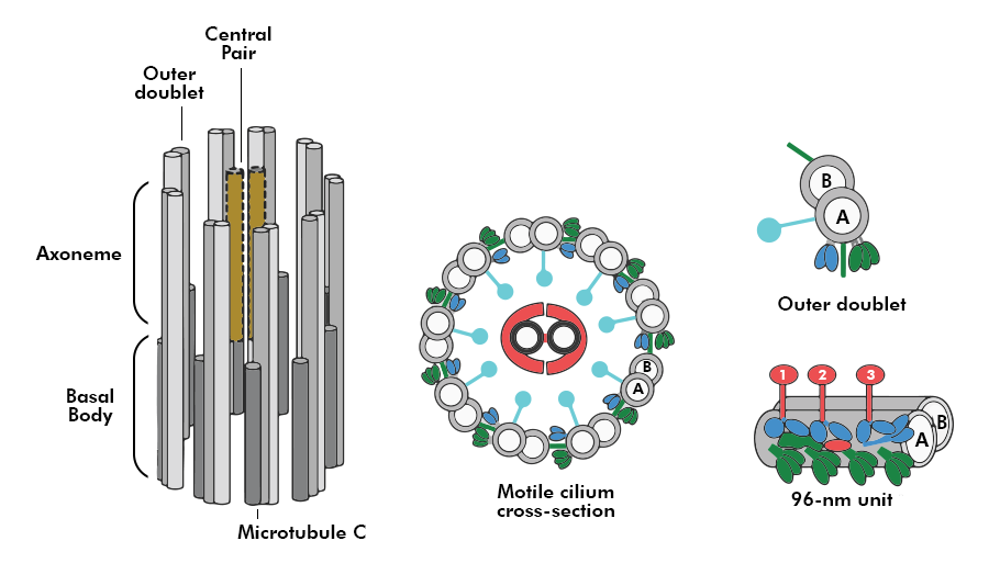
**Cilia and Flagella
Cilia and flagella are hair-like extensions of the cell membrane. Cilia are tiny structures that aid in the circulation of the fluid surrounding the cell or the cell itself. Flagella, which are significantly longer, are what move cells.
The plasma membrane covers the cilium or flagellum. Many microtubules at their center, known as the axoneme, are parallel to the long axis. The axoneme typically has two microtubules in the center and nine doublets of peripheral microtubules organized radially (9+2 array).
A radial spoke connects one of each peripheral doublet's tubules to the central tubules, which are connected by bridges and likewise encased by a central sheath. There are nine radial spokes as a result. Additionally, linkers are used to connect the peripheral doublets. The basal bodies, a centriole-like structure, give rise to both the cilium and flagellum.
 **Centrosome and Centrioles
A centrosome is an organelle that typically has two centrioles, which are cylindrical structures. In a centrosome, both centrioles are perpendicular to one another and are organized similarly to a cartwheel. They are composed of nine peripheral tubulin protein fibrils that are uniformly spaced apart.
The peripheral fibrils are triplets in each. The triplets next door are connected as well. The hub, which connects with tubules of the peripheral triplets by radial spokes likewise formed of protein, is the center portion of the proximal section of the centriole. When animal cells divide, the centrioles create the spindle fibers that become the spindle apparatus as well as the basal body of cilia or flagella.
**Also Read: Centrosome and Centriole

**Nucleus
The cell nucleus is a membrane-bound structure that stores the genetic material of the cell and regulates its growth and procreation.
**Structure of Nucleus
The nuclear membrane, nucleoplasm, chromosomes, and nucleolus are all parts of the nucleus' structure.
- **Nuclear Membrane: This two-layered membrane protects the nucleus's contents. The endoplasmic reticulum is joined to the membrane's outer layer. The nuclear envelope is made up of phospholipids that form a lipid bilayer, just like the cell membrane.
- **Nucleoplasm: The gelatinous material found inside the nuclear membrane is known as nucleoplasm. This semi-aqueous substance, also known as karyoplasm, is similar to the cytoplasm in that it mostly consists of water with dissolved salts, enzymes, and organic molecules suspended inside. Nucleoplasm surrounds the nucleolus and chromosomes.
- **Chromosomes: Chromosomes are an organelle found in the nucleus. DNA, which makes up chromosomes and carries instructions for cell development, growth, and reproduction, is what determines a person's genetic makeup. Histones, a type of protein molecule, and DNA strings, collectively known as chromatin, make up chromosomes.
- **Nucleolus: The nucleolus is a dense, membrane-less structure found inside the nucleus that is made up of RNA and proteins. Nucleolar organizers, which are chromosomal regions having the genes for ribosome synthesis on them, are found in the nucleolus.

**Functions of Nucleus
The nucleus has several crucial functions:
- It regulates an organism's inherited traits.
- Is also in charge of protein synthesis; cell division; growth; and differentiation.
- Messenger RNA (mRNA) is created for protein synthesis at a transcription site in the nucleus.
- The nucleolus is where ribosomes are created.
- Nuclear pores provide for the selective transit of energy molecules and regulatory components.
**Microbodies
Both plan and animal cells include a large number of membrane-bound tiny vesicles called microbodies that contain a variety of enzymes.
Conclusion - Cell the Unit of Life Class 11 Notes
In conclusion, the **cell is the unit of life. Cell organelles play vital roles in maintaining the functions of living organisms. The structure of cell includes various cell organelles and they have specific function which contributes to the overall health and survival of the cell. Understanding the cell structure function helps us get better knowledge of working of the cell as the unit of life.
**Also Read: